. 2009 Apr 20;185(2):357-70.
doi: 10.1083/jcb.200809110. Epub 2009 Apr 13.

FAK alters invadopodia and focal adhesion composition and dynamics to regulate breast cancer invasion

Affiliations

FAK alters invadopodia and focal adhesion composition and dynamics to regulate breast cancer invasion

Keefe T Chan et al. J Cell Biol. .

Abstract

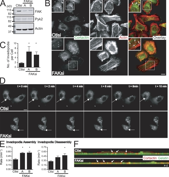
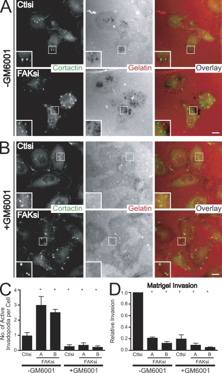
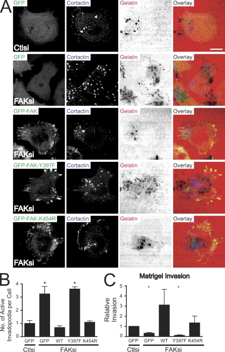
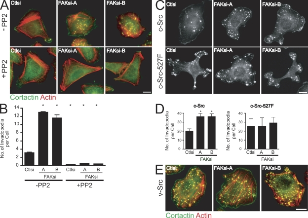
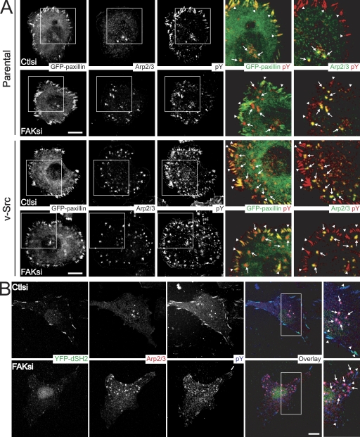
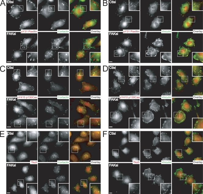
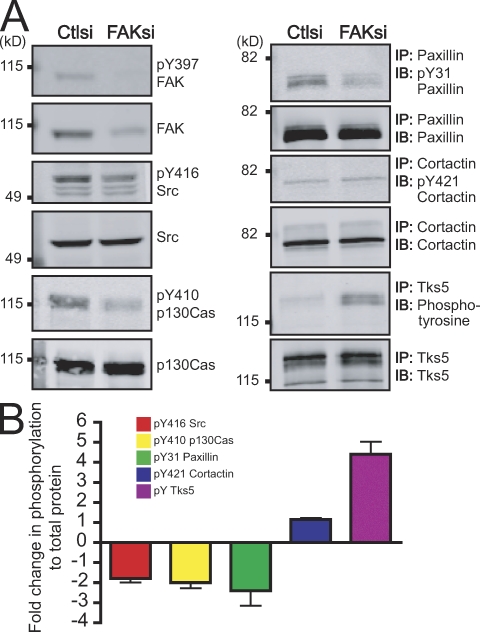
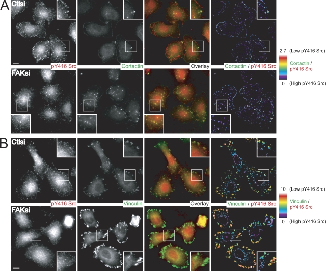
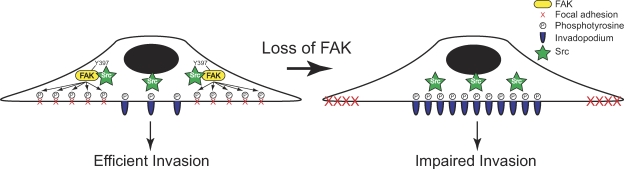
Focal adhesion kinase (FAK) is important for breast cancer progression and invasion and is necessary for the dynamic turnover of focal adhesions. However, it has not been determined whether FAK also regulates the dynamics of invasive adhesions formed in cancer cells known as invadopodia. In this study, we report that endogenous FAK functions upstream of cellular Src (c-Src) as a negative regulator of invadopodia formation and dynamics in breast cancer cells. We show that depletion of FAK induces the formation of active invadopodia but impairs invasive cell migration. FAK-deficient MTLn3 breast cancer cells display enhanced assembly and dynamics of invadopodia that are rescued by expression of wild-type FAK but not by FAK that cannot be phosphorylated at tyrosine 397. Moreover, our findings demonstrate that FAK depletion switches phosphotyrosine-containing proteins from focal adhesions to invadopodia through the temporal and spatial regulation of c-Src activity. Collectively, our findings provide novel insight into the interplay between FAK and Src to promote invasion.


Figures

Figure 1.
Figure 1.
FAK negatively regulates invadopodia formation and dynamics. (A) Cell lysates from MTLn3 cells transiently transfected with control siRNA (Ctlsi) or FAK siRNA (FAKsi A and FAKsi B) were analyzed by Western blotting and probed for FAK or Pyk2. Actin was probed as a loading control. (B) MTLn3 cells were plated on fibronectin-coated coverslips and stained with anticortactin antibody (green) and rhodamine-phalloidin (red). Boxed regions depict regions of invadopodia shown magnified in insets. (C) Quantification of cortactin- and actin-containing invadopodia is expressed as the mean number of invadopodia per cell. (D) GFP-cortactin was transiently cotransfected with control siRNA or FAK siRNA (FAK siRNA B is shown) into MTLn3 cells. Cells were plated on fibronectin-coated glass-bottomed dishes and analyzed by time-lapse fluorescence microscopy. Time-lapse montages demonstrate representative images of the dynamics of the invadopodia marker GFP-cortactin over a period of 10 min. (E) Rate constants for assembly and disassembly were calculated from plots of fluorescence intensities of GFP-cortactin as described in Materials and methods (Videos 1–3). (F) Representative x-z confocal images of invadopodia in MTLn3 cells show cortactin-containing invadopodia (red) projecting into the gelatin matrix (green). Arrows indicate representative invadopodia. Data shown are means ± SEM of three independent experiments. *, P < 0.05 by t test compared with control siRNA. Bars, 10 µm.
Figure 2.
Figure 2.
Enhanced invadopodia formation in FAK-deficient MTLn3 cells requires MMP activity but is not sufficient for invasion. (A) MTLn3 cells transiently transfected with control siRNA (Ctlsi) or FAK siRNA (FAKsi) were cultured on fibronectin–Alexa Fluor 568 gelatin coverslips in the presence of DMSO vehicle control (−GM6001) and stained with anticortactin antibody (green). (B) MTLn3 cells transiently transfected with control siRNA or FAK siRNA were cultured on fibronectin–Alexa Fluor 568 gelatin-coated coverslips in the presence of 50 µM GM6001 (+GM6001) and stained with anticortactin antibody (green). Boxed regions in A and B depict regions of invadopodia shown magnified in insets. (C) Quantification of active invadopodia, defined as colocalizing areas of cortactin staining and matrix degradation, is expressed as the mean number of active invadopodia per cell. (D) MTLn3 cells transiently transfected with control siRNA or FAK siRNA were cultured in the presence of vehicle control (−GM6001) or 50 µM GM6001 (+GM6001) and were assayed for their ability to invade through Matrigel-coated membranes. Relative invasion was determined by normalizing the number of cells invaded to those transfected with control siRNA (−GM6001). Data shown are means ± SEM of three independent experiments. *, P < 0.05 by one-way ANOVA compared with control siRNA (−GM6001). Bars, 10 µm.
Figure 3.
Figure 3.
Expression of wild-type but not Y397F-FAK restores enhanced matrix-degrading invadopodia and impaired invasion in FAK-deficient MTLn3 cells. (A) MTLn3 cells stably expressing GFP, GFP-FAK (wild type), GFP-FAK-Y397F, and GFP-FAK-K454R were transiently transfected with control siRNA (Ctlsi) or FAK siRNA (FAKsi), cultured on fibronectin–Alexa Fluor 568 gelatin coverslips, and stained with anticortactin antibody (blue). Bar, 10 µm. (B) Quantification of active invadopodia, defined as colocalizing areas of cortactin staining and matrix degradation, is expressed as the mean number of active invadopodia per cell. (C) MTLn3 cells treated as in A were assayed for their ability to invade through Matrigel-coated membranes. Relative invasion was determined by normalizing the number of cells invaded to those transfected with control siRNA. WT, wild type. Data shown are means ± SEM of three independent experiments. *, P < 0.05 by t test compared with GFP-expressing cells transfected with control siRNA.
Figure 4.
Figure 4.
FAK functions upstream of Src kinase to regulate invadopodia formation. (A) MTLn3 cells transiently transfected with control siRNA (Ctlsi) or FAK siRNA (FAKsi-A and FAKsi-B) were cultured on fibronectin-gelatin coverslips in the presence of vehicle control (−PP2) or 2 µM PP2 (+PP2). Cells were stained with anticortactin antibody (green) and rhodamine-phalloidin (red). (B) Quantification of cortactin- and actin-containing invadopodia is expressed as the mean number of invadopodia per cell. (C) MTLn3 cells expressing c-Src or c-Src–527F were transiently transfected with control siRNA or FAK siRNA, cultured on fibronectin-gelatin coverslips, and stained with anticortactin antibody. (D) Quantification of cortactin-containing invadopodia in c-Src– or c-Src–527F-expressing cell lines is expressed as the mean number of invadopodia per cell. (E) MTLn3 cells expressing v-Src were transiently transfected with control siRNA or FAK siRNA and cultured on fibronectin-gelatin coverslips. Cells were stained with anticortactin antibody (green) and rhodamine-phalloidin (red). Data shown are means ± SEM of three independent experiments. *, P < 0.05 by one-way ANOVA compared with control siRNA (−PP2). Bars, 10 µm.
Figure 5.
Figure 5.
FAK differentially regulates tyrosine phosphorylation at focal adhesions and invadopodia. (A) MTLn3 (parental) cells or v-Src–transformed MTLn3 cells transiently transfected with control siRNA (Ctlsi) or FAK siRNA (FAKsi) and cotransfected with GFP-paxillin were cultured on fibronectin-gelatin coverslips and stained with anti-p34Arc (Arp2/3 subunit) antibody and antiphosphotyrosine antibody. Two sets of overlays are depicted to illustrate localization of GFP-paxillin or Arp2/3 (green) with phosphotyrosine (pY) staining (red). (B) MTLn3 cells transiently transfected with control siRNA or FAK siRNA and cotransfected with YFP-dSH2 were cultured on fibronectin-gelatin coverslips and stained with anti-p34Arc antibody (red) and antiphosphotyrosine antibody (blue). Regions outlined by boxes indicate images of invadopodia and focal adhesions. Arrows indicate representative invadopodia, and arrowheads indicate representative focal adhesions. Bars, 10 µm.
Figure 6.
Figure 6.
FAK modulates the localization of pY31-paxillin, pY410 p130Cas, and Tks5 in MTLn3 cells. (A–F) MTLn3 cells transiently transfected with control siRNA (Ctlsi) or FAK siRNA (FAKsi) were cultured on fibronectin-gelatin coverslips and stained with anti-pY31 paxillin (A and B), anti-pY410 p130Cas (C and D), or anti-Tks5/FISH antibody (E and F; red). Cells were costained with the invadopodia marker cortactin (A, C, and E; green) or the focal adhesion marker vinculin (B, D, and F; green). Regions outlined by boxes correspond to magnified images of pY31-paxillin, pY410 p130Cas, or Tks5 localization at focal adhesions and/or invadopodia shown in insets. Bars, 10 µm.
Figure 7.
Figure 7.
FAK regulates a switch in tyrosine phosphorylation at focal adhesions and invadopodia. (A) Cell lysates from MTLn3 cells transiently transfected with control siRNA (Ctlsi) or FAK siRNA (FAKsi) were analyzed by immunoblotting (IB) or immunoprecipitation (IP)/immunoblotting and probed with phospho-specific antibodies anti-pY397 FAK, anti-pY416 Src, anti-pY410 p130Cas, anti-pY31 paxillin, anti-pY421 cortactin, or antiphosphotyrosine and antibodies to total proteins anti-FAK, anti-Src, anti-p130Cas, antipaxillin, anticortactin, or anti-Tks5/FISH. (B) Quantification of immunoblots or immunoprecipitation/immunoblots is expressed as fold change in phosphorylation to total protein. Fold change was determined by the ratio of normalized phosphorylation to total protein in FAK siRNA compared with control siRNA. Data shown are means ± SEM of three independent experiments.
Figure 8.
Figure 8.
Recruitment of active Src to focal adhesions but not invadopodia requires FAK. (A and B) MTLn3 cells transiently transfected with control siRNA (Ctlsi) or FAK siRNA (FAKsi) were cultured on fibronectin-gelatin coverslips and stained with anti-pY416 Src antibody (red) and anticortactin antibody (A, green) or antivinculin antibody (B, green). (A) Ratiometric images are presented as a ratio of cortactin/pY416 Src. (B) Ratiometric images are presented as a ratio of vinculin/pY416 Src. Regions outlined by boxes correspond to magnified images of pY416 Src localization at focal adhesions and invadopodia shown in insets. The color bars indicate the ratio on a scale of black = 0 (high pY416 Src) to white = 10 (low pY416 Src). Bars, 10 µm.
Figure 9.
Figure 9.
A model for FAK in invadopodia regulation. FAK and its phosphorylation at Tyr-397 recruit a pool of active Src to mediate localized tyrosine phosphorylation of substrates at focal adhesions. A balance between tyrosine phosphorylation at focal adhesions and invadopodia is required for proper dynamics and cell migration. Depletion of FAK releases active Src to mediate enhanced phosphorylation of substrates at invadopodia, which impairs focal adhesion dynamics but enhances the formation and dynamics of invadopodia; however, this is not sufficient for effective invasion.

References

    1. Artym V.V., Zhang Y., Seillier-Moiseiwitsch F., Yamada K.M., Mueller S.C. 2006. Dynamic interactions of cortactin and membrane type 1 matrix metalloproteinase at invadopodia: defining the stages of invadopodia formation and function.Cancer Res. 66:3034–3043 - PubMed
    1. Bellis S.L., Miller J.T., Turner C.E. 1995. Characterization of tyrosine phosphorylation of paxillin in vitro by focal adhesion kinase.J. Biol. Chem. 270:17437–17441 - PubMed
    1. Benlimame N., He Q., Jie S., Xiao D., Xu Y.J., Loignon M., Schlaepfer D.D., Alaoui-Jamali M.A. 2005. FAK signaling is critical for ErbB-2/ErbB-3 receptor cooperation for oncogenic transformation and invasion.J. Cell Biol. 171:505–516 - PMC - PubMed
    1. Bowden E.T., Onikoyi E., Slack R., Myoui A., Yoneda T., Yamada K.M., Mueller S.C. 2006. Co-localization of cortactin and phosphotyrosine identifies active invadopodia in human breast cancer cells.Exp. Cell Res. 312:1240–1253 - PubMed
    1. Brabek J., Constancio S.S., Siesser P.F., Shin N.Y., Pozzi A., Hanks S.K. 2005. Crk-associated substrate tyrosine phosphorylation sites are critical for invasion and metastasis of SRC-transformed cells.Mol. Cancer Res. 3:307–315 - PubMed

Publication types

MeSH terms