. 2009 Jun 11;113(24):6094-101.
doi: 10.1182/blood-2008-06-165225. Epub 2009 Apr 13.

Human embryonic stem cells differentiate into a homogeneous population of natural killer cells with potent in vivo antitumor activity

Affiliations

Human embryonic stem cells differentiate into a homogeneous population of natural killer cells with potent in vivo antitumor activity

Petter S Woll et al. Blood. .

Abstract

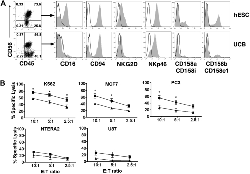
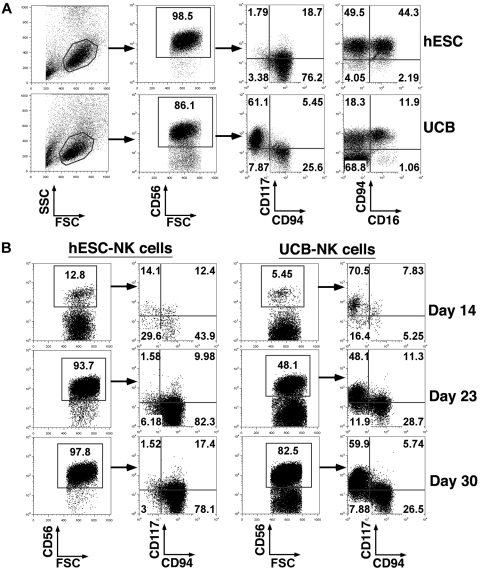
Natural killer (NK) cells serve as important effectors for antitumor immunity, and CD56+CD45+ NK cells can be routinely derived from human embryonic stem cells (hESCs). However, little is know about the ability of hESC-derived NK cells to mediate an effective in vivo antitumor response. Using bioluminescent imaging, we now demonstrate that H9 line hESC-derived NK cells mediate effective clearance of human tumor cells in vivo. In addition to increased in vitro killing of diverse tumor targets, the in vivo tumor clearance by H9 hESC-derived NK cells was more effective compared with NK cells derived from umbilical cord blood (UCB). Phenotypic analysis demonstrates the hESC-derived NK cells are uniformly CD94+CD117(low/-), an NK-cell population characterized by potent cytolytic activity and thus more competent to mediate tumor clearance. These studies demonstrate that hESCs provide an important model to study human lymphocyte development and may serve as a novel source for antitumor immunotherapy.


Figures

Figure 1
Figure 1
Derivation of NK cells from hESC capable of direct cytolysis of diverse human tumor cell lines. (A) Flow cytometric identification of CD56+CD45+ NK cells derived from hESC and UCB progenitor cells cultured in NK-cell conditions for 35 days. Histograms demonstrate that CD56+CD45+-gated NK cells express a repertoire of receptors important for regulating NK-cell activity, including CD16, C-type lectin-like receptors (CD94 and NKG2D), natural cytotoxicity receptors (NKp46), and killer-cell Ig-like receptors (CD158). (B) In vitro direct cytolysis was evaluated by standard 4-hour 51Cr-release assay after 35 days in NK-cell culture. NK cell–mediated killing was assessed by incubating effector cells with tumor targets from K562 (n = 7), MCF7 (n = 3), NTERA2 (n = 3), PC3 (n = 3), and U87 (n = 3) at indicated effector-to-target cell ratios. hESC-derived NK cells (■) demonstrate a significantly higher cytolytic activity against K562, MCF7, and PC3 tumor cells compared with UCB-derived NK cells (▲). Mean ± SEM is shown. *P < .05.
Figure 2
Figure 2
hESC-derived NK cells demonstrate clearance of established K562 tumors xenografted in mice with higher efficacy compared with NK cells generated from UCB. NOD/SCID mice were inoculated with luc+ K562 tumor cells, and cells were allowed to engraft for 3 days before animals were given 1 systemic (intravenous) infusion of NK cells. All hESC- and UCB-derived NK cells were injected after 30 to 35 days of culture. Mice were monitored by bioluminescent imaging at days 0, 4, 7, 14, and 21. In addition, some mice demonstrating tumor regression were monitored long-term (up to 8 weeks) for tumor recurrence. (A) Representative in vivo bioluminescent images of animals at the day of tumor inoculation and 21 days after tumor inoculation. Non-NK cell–treated control animals did receive the IL-2/IL-15 cytokine regimen and typically developed large tumors as indicated by luciferase expression. Mice treated with UCB-derived NK (UCB-NK) cells typically display slower tumor progression. All mice treated with hESC-derived NK (hESC-NK) cells displayed a complete clearance of tumor cells. (B) Analysis of luciferase activity (photons/second) of individual mice and the mean activity in each treatment group. Luciferase activity was analyzed from the site of tumor inoculation in control (n = 20, ■), UCB-NK treated (n = 13, [formula image]), and hESC-NK–treated (n = 13, ●) mice. Error bars indicated SEM. *P < .01 for hESC-NK vs UCB-NK and control. (C) CD56+ NK cells were magnetically sorted and injected intravenously into tumor-bearing mice. Mice treated with either sorted CD56+ hESC-NK cells (n = 3, —) or sorted CD56+ UCB-NK cells (n = 2, formula image) demonstrate tumor regression similar to mice treated with unsorted hESC- or UCB-NK cells.
Figure 3
Figure 3
hESC-derived NK cells protect against K562 metastasis. (A) Representative images of micrometastasis foci in liver, lungs, spleen, and kidneys as identified by bioluminescent imaging. (B) Quantification of luciferase activity (p/s) from isolated organs. Luciferase intensity from each well was quantified using LivingImage software. Mean ± SEM from indicated organs is shown.
Figure 4
Figure 4
Development of homogeneously mature NK cells from hESCs. (A) Phenotypic analysis of hESC-NK cells compared with UCB-NK cells. hESC- and UCB-derived progenitors cultured in NK-cell conditions for 30 days were evaluated by flow cytometry for surface antigen expression. Two distinct populations, CD117+CD94 and CD117−/lowCD94+, are found in UCB-derived cells, whereas almost all hESC-derived cells are CD117−/lowCD94+. Additional analysis for CD94 and CD16 expression on the CD56+ cell population also demonstrates distinct differences between the hESC and UBC-NK cell populations. (B) Time-course analysis for expression of CD117 and CD94 on CD56+ NK cells. Cells were harvested after 14, 23, and 30 days in NK culture. FSC indicates forward scatter; and SSC, side scatter.
Figure 5
Figure 5
Phenotypic and functional analysis of hESC-NK cells compared with UCB-NK cells. (A) Flow cytometric analysis for 4 individual KIRs demonstrates a higher percentage of hESC-NK cells expressing these regulatory receptors compared with what is found on UCB-NK cells. (B) Expression of CD161 and NKp44 on hESC- and UCB-NK cells. (C) Expression of perforin and granzyme B on CD56+ NK cells was evaluated by intracellular flow cytometric straining. Histograms of CD56+-gated cells are shown. Mean fluorescent intensity (MFI) was analyzed from all hESC-derived CD56+ cells and from perforin and granzyme B-positive CD56+ cells from UCB. Isotype control is indicated in open histograms. (D) Cytolytic activity against K562, PC3, and MCF7 tumor cells was compared between purified CD117−/lowCD94+ NK cells derived from hESCs (□) and UCB (formula image
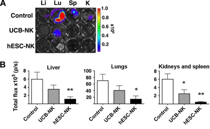
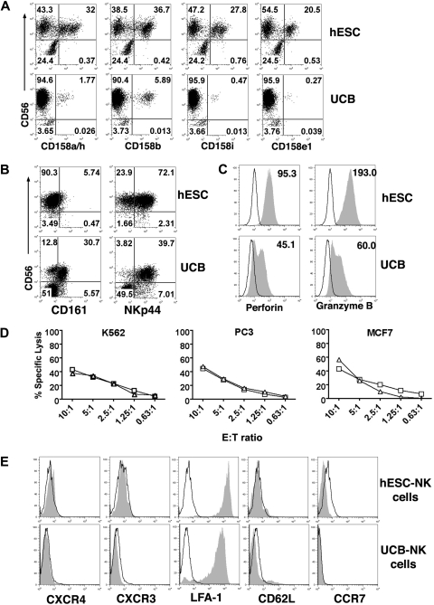
) at the indicated effector-to-target cell ratios. Representative results from 2 separate experiments are shown. (E) Flow cytometric analysis of cell trafficking molecules on hESC- and UCB-derived NK cells. Histograms of CD56+-gated NK cells are shown. Open histogram indicates isotype control.

References

    1. Dudley ME, Wunderlich JR, Robbins PF, et al. Cancer regression and autoimmunity in patients after clonal repopulation with antitumor lymphocytes. Science. 2002;298:850–854. - PMC - PubMed
    1. Morgan RA, Dudley ME, Wunderlich JR, et al. Cancer regression in patients after transfer of genetically engineered lymphocytes. Science. 2006;314:126–129. - PMC - PubMed
    1. Dudley ME, Wunderlich JR, Yang JC, et al. Adoptive cell transfer therapy following non-myeloablative but lymphodepleting chemotherapy for the treatment of patients with refractory metastatic melanoma. J Clin Oncol. 2005;23:2346–2357. - PMC - PubMed
    1. Khong HT, Restifo NP. Natural selection of tumor variants in the generation of “tumor escape” phenotypes. Nat Immunol. 2002;3:999–1005. - PMC - PubMed
    1. Smyth MJ, Hayakawa Y, Takeda K, Yagita H. New aspects of natural-killer-cell surveillance and therapy of cancer. Nat Rev Cancer. 2002;2:850–861. - PubMed

Publication types

MeSH terms