. 2009:15:685-99.
Epub 2009 Apr 3.

Loss of AND-34/BCAR3 expression in mice results in rupture of the adult lens

Affiliations

Loss of AND-34/BCAR3 expression in mice results in rupture of the adult lens

Richard I Near et al. Mol Vis. 2009.

Abstract

Purpose: AND-34/BCAR3 (Breast Cancer Anti-Estrogen Resistance 3) associates with the focal adhesion adaptor protein, p130CAS/BCAR1. Expression of AND-34 regulates epithelial cell growth pattern, motility, and growth factor dependence. We sought to establish the effects of the loss of AND-34 expression in a mammalian organism.

Methods: AND-34(-/-) mice were generated by homologous recombination. Histopathology, in situ hybridization, and western blotting were performed on murine tissues.

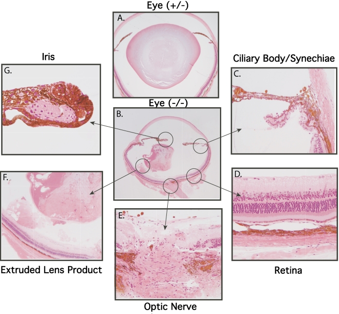
Results: Western analyses confirmed total loss of expression in AND-34(-/-) splenic lymphocytes. Mice lacking AND-34 are fertile and have normal longevity. While AND-34 is widely expressed in wild type mice, histologic analysis of multiple organs in AND-34(-/-) mice is unremarkable and analyses of lymphocyte development show no overt changes. A small percentage of AND-34(-/-) mice show distinctive small white eye lesions resulting from the migration of ruptured cortical lens tissue into the anterior chamber. Following initial vacuolization and liquefaction of the lens cortex first observed at postnatal day three, posterior lens rupture occurs in all AND-34(-/-) mice, beginning as early as three weeks and seen in all mice at three months. Western blot analysis and in situ hybridization confirmed the presence of AND-34 RNA and protein in lens epithelial cells, particularly at the lens equator. Prior data link AND-34 expression to the activation of Akt signaling. While Akt Ser 473 phosphorylation was readily detectable in AND-34(+/+) lens epithelial cells, it was markedly reduced in the AND-34(-/-) lens epithelium. Basal levels of p130Cas phosphorylation were higher in AND-34(+/+) than in AND-34(-/-) lens epithelium.

Conclusions: These results demonstrate the loss of AND-34 dysregulates focal adhesion complex signaling in lens epithelial cells and suggest that AND-34-mediated signaling is required for maintenance of the structural integrity of the adult ocular lens.


Figures

Figure 1
Figure 1
AND-34 targeting. A: Exon structure of AND-34 cDNA is shown. B: Production of the recombinant ES clone is demonstrated. A genomic phage clone containing exons 4 and 5 was mapped as shown. The area between the indicated SmaI sites (3.5 kb) was deleted, and the two surrounding “arms”, 5.3 kb of the 5′ sequence and 1.8 kb of the 3′ sequence, were cloned into the pPNT vector. Location of wild type and knockout PCR primers are shown as well as the hybridization probe used for Southern blotting. C: Screening for homologous recombination. DNA prepared from ES clones was purified, aliquots from four clones per lane were pooled, and PCR was performed to detect the recombined KO allele. The first lane shows a pool that contains an ES clone that has the KO allele (arrows indicate lanes of PCRs with clones that contain no AND-34−/− allele). The control PCR used primers specific for Neo. Homologous recombination was confirmed by Southern blot analysis as shown. DNA from individual clones was cleaved with BamHI and analyzed with the indicated probe. Clones 165 and 169 display the recombinant allele. Wild type 129 mouse DNA serves as a control. M=marker lane.
Figure 2
Figure 2
Exons 4 and 5 of AND-34−/− mice lack AND-34 protein. Immunoblots of whole cell lysates from splenic lymphocytes from wild type (+/+) and knockout (−/−) mice were performed with two different polyclonal anti-AND-34 antisera. As positive and negative controls, lysates of 293T cells transfected with an AND-34 expression plasmid (pAND) or vector alone (pcDNA) were also included. A: AND-34−/− mice in which exon 2 had been targeted were assessed with antiserum to peptide cYEKQLKPFSKLLHEGREST. The presence of residual truncated AND-34 protein product is noted in knockout splenic lymphocytes. B: AND-34−/− mice in which exons 4 and 5 had been targeted were examined with antiserum to peptide KSPLAERRTDAYQDVSIc. The AND-34 protein is absent in knockout splenic lymphocytes. A non-specific band (N.S.) is detected with this antiserum.
Figure 3
Figure 3
AND-34 expression in mouse tissues. A: Western blot analysis of wild type mouse tissues is shown. AND-34 was immunoprecipitated directly from lysates of mouse organs with polyclonal anti-AND-34 antibody whereas p130CAS was detected directly from whole cell lysates with a monoclonal anti-p130CAS antibody that is also able to detect HEF1. B: In situ hybridization for AND-34 transcripts from lymph nodes and spleens of AND-34+/+ is displayed. The tissues were probed with sense or antisense hybridization probes as shown in dark-field microscopy. With dark-field illumination, the positive hybridization signal of the silver grains in the autoradiographic emulsion appears as white dots over a black background. C: In situ hybridization for AND-34 transcripts in the lungs of AND-34+/+ mice is shown in bright-field microscopy. With bright-field illumination, the positive hybridization signal appears as black dots. Hybridization was detected in a subset of alveolar cells with the morphology of lung fibroblasts. Arrows indicate a strong hybridization signal.
Figure 4
Figure 4
Ocular phenotype in AND-34−/− mice. A: The lens cortex was observed in the anterior chamber of the eye in a subset of AND-34−/− mice (four-month-old mouse shown on right). Comparable abnormalities were never observed in AND-34-/+ or AND-34+/+ mouse eyes (left). B: Dissected whole lens from a three-month-old AND-34−/− 129 strain mouse was compared with a three-month-old AND-34+/+ lens as viewed under a dissecting microscope. The AND-34+/+ lens was a uniform translucent, smooth, spherical structure whereas the lens from the AND-34−/− mouse eye was a partially opaque, irregular structure that had clearly lost a portion of its internal contents.
Figure 5
Figure 5
Paraffin-embedded histopathology of AND-34−/− mouse eyes. Paraffin tissue sections of four-month-old AND-34−/− eyes are compared with a four-month-old AND-34+/+ eye. Panel B shows a fragment of anterior chamber lens cortex (short arrow) in front of the iris (long arrow). Panel C shows a fragment of lens cortex (short arrow) behind the iris (long arrow). Panel D shows that the fragment of lens cortex lacks both epithelial cells and a lens capsule. The lens capsule is visible in the lens itself on the left side of this panel. In panels A and B, the loss of lens fiber material from the lens is due to artifactual shearing during the cutting of the paraffin section.
Figure 6
Figure 6
Plastic-embedded histopathology of AND-34−/− mouse eyes. Tissue sections from three month-old mature heterozygous AND-34+/− and homozygous knockout AND-34−/− eyes are shown in Panels A and B, respectively. Abnormalities in the indicated regions of the AND-34−/− eye are described in the text.
Figure 7
Figure 7
Lens rupture in AND-34−/− mice occurs postnatally following initial development of lens vacuolization. AND-34−/− eyes from 3-, 24-, and 33-day-old AND-34−/− mice (P3, P24, and P33) were fixed, plastic embedded, and sectioned. Vacuolization of the cortical lens fibers but no examples of lens rupture were identified in the eyes of three-day-old mice (upper panels). Lens rupture with extrusion of cortical fiber material was first detected in 24- and 33-day old mice (lower panels).
Figure 8
Figure 8
Expression of AND-34 transcript in lens tissue. Eyes from AND-34+/+ mice (two months old) were fixed and probed with sense or antisense hybridization probes for AND-34 transcripts. Expression of AND-34 was detected by the antisense probe in the lens epithelial cells at the lens equator (lower panels) but was not reproducibly observed in the anterior epithelial cells (upper panels).
Figure 9
Figure 9
AND-34 expression regulates p130Cas and Akt signaling in lens epithelial cells. Lenses were removed from the eyes of AND-34−/− and AND-34+/+ mice (four months old), and the capsular epithelial layer (EPI) was separated from the lens fibers (FIB) by dissection. A: The expression of AND-34/NSP2 and CHAT/NSP3 were examined in the lens tissues of AND-34+/+ mice by immunoblot. Lysates from HEK293T cells that were transfected with AND-34 (pAND) or CHAT (pCHAT) expression constructs were used as controls. Expression of crystallin, a component of lens fibers, and tubulin were also examined. To detect low level expression of AND-34 in lens fibers, fivefold more of the lens fiber sample was loaded in the first lane (Fib-5X) than in the fifth lane (Fib-1X). B: The phosphorylation status of p130Cas and AKT Ser 473 was examined in AND-34−/− and AND-34+/+ lens epithelial and lens fiber samples by western blot analysis. Phosphorylated p130Cas is characterized by reduced PAGE migration. Total levels of AKT expression were also assessed as a control. Similar results were obtained in three experiments.

References

    1. Lerner A, Clayton LK, Mizoguchi E, Ghendler Y, van Ewijk W, Koyasu S, Bhan AK, Reinherz EL. Cross-linking of T cell receptors on double positive thymocytes induces a cytokine-mediated stromal activation process linked to cell death. EMBO J. 1996;15:5876–87. - PMC - PubMed
    1. Cai D, Clayton LK, Smolyar A, Lerner A. AND-34, a novel p130Cas-binding thymic stromal cell protein regulated by adhesion and inflammatory cytokines. J Immunol. 1999;163:2104–12. - PubMed
    1. Gotoh T, Cai D, Tian X, Feig L, Lerner A. p130Cas regulates the activity of AND-34, a novel Ral, Rap1 and R-Ras guanine nucleotide exchange factor. J Biol Chem. 2000;275:30118–23. - PubMed
    1. Cai D, Felekkis KN, Near RI, O'Neill GM, van Seventer JM, Golemis EA, Lerner A. The GDP exchange factor AND-34 is expressed in B cells, associates with HEF1, and activates Cdc42. J Immunol. 2003;170:969–78. - PubMed
    1. van Agthoven T, van Agthoven TL, Dekker A, van der Spek PJ, Vreede L, Dorssers LC. Identification of BCAR3 by a random search for genes involved in antiestrogen resistance of human breast cancer cells. EMBO J. 1998;17:2799–808. - PMC - PubMed

Publication types

MeSH terms