Effect of filler level and particle size on dental caries-inhibiting Ca-PO(4) composite
- PMID: 19365616
- PMCID: PMC3056554
- DOI: 10.1007/s10856-009-3740-2
Effect of filler level and particle size on dental caries-inhibiting Ca-PO(4) composite
Abstract
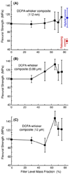
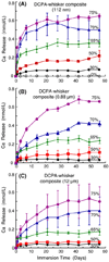
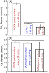
Secondary caries and restoration fracture are common problems in restorative dentistry. The aim of this study was to develop Ca-PO(4) nanocomposite having improved stress-bearing properties and Ca and PO(4) ion release to inhibit caries, and to determine the effects of filler level. Nanoparticles of dicalcium phosphate anhydrous (DCPA), two larger DCPA powders, and reinforcing whiskers were incorporated into a resin. A 6 x 3 design was tested with six filler mass fractions (0, 30, 50, 65, 70, and 75%) and three DCPA particle sizes (112 nm, 0.88 mum, 12.0 mum). The DCPA nanocomposite at 75% fillers had a flexural strength (mean +/- SD; n = 6) of 114 +/- 23 MPa, matching the 112 +/- 22 MPa of a commercial non-releasing, hybrid composite (P > 0.1). This was 2-fold of the 60 +/- 6 MPa of a commercial releasing control. Decreasing the particle size increased the ion release. Increasing the filler level increased the ion release at a rate faster than being linear. The amount of ion release from the nanocomposite matched or exceeded those of previous composites that released supersaturating levels of Ca and PO(4) and remineralized tooth lesions. This suggests that the much stronger nanocomposite may also be effective in remineralizing tooth lesion and inhibiting caries. In summary, combining calcium phosphate nanoparticles with reinforcing co-fillers in the composite provided a way to achieving both caries-inhibiting and stress-bearing capabilities. Filler level and particle size can be tailored to achieve optimal composite properties.
Figures


Similar articles
-
Nanocomposites with Ca and PO4 release: effects of reinforcement, dicalcium phosphate particle size and silanization.Dent Mater. 2007 Dec;23(12):1482-91. doi: 10.1016/j.dental.2007.01.002. Epub 2007 Mar 6. Dent Mater. 2007. PMID: 17339048 Free PMC article.
-
Calcium and phosphate ion releasing composite: effect of pH on release and mechanical properties.Dent Mater. 2009 Apr;25(4):535-42. doi: 10.1016/j.dental.2008.10.009. Epub 2008 Dec 20. Dent Mater. 2009. PMID: 19101026 Free PMC article.
-
Nanocomposite containing amorphous calcium phosphate nanoparticles for caries inhibition.Dent Mater. 2011 Aug;27(8):762-9. doi: 10.1016/j.dental.2011.03.016. Epub 2011 Apr 22. Dent Mater. 2011. PMID: 21514655 Free PMC article.
-
Strong nanocomposites with Ca, PO(4), and F release for caries inhibition.J Dent Res. 2010 Jan;89(1):19-28. doi: 10.1177/0022034509351969. J Dent Res. 2010. PMID: 19948941 Free PMC article. Review.
-
Direct resin-based composites: current recommendations for optimal clinical results.Compend Contin Educ Dent. 2005 Jul;26(7):481-2, 484-90; quiz 492, 527. Compend Contin Educ Dent. 2005. PMID: 16060378 Review.
Cited by
-
Compositional boundaries for functional dental composites containing calcium orthophosphate particles.J Mech Behav Biomed Mater. 2023 Aug;144:105928. doi: 10.1016/j.jmbbm.2023.105928. Epub 2023 Jun 7. J Mech Behav Biomed Mater. 2023. PMID: 37302206 Free PMC article.
-
Recent advances and developments in composite dental restorative materials.J Dent Res. 2011 Apr;90(4):402-16. doi: 10.1177/0022034510381263. Epub 2010 Oct 5. J Dent Res. 2011. PMID: 20924063 Free PMC article. Review.
-
Biocompatible Nanocomposite Enhanced Osteogenic and Cementogenic Differentiation of Periodontal Ligament Stem Cells In Vitro for Periodontal Regeneration.Materials (Basel). 2020 Nov 4;13(21):4951. doi: 10.3390/ma13214951. Materials (Basel). 2020. PMID: 33158111 Free PMC article.
-
Novel Bioactive and Therapeutic Root Canal Sealers with Antibacterial and Remineralization Properties.Materials (Basel). 2020 Mar 1;13(5):1096. doi: 10.3390/ma13051096. Materials (Basel). 2020. PMID: 32121595 Free PMC article. Review.
-
Therapeutic polymers for dental adhesives: loading resins with bio-active components.Dent Mater. 2014 Jan;30(1):97-104. doi: 10.1016/j.dental.2013.06.003. Epub 2013 Jul 27. Dent Mater. 2014. PMID: 23899387 Free PMC article.
References
-
- Söderholm KJ, Zigan M, Ragan M, Fischlschweiger W, Bergman M. Hydrolytic degradation of dental composites. J Dent Res. 1984;63:1248–1254. - PubMed
-
- Goldberg AJ, Burstone CJ, Hadjinikolaou I, Jancar J. Screening of matrices and fibers for reinforced thermoplastics intended for dental applications. J Biomed Mater Res. 1994;28:167–173. doi: 10.1002/jbm.820280205. - DOI - PubMed
-
- Ferracane JL, Berge HX, Condon JR. In vitro aging of dental composites in water–effect of degree of conversion, filler volume, and filler/matrix coupling. J Biomed Mater Res. 1998;42:465–472. doi: 10.1002/(SICI)1097-4636(19981205)42:3<465::AID-JBM17>3.0.CO;2-F. - DOI - PubMed
-
- Krämer N, García-Godoy F, Reinelt C, Frankenberger R. Clinical performance of posterior compomer restorations over 4 years. Am J Dent. 2006;19:61–66. - PubMed
-
- Sarrett DC. Clinical challenges and the relevance of materials testing for posterior composite restorations. Dent Mater. 2005;21:9–20. doi: 10.1016/j.dental.2004.10.001. - DOI - PubMed
Publication types
MeSH terms
Substances
Grants and funding
LinkOut - more resources
Full Text Sources
Medical

