. 2009 Sep;10(3):355-67.
doi: 10.1007/s10162-009-0170-2. Epub 2009 Apr 14.

Enhanced survival of spiral ganglion cells after cessation of treatment with brain-derived neurotrophic factor in deafened guinea pigs

Affiliations

Enhanced survival of spiral ganglion cells after cessation of treatment with brain-derived neurotrophic factor in deafened guinea pigs

Martijn J H Agterberg et al. J Assoc Res Otolaryngol. 2009 Sep.

Abstract

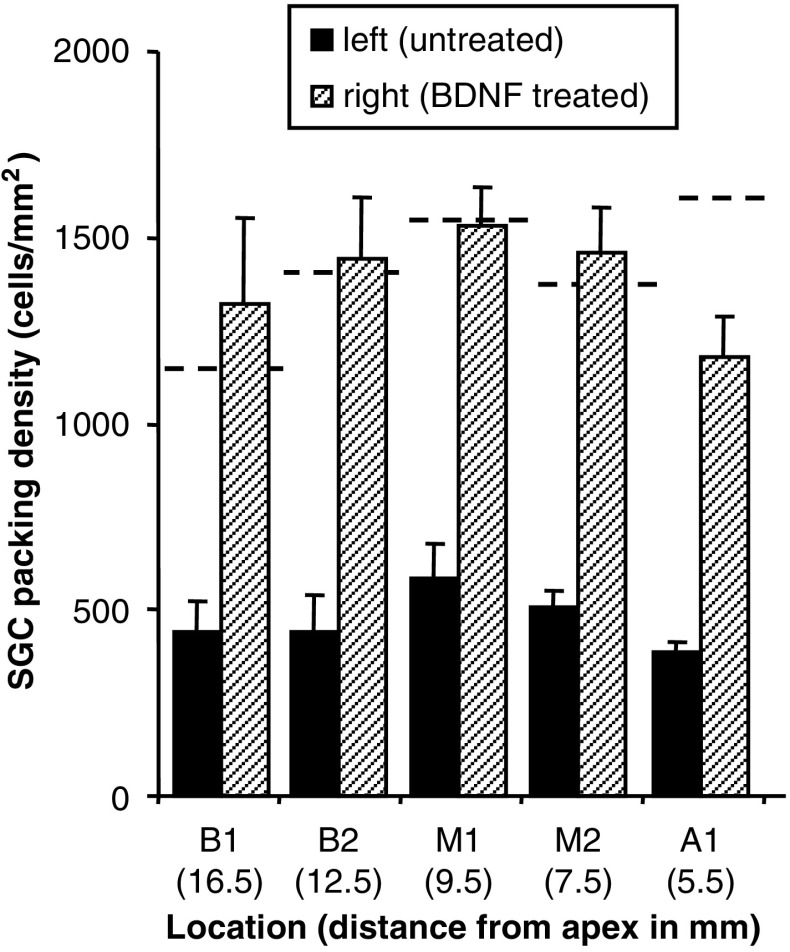
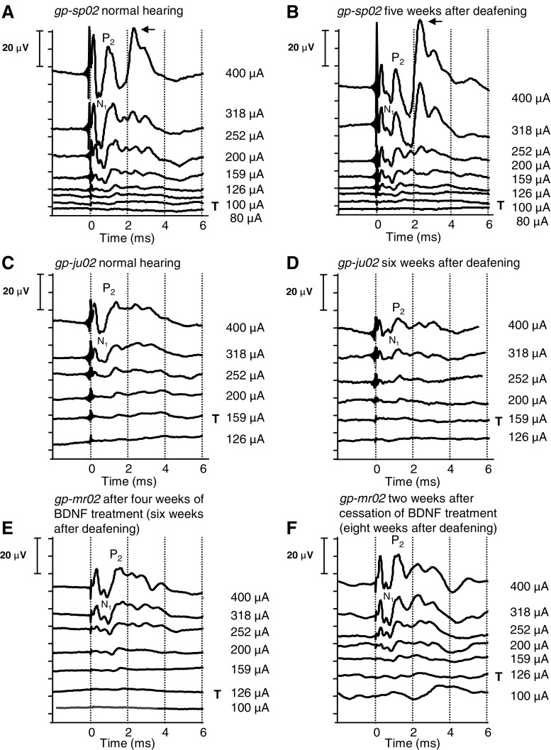
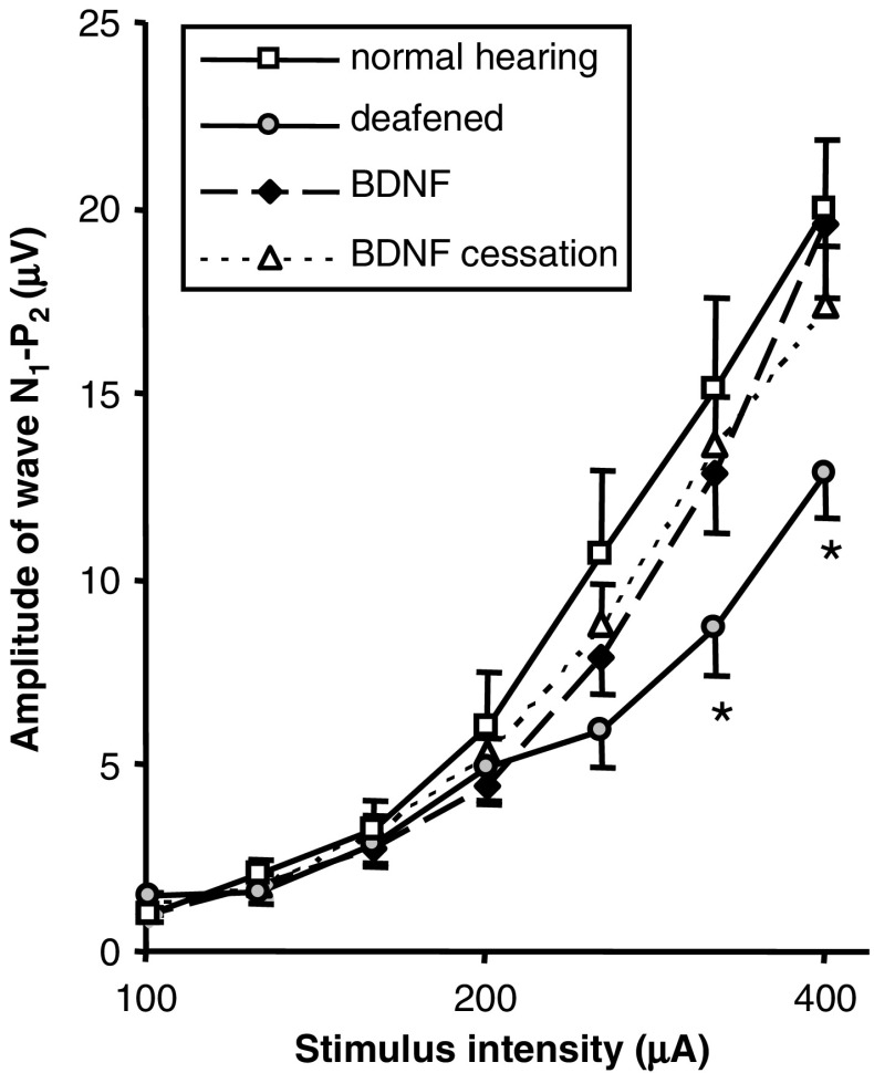
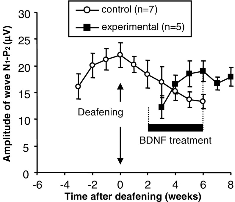
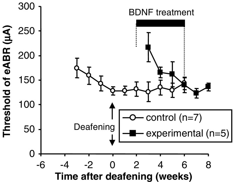
Exogenous delivery of neurotrophic factors into the cochlea of deafened animals rescues spiral ganglion cells (SGCs) from degeneration. To be clinically relevant for human cochlear implant candidates, the protective effect of neurotrophins should persist after cessation of treatment and the treated SGCs should remain functional. In this study, the survival and functionality of SGCs were investigated after temporary treatment with brain-derived neurotrophic factor (BDNF). Guinea pigs in the experimental group were deafened, and 2 weeks later, the right cochleae were implanted with an electrode array and drug delivery cannula. BDNF was administered to the implanted cochleae during a 4-week period via a mini-osmotic pump. After completion of the treatment, the osmotic pumps were removed. Two weeks later, the animals were killed and the survival of SGCs was analyzed. To monitor the functionality of the auditory nerve, electrically evoked auditory brainstem responses (eABRs) were recorded in awake animals throughout the experiment. BDNF treatment resulted in enhanced survival of SGCs 2 weeks after cessation of the treatment and prevented the decreases in size and circularity that are seen in the untreated contralateral cochleae. The amplitude of the suprathreshold eABR response in BDNF-treated animals was significantly larger than in deafened control animals and comparable to that in normal-hearing control animals. The amplitude in the BDNF-treated group did not decrease significantly after cessation of treatment. The eABR latency in BDNF-treated animals was longer than normal and comparable to that in deafened control animals. These morphological and functional findings demonstrate that neurotrophic intervention had a lasting effect, which is promising for future clinical application of neurotrophic factors in implanted human cochleae.


Figures

FIG. 1
FIG. 1
Treatment schedule of four different animal cohorts (AD). A Deafened and 2 weeks later implanted and treated with BDNF; B deafened and 2 weeks later implanted; C first implanted and 4 weeks later deafened; D only implanted. Deafening was performed systemically affecting both ears. Cochlear implantation (AD) and BDNF treatment (A) was applied to the right ear. After implantation, eABRs were regularly recorded in each group (by electrically stimulating the implanted right ear). For electrophysiological analysis, eABRs of the BDNF-treated animals (A) were compared to eABRs of normal-hearing animals (C before deafening, D) and to eABRs of deafened animals (B, C after deafening). Note that data in normal-hearing and deafened conditions were obtained in the same animals (C). For histological analysis, the main comparison was made within the animals treated with BDNF (A): BNDF-treated right ears were compared to untreated left ears.
FIG. 2
FIG. 2
A typical example of an aABR (top) evoked in a normal-hearing animal, with clicks of 45 dB nSL and an eABR (bottom) evoked with 400-μA pulses. Stimulus onset was at time 0. Note the large stimulus artifact in the eABR before peak P1. The peak-to-peak amplitude, N1–P2, of the eABR was measured to assess the functional status of the auditory nerve. When the N1 and/or P2 consisted of more than one subpeak, the first subpeak on each site of the fast rising part of the eABR complex (indicated with the arrows) is used in the analysis.
FIG. 3
FIG. 3
Light micrograph of a midmodiolar section (1 μm) of a normal guinea pig cochlea showing the different locations at which SGCs were examined. The distance from the apex of the locations B1 through A3 are specified in the table. n. VIII cochlear nerve.
FIG. 4
FIG. 4
Light micrographs of Rosenthal’s canal at location B2 (upper basal turn) with the distribution of SGCs (arrowheads) and nerve fibers (arrows). SGCs and nerve fibers in the spiral ganglion from A a normal cochlea, B a cochlea 2 weeks after deafening, C a left cochlea 6 weeks after deafening, D the right implanted cochlea from the same animal as in (C), E the left cochlea of an animal 8 weeks after deafening, and F the right implanted cochlea treated with BDNF from the same animal as in (E) (2 weeks after cessation of treatment).
FIG. 5
FIG. 5
Mean SGC packing densities at cochlear locations B1, B2, M1, M2, and A1 in the left (untreated) and right (BDNF-treated) cochlea of deafened animals (n = 6). Dashed lines represent SGC densities at locations B1–A1 in normal cochleae. Error bars: SEM.
FIG. 6
FIG. 6
Perikaryal area (A) and cell circularity (B) of SGCs in left untreated versus BDNF-treated cochleae. Only SGCs with an obvious nucleus in basal location B2 were included. Data were obtained by averaging all individual SGC measurements within one spiral ganglion, followed by averaging across cochleae. Dashed lines represent measurements in normal-hearing animals. n the number of SGCs measured. Error bars: SEM. *p < 0.05.
FIG. 7
FIG. 7
Recordings of representative eABRs evoked with current pulses of 400 μA down to below threshold in two control animals (gp-sp02 and gp-ju02) and one experimental animal (gp-mr02). Recordings of two control animals are depicted to illustrate the inter-animal variability. A, C Recorded in normal-hearing condition, prior to deafening; B, D recorded 5 and 6 weeks after deafening, respectively. E Recorded after 4 weeks of BDNF treatment and F 2 weeks after cessation of the treatment. T indicates the threshold of wave N1–P2. The arrow indicates the peak that probably reflects the digastric muscle response.
FIG. 8
FIG. 8
Mean input–output functions of eABRs recorded in control animals before deafening (normal hearing, n = 12) and 6 weeks after deafening (n = 9), and in the experimental animals immediately after 4 weeks of BDNF treatment (n = 5) and 2 weeks after cessation of BDNF treatment (n = 5). Error bars: SEM. *p < 0.05 indicates statistical significant difference between the untreated animals 6 weeks after deafening and each of the other data points.
FIG. 9
FIG. 9
Mean N1–P2 amplitudes of eABRs recorded to 400-μA pulses in control (n = 7) and experimental (n = 5) animals. The control animals were implanted at −4 weeks and deafened at 0 weeks. The experimental animals were deafened at 0 weeks and implanted in the right cochlea at 2 weeks. Following implantation, the experimental animals received BDNF for a 4-week period (indicated with a horizontal bold bar). Error bars: SEM.
FIG. 10
FIG. 10
Mean eABR thresholds observed for wave N1–P2 in control (n = 7) and experimental (n = 5) animals. The experimental animals were deafened at 0 weeks and implanted in the right cochlea at 2 weeks. Following implantation, the experimental animals received BDNF for a 4-week period (indicated with a horizontal bold bar). Threshold criterion 2 μV; error bars: SEM.
FIG. 11
FIG. 11
Mean N1 latencies of eABRs evoked with 400-μA pulses, observed in control (n = 7) and experimental (n = 5) animals. The experimental animals were deafened at 0 weeks and implanted in the right cochlea at 2 weeks. Following implantation, the experimental animals received BDNF for a 4-week period (indicated with a horizontal bold bar). Statistical analyses within each group are not shown, but are described in the text. Error bars: SEM.

References

    1. Agterberg MJH, Versnel H, de Groot JCMJ, Smoorenburg GF, Albers FWJ, Klis SFL. Morphological changes in spiral ganglion cells after intracochlear application of brain-derived neurotrophic factor in deafened guinea pigs. Hear. Res. 2008;244:25–34. doi: 10.1016/j.heares.2008.07.004. - DOI - PubMed
    1. Blamey P. Are spiral ganglion cell numbers important for speech perception with a cochlear implant? Am. J. Otol. 1997;18(suppl):11–12. - PubMed
    1. Brown JN, Miller JM, Altschuler RA, Nuttall AL. Osmotic pump implant for chronic infusion of drugs into the inner ear. Hear. Res. 1993;70:167–172. doi: 10.1016/0378-5955(93)90155-T. - DOI - PubMed
    1. Chikar JA, Colesa DJ, Swiderski DL, Polo AD, Raphael Y, Pfingst BE. Over-expression of BDNF by adenovirus with concurrent electrical stimulation improves cochlear implant thresholds and survival of auditory neurons. Hear. Res. 2008;245:24–34. doi: 10.1016/j.heares.2008.08.005. - DOI - PMC - PubMed
    1. De Groot JCMJ, Veldman JE, Huizing EH. An improved fixation method for guinea pig cochlear tissues. Acta Otolaryngol. 1987;104:234–242. doi: 10.3109/00016488709107323. - DOI - PubMed

Publication types

MeSH terms

Substances