. 2009 Nov;30(11):3495-508.
doi: 10.1002/hbm.20773.

Altered somatosensory processing in trigeminal neuralgia

Affiliations

Altered somatosensory processing in trigeminal neuralgia

Maria Blatow et al. Hum Brain Mapp. 2009 Nov.

Abstract

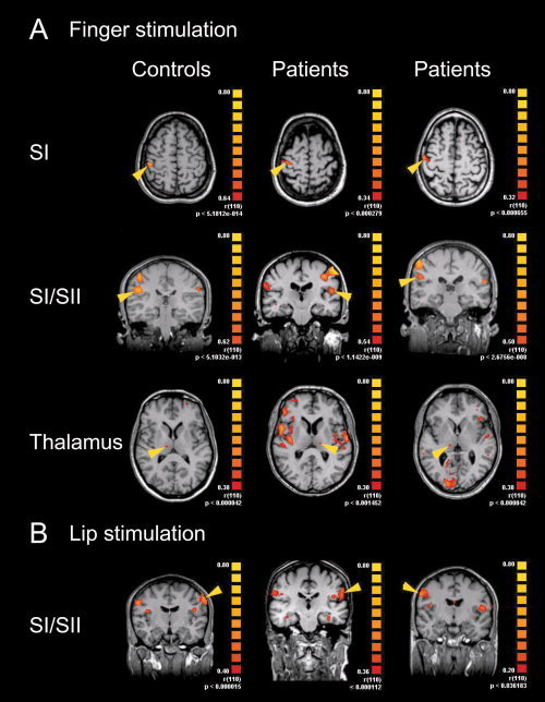
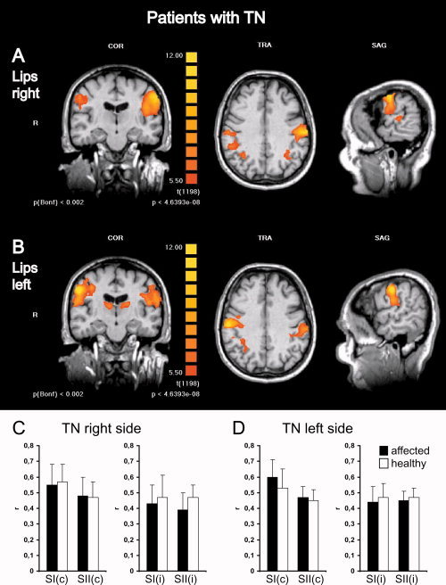
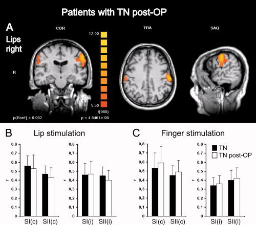
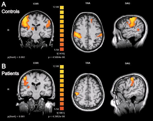
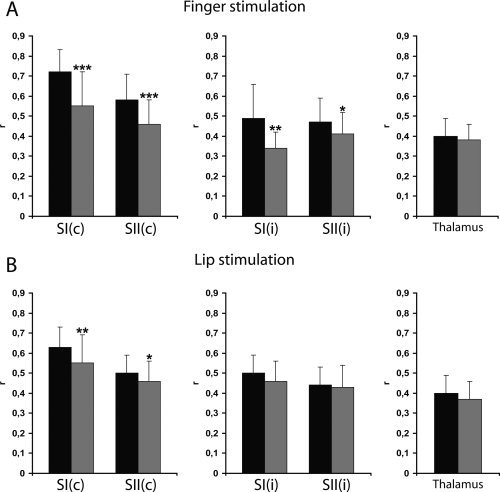
Trigeminal neuralgia (TN) is a pain state characterized by intermittent unilateral pain attacks in one or several facial areas innervated by the trigeminal nerve. The somatosensory cortex is heavily involved in the perception of sensory features of pain, but it is also the primary target for thalamic input of nonpainful somatosensory information. Thus, pain and somatosensory processing are accomplished in overlapping cortical structures raising the question whether pain states are associated with alteration of somatosensory function itself. To test this hypothesis, we used functional magnetic resonance imaging to assess activation of primary (SI) and secondary (SII) somatosensory cortices upon nonpainful tactile stimulation of lips and fingers in 18 patients with TN and 10 patients with TN relieved from pain after successful neurosurgical intervention in comparison with 13 healthy subjects. We found that SI and SII activations in patients did neither depend on the affected side of TN nor differ between operated and nonoperated patients. However, SI and SII activations, but not thalamic activations, were significantly reduced in patients as compared to controls. These differences were most prominent for finger stimulation, an area not associated with TN. For lip stimulation SI and SII activations were reduced in patients with TN on the contra- but not on the ipsilateral side to the stimulus. These findings suggest a general reduction of SI and SII processing in patients with TN, indicating a long-term modulation of somatosensory function and pointing to an attempt of cortical adaptation to potentially painful stimuli.


Figures

Figure 1
Figure 1
Pneumatic stimulation device for fMRI. Fully automated nonpainful pneumatically‐driven tactile stimulation using high resistance pneumatic tubes and finger clips/face mask with a flexible membrane was applied for each body side and location separately. [Color figure can be viewed in the online issue, which is available at www.interscience.wiley.com.]
Figure 2
Figure 2
Representative fMRI of SI, SII, and thalamic activations during finger and lip stimulations in individual subjects. Unilateral stimulation of left or right fingers (A) or lips (B) elicited BOLD‐activations in SI and SII areas and thalamus in controls (left panel), patients with TN (middle panel) and patients with TN after neurosurgical intervention (right panel). Representative examples are chosen for left or right stimulations in transversal or coronal views, yellow arrowheads point to activations contralateral to the stimulus. For display of individual data colors depicting the r‐statistic with uncorrected P‐values for the entire activation map were chosen to facilitate comparison with r‐values presented in this study. Each cluster fulfilled criteria of significance with a lower threshold of r > 0.25 and P < 0.05 (Bonferoni corrected; see methods section).
Figure 3
Figure 3
Somatosensory activations in patients with TN show no differences depending on stimulation of the affected or unaffected body side. Group level fMRI of bilateral somatosensory activations in the postcentral gyrus and parietal operculum in patients with TN on the right side are shown for stimulation of the right (A) and left (B) lips. To conserve anatomical details, group level BOLD‐activation maps (n = 11) were overlaid onto anatomical images of one individual subject. Left panels show a coronal view (COR) through the plane of the postcentral gyrus, middle panels a transversal view (TRA) through the plane of SI activations and left panels a sagittal view (SAG) through the plane of contralateral activations. R, right, L, left. For display of group data (different from Fig. 2) colors depicting the t‐statistic with uncorrected and Bonferoni corrected P‐values were chosen to facilitate comparison with other studies where mostly t‐values are presented. Each activation map had a false discovery rate (FDR) < 0.001. Statistical thresholds were deliberately kept equal to allow comparison of activation maps. No major differences in activation levels can be observed depending on the stimulation side. Note bilateral thalamic activations in (B) (left panel) and coactivations in the superior parietal lobules in (A) and (B) (middle panel). Histograms depict quantitative analyses of r‐values (mean ± SD) comparing SI and SII activations elicited by lip stimulation on the affected (black bars) or the healthy side (white bars) for patients with TN on the right side (C) and left side (D) separately. c, contralateral; i, Ipsilateral; r, correlation of the measured BOLD‐signal to the applied hemodynamic reference function.
Figure 4
Figure 4
Somatosensory activations in patients with TN show no differences before and after neurosurgical treatment and prolonged pain relief. (A) Group level fMRI of bilateral somatosensory activations in the postcentral gyrus and parietal operculum in patients with TN on the right side after successful microvascular decompression by Jannetta technique are shown for stimulation of the right lips. To conserve anatomical details, group level BOLD‐activation maps (n = 10) were overlaid onto anatomical images of one individual subject. Left panels show a coronal view (COR) through the plane of the postcentral gyrus, middle panels a transversal view (TRA) through the plane of SI activations and left panels a sagittal view (SAG) through the plane of contralateral activations. R, right; L, left. Colors depict the t‐statistic with uncorrected and Bonferoni corrected P‐values. Statistical thresholds were chosen as in Figure 3 to allow comparison of activation maps. Histograms depict quantitative analyses of r‐values (mean ± SD) comparing SI and SII activations in patients with TN before (black bars) and after pain relief (white bars) for lip (B) and finger (C) stimulations. c, contralateral; i, ipsilateral; r, correlation of the measured BOLD‐signal to the applied hemodynamic reference function.
Figure 5
Figure 5
Somatosensory activations are significantly reduced in patients with TN as compared to healthy subjects. Group level fMRI of bilateral somatosensory activations in the postcentral gyrus and parietal operculum in control subjects (n = 13) (A) and patients with TN (n = 18) (B) are shown for stimulation of the right fingers. Activation maps are presented as in Figures 3 and 4 and statistical thresholds for image display were kept constant for easier comparison. BOLD‐activation is visibly reduced in patients versus controls; at the chosen threshold no ipsilateral SI/SII activation is displayed in the patients' fMRI. Note contralateral thalamic activation in (A) (left panel).
Figure 6
Figure 6
Quantitative assessment of somatosensory activations in patients with TN and healthy subjects. Correlations of BOLD‐signals to the applied hemodynamic reference function (r; mean ± SD) are plotted for contra‐ and ipsilateral SI and SII and contralateral thalamic activations for finger (A) and lip (B) stimulations in healthy controls (black bars) and patients with TN (gray bars). c, contralateral; i, ipsilateral. Levels of statistical significance: ***P < 0.0001, **P < 0.01, *P < 0.05.

References

    1. Annett M ( 1970): A classification of hand preference by association analysis. Br J Psychol 61: 303–321. - PubMed
    1. Apkarian AV,Bushnell MC,Treede RD,Zubieta JK ( 2005): Human brain mechanisms of pain perception and regulation in health and disease. Eur J Pain 9: 463–484. - PubMed
    1. Barker FG II,Jannetta PJ,Bissonette DJ,Larkins MV,Jho HD ( 1996): The long‐term outcome of microvascular decompression for trigeminal neuralgia. N Engl J Med 334: 1077–1083. - PubMed
    1. Basbaum AI,Jessell TM ( 2000): The perception of pain In: Kandel ER, Schwartz JH, Jessell TM, editors. Principles of Neural Science. New York: McGraw‐Hill; pp 472–491.
    1. Becerra L,Morris S,Bazes S,Gostic R,Sherman S,Gostic J,Pendse G,Moulton E,Scrivani S,Keith D,Chizh B,Borsook D ( 2006): Trigeminal neuropathic pain alters responses in CNS circuits to mechanical (brush) and thermal (cold and heat) stimuli. J Neurosci 26: 10646–10657. - PMC - PubMed

Publication types

MeSH terms