. 2009 Jun 2;48(21):4642-51.
doi: 10.1021/bi802171c.

Regulatory autophosphorylation sites on protein kinase C-delta at threonine-141 and threonine-295

Affiliations

Regulatory autophosphorylation sites on protein kinase C-delta at threonine-141 and threonine-295

Vitalyi O Rybin et al. Biochemistry. .

Abstract

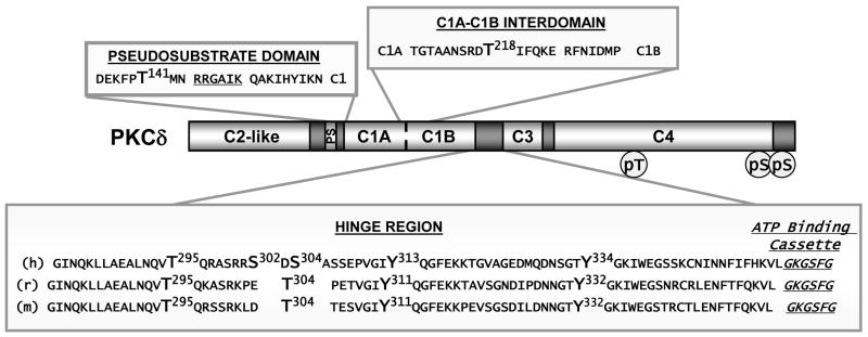
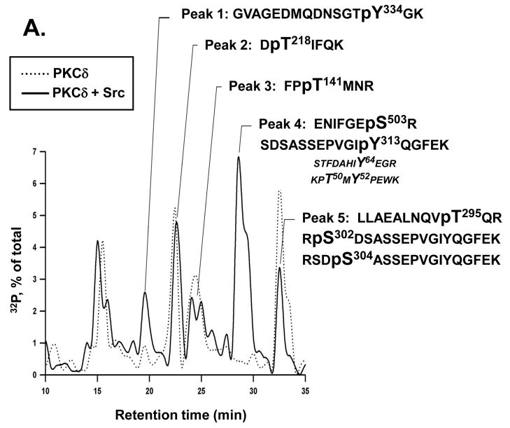
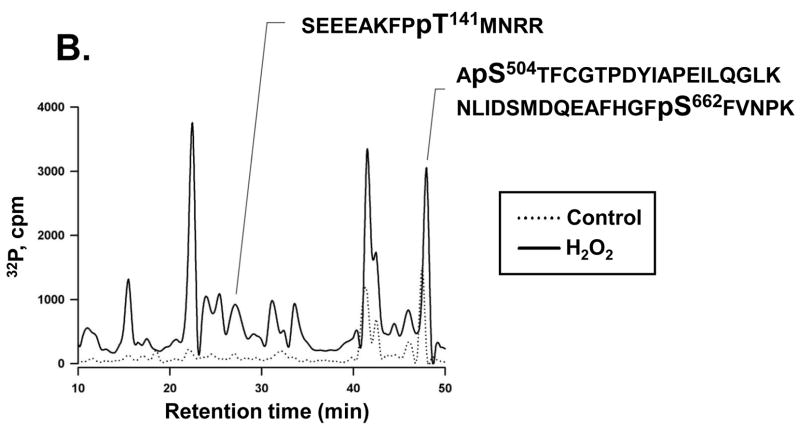
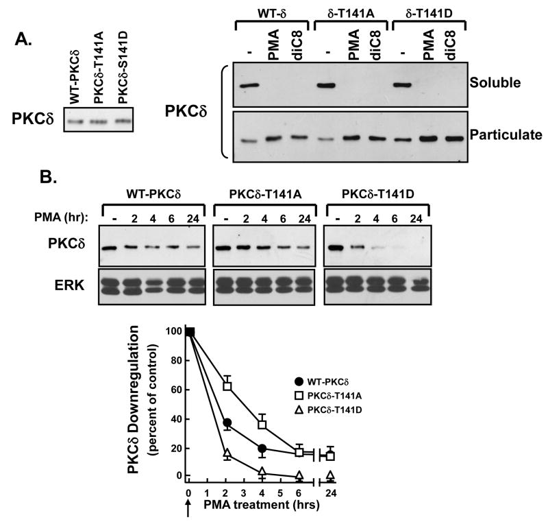
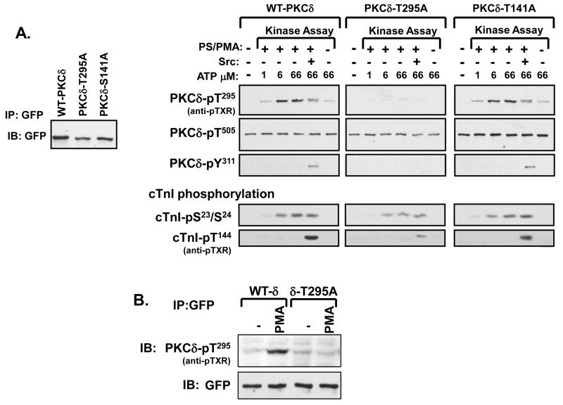
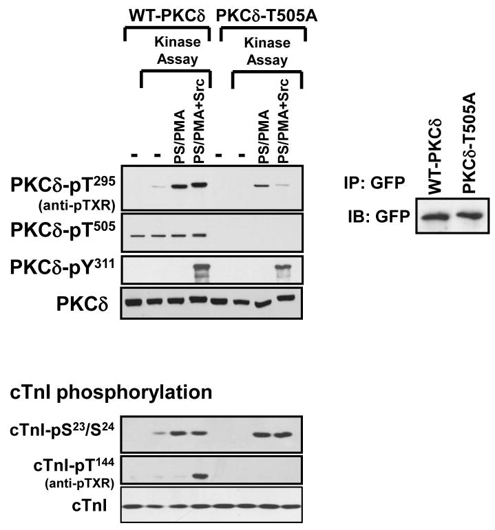
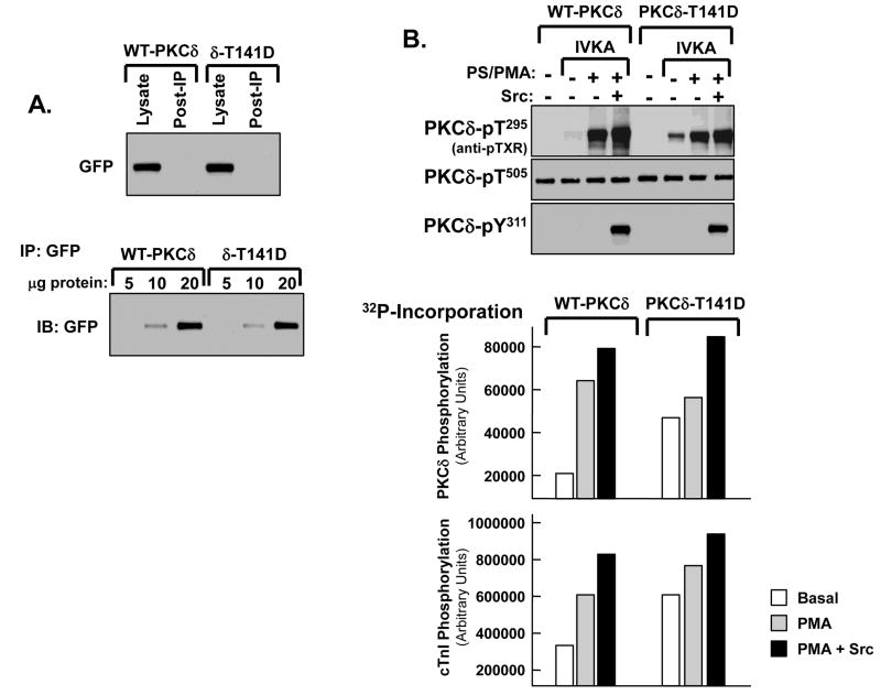
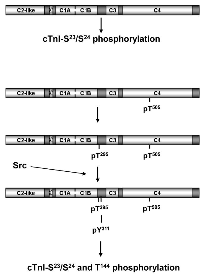
Protein kinase C-delta (PKCdelta) is a Ser/Thr kinase that regulates a wide range of cellular responses. This study identifies novel in vitro PKCdelta autophosphorylation sites at Thr(141) adjacent to the pseudosubstrate domain, Thr(218) in the C1A-C1B interdomain, Ser(295), Ser(302), and Ser(304) in the hinge region, and Ser(503) adjacent to Thr(505) in the activation loop. Cell-based studies show that Thr(141) and Thr(295) also are phosphorylated in vivo and that Thr(141) phosphorylation regulates the kinetics of PKCdelta downregulation in COS7 cells. In vitro studies implicate Thr(141) and Thr(295) autophosphorylation as modifications that regulate PKCdelta activity. A T141D substitution markedly increases basal lipid-independent PKCdelta activity; the PKCdelta-T141D mutant is only slightly further stimulated in vitro by PMA treatment, suggesting that Thr(141) phosphorylation relieves autoinhibitory constraints that limit PKCdelta activity. Mutagenesis studies also indicate that a phosphorylation at Thr(295) contributes to the control of PKCdelta substrate specificity. We previously demonstrated that PKCdelta phosphorylates the myofilament protein cardiac troponin I (cTnI) at Ser(23)/Ser(24) when it is allosterically activated by lipid cofactors and that the Thr(505)/Tyr(311)-phosphorylated form of PKCdelta (that is present in assays with Src) acquires as additional activity toward cTnI-Thr(144). Studies reported herein show that a T505A substitution reduces PKCdelta-Thr(295) autophosphorylation and that a T295A substitution leads to a defect in Src-dependent PKCdelta-Tyr(311) phosphorylation and PKCdelta-dependent cTnI-Thr(144) phosphorylation. These results implicate PKCdelta-Thr(295) autophosphorylation as a lipid-dependent modification that links PKCdelta-Thr(505) phosphorylation to Src-dependent regulation of PKCdelta catalytic function. Collectively, these studies identify novel regulatory autophosphorylations on PKCdelta that serve as markers and regulators of PKCdelta activity.


Figures

Fig. 1
Fig. 1. Schematic of autophosphorylation sites adjacent to the pseudosubstrate domain, in the C1A–C1B interdomain region and in the hinge region of PKCδ
Domain structure of PKCδ with conserved regions (C2-like and C1A–C1B) regions in the regulatory domain, conserved (C3 and C4) regions in the catalytic domain. The PKCδ autophosphorylation sites identified in this study are located N-terminal to the pseudosubstrate (PS) domain sequence (which is underlined in the figure), in the C1A–C1B interdomain and in the hinge region. PKCδ tyrosine phosphorylation sites at position Tyr155 (C-terminal to the pseudosubstrate domain sequence) and at tyrosine residues in the hinge region also are indicated. Pseudosubstrate and C1A–C1B intedomain regions of PKCδ are evolutionarily conserved; species differences in the hinge region sequences of human (h), rat (r), and mouse (m) PKCδ are provided.
Fig. 2
Fig. 2. PKCδ autophosphorylation at Thr141, Thr218, Thr295, Ser302, Ser304, and Thr503
Panel A: PKCδ was incubated in a kinase buffer containing [32γP]-ATP without and with active Src and PKCδ phosphorylation was tracked by and ESI-LC/MS/MS analysis according to Experimental Procedures. Panel B: Cardiomyocytes cultures were metabolically labeled with 32P and then treated for 10 min with vehicle or 5 mM H2O2. Heterologously overexpressed mouse PKCδ was immunoprecipitated from the cultures and then subjected to phosphopeptide mapping analysis as described in Experimental Procedures.
Fig. 3
Fig. 3. An autophosphorylation reaction at Thr141 regulates the kinetics of PKCδ activation and downregulation in COS7 cells
Panel A: COS7 cells that heterologously overexpress GFP-tagged WT-PKCδ, PKCδ-T141A, and PKCδ-T141D were treated for 20 min with vehicle, 200 nM PMA or 200 nM diC8. A small aliquot of cell lysate was retained for Western blotting studies to validate that WT-PKCδ, PKCδ-T141A, and PKCδ-T141D expression levels are similar in resting COS7 cells (left). The remainder of the cell lysate was then partitioned into soluble and particulate fractions according to Experimental Procedures and then immunoblotting studies were performed to track PKCδ fractionation between soluble and particulate fractions (right). Since PKCδ was expressed as a fusion protein with EGFP, PKCδ was detected with an anti-GFP antibody; no band is identified in uninfected cultures (data not shown). Results are typical of data obtained in 5 separate experiments. Panel B: COS7 cells that heterologously overexpress WT-PKCδ, PKCδ-T141A, and PKCδ-T141D were treated with vehicle for 24 hr or with 200 nM PMA for the indicated intervals; short incubations with PMA were initiated with a delay, so that all samples were harvested simultaneously (and the total treatment interval was similar for all samples). The kinetics of PKCδ protein downregulation was tracked by immunoblot analysis; immunoblotting for ERK is included to control for any minor differences in sample loading. The immunoblotting data and quantification are from a single experiment, with similar results in two separate experiments.
Fig 4
Fig 4. PKCδ autophosphorylation at Thr295 is required for Src-dependent PKCδ-Tyr311 phosphorylation and PKCδ-dependent cTnI phosphorylation at Thr144
Panel A: COS7 cells were transfected with plasmids that drive expression of WT- and T295A- or T141A-substituted forms of PKCδ fused to GFP. PKCδ was immunoprecipitated with anti-GFP, subjected to immunoblotting with anti-GFP to validate equal protein recovery (left). Equal amounts of enzyme were then subjected to immuno-complex kinase assays without and with lipid cofactors, Src, or different ATP concentrations as indicated (right). Immunoblot analysis was used to track PKCδ phosphorylation at Thr505, Thr295 (detected with the anti-pTXR PSSA), and Tyr311 as well as cTnI phosphorylation at Ser23/Ser24 and Thr144 (detected with the anti-pTXR PSSA). Panel B: COS7 cells were transfected with plasmids that drive expression of WT-PKCδ or PKCδ-T295A fused to GFP. PKCδ was immunoprecipitated with anti-GFP and subjected to immunoblotting with anti-GFP to validate equal protein recovery (bottom) and anti-TXR immunoreactivity (to track Thr295 phosphorylation).
Fig 5
Fig 5. The PKCδ-T505A mutant displays a defect in PKCδ autophosphorylation at Thr295 and PKCδ-dependent cTnI phosphorylation at Thr144
Panel A: COS7 cells were transfected with plasmids that drive expression of WT- and T505A-substituted forms of PKCδ fused to GFP. PKCδ was immunoprecipitated with anti-GFP, subjected to immuno-complex kinase assays without and with lipid cofactors or Src (top) and immunoblotting with anti-GFP to validate equal protein recovery (bottom). PKCδ and cTnI phosphorylation were detected as described in the legend to Fig 4. Results were replicated in two additional separate experiments.
Fig 6
Fig 6. A T141D substitution activates PKCδ
COS7 cells were transfected with plasmids that drive expression of WT-PKCδ or PKCδ-T141D fused to GFP; PKCδ was then immunoprecipitated with anti-GFP. Panel A: Immunoblotting of cell lysates to show that WT-PKCδ and PKCδ-T141D expression levels are similar and that the anti-GFP immunoprecipitation protocol clears both enzymes from cell lysates (top). Immunoblotting was performed on increasing amounts of protein recovered in anti-GFP pull-downs, to enhance our ability to detect even minor differences in WT-PKCδ versus PKCδ-T141D protein recovery. The Western blot in Fig 6A (bottom) shows that equivalent amounts of WT-PKCδ and PKCδ-T141D were recovered in the anti-GFP pull-downs. Panel B: Equal amounts of WT-PKCδ and PKCδ-T141D were subjected to immuno-complex kinase assays without and with lipid cofactors or Src. Immunoblot analysis was used to track PKCδ phosphorylation at Thr505, Thr295 (detected with the anti-pTXR PSSA), and Tyr311 (top). 32P-incorporation into PKCδ and cTnI also was tracked by PhosphorImager analysis (bottom). Results are from a single experiment and are representative of three independent experiments with similar results.
Fig 7
Fig 7. Schematic of the controls of PKCδ phosphorylation and activity toward cTnI
See text.

Similar articles

Cited by

References

    1. Steinberg SF. Distinctive activation mechanisms and functions for protein kinase C-δ. Biochem J. 2004;384:449–459. - PMC - PubMed
    1. Rybin VO, Guo J, Gertsberg Z, Feinmark SJ, Steinberg SF. Phorbol 12-myristate 13-acetate-dependent protein kinase C-δ-Tyr311 phosphorylation in cardiomyocyte caveolae. J Biol Chem. 2008;283:17777–17788. - PMC - PubMed
    1. Newton AC. Diacylglycerol’s affair with protein kinase C turns 25. Trends Pharmacol Sci. 2004;25:175–177. - PubMed
    1. Sumandea MP, Rybin VO, Hinken AC, Wang C, Kobayashi T, Harleton E, Sievert G, Balke CW, Feinmark SJ, Solaro RJ, Steinberg SF. Tyrosine phosphorylation modifies PKCδ-dependent phosphorylation of cardiac troponin I. J Biol Chem. 2008;283:22680–22689. - PMC - PubMed
    1. Newton AC, Koshland DE., Jr Protein kinase C autophosphorylates by an intrapeptide reaction. J Biol Chem. 1987;262:10185–10188. - PubMed

Publication types