

Cell tracing dyes significantly change single cell mechanics
- PMID: 19366241
- PMCID: PMC2698996
- DOI: 10.1021/jp8103358
Cell tracing dyes significantly change single cell mechanics
Abstract
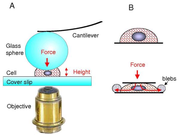
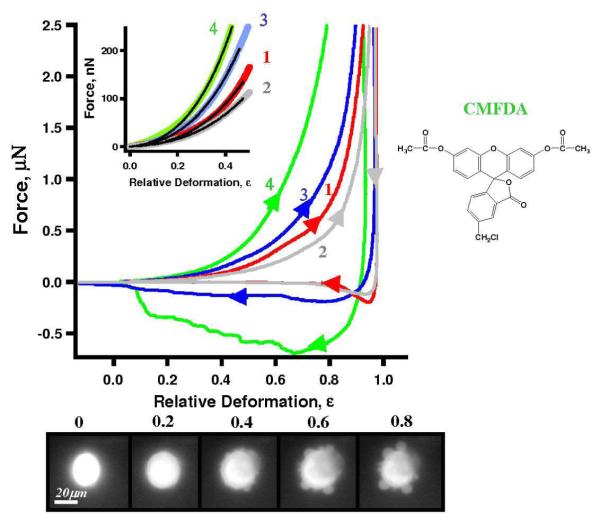
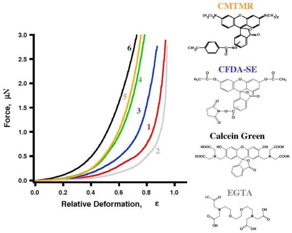
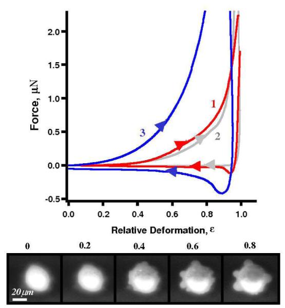
Cell tracing dyes are very frequently utilized in cellular biology research because they provide highly sensitive fluorescent tags that do not compromise cellular functions such as growth and proliferation. In many investigations concerning cellular adhesion and mechanics, fluorescent dyes have been employed with the assumption of little impact on the results. Using the single cell compression technique developed by our team, the single cell mechanics of MDA-MB-468 and MLC-SV40 cells were investigated as a function of dye uptake. Cell tracing dyes increase living cell stiffness 3-6 times and cell-to-probe adhesion up to 7 times. These results suggest a more significant effect than toxins, such as thrombin. A simple analytical model was derived to enable the extraction of the Young's moduli of the cell membrane and cytoskeleton from the force-deformation profiles measured for individual cells. The increase in Young's modulus of the membrane is 3-7 times, which is more significant than that of the cytoskeleton (1.1-3.4 times). We propose that changes in cell mechanics upon the addition of fluorescent tracing dye are primarily due to the incorporation of amphiphilic dye molecules into the cellular plasma membrane, which increases the lateral interaction among phospholipid chains and thus enhances their rigidity and adhesion.
Figures

References
-
- Lichtenfels R, Biddison WE, Schulz H, Vogt AB, Martin R. Care-lass (calcein-release-assay), an improved fluorescence-based test system to measure cytotoxic T-lymphocyte activity. Journal of Immunological Methods. 1994;172(2):227–239. - PubMed
-
- Wang XM, Terasaki PI, Rankin GW, Chia D, Zhong HP, Hardy S. A new microcellular cytotoxicity test based on calcein AM release. Human Immunology. 1993;37(4):264–270. - PubMed
-
- Lyons AB. Divided we stand: Tracking cell proliferation with carboxyfluorescein diacetate succinimidyl ester. Immunology and Cell Biology. 1999;77(6):509–515. - PubMed
-
- Declerck LS, Bridts CH, Mertens AM, Moens MM, Stevens WJ. Use of fluorescent dyes in the determination of adherence of human-leukocytes to endothelial-cells and the effect of fluorochromes on cellular function. Journal of Immunological Methods. 1994;172(1):115–124. - PubMed
-
- Vankessel KPM, Park CT, Wright SD. A fluorescence microassay for the quantitation of iIntegrin-mediated adhesion of neutrophil. Journal of Immunological Methods. 1994;172(1):25–31. - PubMed
Publication types
MeSH terms
Substances
Grants and funding
LinkOut - more resources
Full Text Sources
Other Literature Sources
Miscellaneous
