

Adrenal schwannoma
Abstract
Background: Adrenal schwannomas are very rare tumors that are difficult to diagnose preoperatively. We report the case of a left adrenal schwannoma incidentally discovered in a 55-year-old man during a postoperative checkup for a cutaneous malignant melanoma.
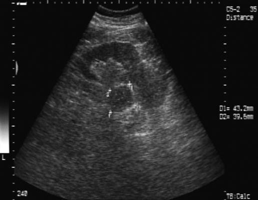
Methods: The biological evaluation was unremarkable, and the radiological examination revealed the adrenal mass that was first considered a metastatic lesion. Adrenalectomy was performed by the laparoscopic approach.
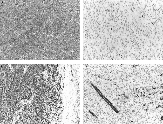
Results: The postoperative course was uneventful. Histological examination established the correct diagnosis of schwannoma, which was also confirmed by immunohistochemical staining.
Conclusions: A nonsecreting adrenal mass can be easily misjudged, especially in the context of a recently operated on malignancy. Unilateral adrenal metastasis needs pathological confirmation, as it can dramatically affect prognosis. Unusual tumors of the adrenal gland may be found incidentally, and a malignant context will generate difficulties in establishing the right management. Complete laparoscopic excision is the treatment of choice whenever feasible and will also clarify pathology.
Figures
References
-
- Rajaratnam A, Waugh J. Adrenal metastases of malignant melanoma: characteristic computed tomography appearances. Australas Radiol. 2005;49(4):325–329 - PubMed
-
- Potente G, Cantisani C, Cantisani V, et al. Valutazione con tomografia computerizzata delle metastasi da melanoma cutaneo. Radiol Med (Torino). 2001;101(4):275–280 - PubMed
-
- Quayle FJ, Spitler JA, Pierce RA, et al. Needle biopsy of incidentally discovered adrenal masses is rarely informative and potentially hazardous. Surgery. 2007;142(4):497–504 - PubMed
-
- Carlini M, Lonardo MT, Boschetto A, et al. Adrenal glands metastases from malignant melanoma. Laparoscopic bilateral adrenalectomy. J Exp Clin Cancer Res. 2003;22(1):141–145 - PubMed
-
- Saraiva P, Rodrigues H, Rodrigues P. Port site recurrence after laparoscopic adrenalectomy for metastatic melanoma. Int Braz J Urol. 2003;29(6):520–521 - PubMed
Publication types
MeSH terms
LinkOut - more resources
Full Text Sources
Medical