Review
. 2009 Jun;1787(6):574-83.
doi: 10.1016/j.bbabio.2009.01.012. Epub 2009 Feb 7.

Architecture of complex I and its implications for electron transfer and proton pumping

Affiliations
Review

Architecture of complex I and its implications for electron transfer and proton pumping

Volker Zickermann et al. Biochim Biophys Acta. 2009 Jun.

Abstract

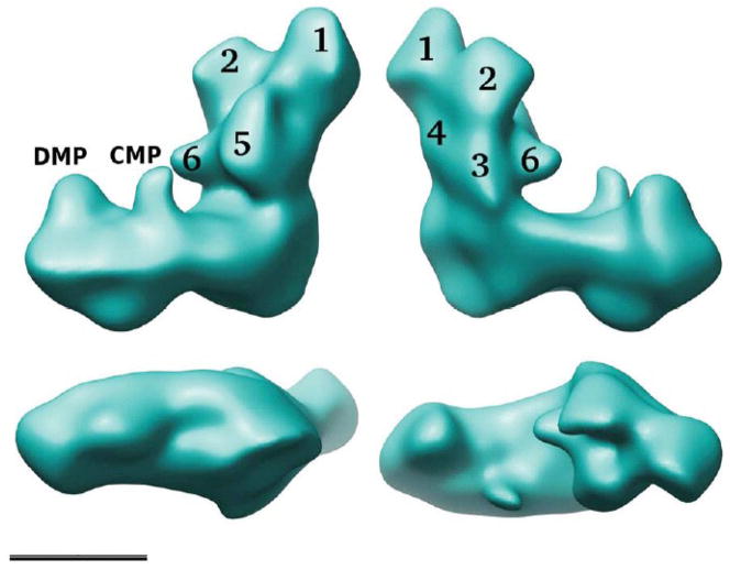
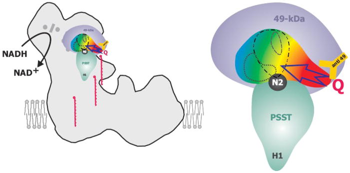
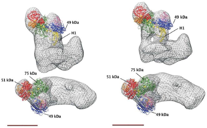
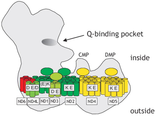
Proton pumping NADH:ubiquinone oxidoreductase (complex I) is the largest and remains by far the least understood enzyme complex of the respiratory chain. It consists of a peripheral arm harbouring all known redox active prosthetic groups and a membrane arm with a yet unknown number of proton translocation sites. The ubiquinone reduction site close to iron-sulfur cluster N2 at the interface of the 49-kDa and PSST subunits has been mapped by extensive site directed mutagenesis. Independent lines of evidence identified electron transfer events during reduction of ubiquinone to be associated with the potential drop that generates the full driving force for proton translocation with a 4H(+)/2e(-) stoichiometry. Electron microscopic analysis of immuno-labelled native enzyme and of a subcomplex lacking the electron input module indicated a distance of 35-60 A of cluster N2 to the membrane surface. Resolution of the membrane arm into subcomplexes showed that even the distal part harbours subunits that are prime candidates to participate in proton translocation because they are homologous to sodium/proton antiporters and contain conserved charged residues in predicted transmembrane helices. The mechanism of redox linked proton translocation by complex I is largely unknown but has to include steps where energy is transmitted over extremely long distances. In this review we compile the available structural information on complex I and discuss implications for complex I function.


Figures

Figure 1
Figure 1. Reconstruction of complex I from Y. lipolytica
The overall structure combining the five major classes at 24 Å resolution is shown [29]; top, two side views; bottom left, a view of the intermembrane space side of the membrane arm; bottom right, view from the matrix side; scale bar 10 nm. In the peripheral arm six domains labelled 1–6 can be discerned; DMP, distal membrane arm protrusion, CMP, central membrane arm protrusion.
Figure 2
Figure 2. Schematic drawing of the ubiquinone binding pocket
The 49-kDa subunit (in violet) and the PSST subunit (in cyan) assemble the ubiquinone binding pocket located next to iron-sulfur cluster N2 (in black). The position of the subunits in the peripheral arm has been modelled according to fit 1 [51] shown in figure 3. The pocket is coloured with a color gradient indicating the severity of point-mutations on complex I activity compared to the parental strain. Red: Almost complete loss of complex I activity for all mutations in this region. Yellow: Strong decrease of complex I activity. Green: Some decrease of complex I activity. Blue: essentially no effects on activity for any mutation in this region. A possible ubiquinone access path in the red region is indicated by an arrow. Within the ubiquinone binding cavity, binding sites of different classes of complex I inhibitors are indicated: ··· C12E8; -·· - rotenone and DQA; --- DQA only. The orange Y represents the anti 49-kDa antibody and marks the position of the corresponding epitope. Ubiquinone-9 molecules are depicted in red at various intermediate positions along a hypothetical “access ramp”, illustrating that the hydrophobic substrate has to leave the membrane bilayer to gain access to its reduction site.
Figure 3
Figure 3. X-ray structure of the peripheral arm fragment from T. thermophilus fitted to 3D reconstructions of complex I from Y. lipolytica, fit 1 from [51]
Left panel, Y. lipolytica complex I at 24 Å resolution as shown in figure 1; right panel, Y. lipolytica complex I class 8_2 with 16.5 Å resolution. At higher resolution the connection of the peripheral arm to the membrane arm via two separate stalks becomes visible. The positions of the largest central hydrophilic subunits and of helix H1 of the PSST subunit are indicated (see text), scale bar 10 nm.
Figure 4
Figure 4. Subcomplexes and central hydrophobic subunits of the membrane arm
Subunits assigned to subcomplex Iα, red; Iγ green; Iβ, yellow. Note that the structural integrity of subcomplex Iγ is a matter of debate (indicated by different shades of green). Ellipses denote putative extramembraneous domains on the inside (note that the topology for ND3 is a matter of debate); highly conserved charged residues in transmembrane helices are assigned to individual subunits (see text), Brackets indicate that residues are close to the ends of putative membrane spanning segments.

References

    1. Brandt U. Energy converting NADH:quinone oxidoreductase (Complex I) Annu Rev Biochem. 2006;75:69–92. - PubMed
    1. Vinogradov AD. Catalytic properties of the mitochondrial NADH-ubiquinone oxidoreductase (Complex I) and the pseudo-reversible active/inactive enzyme transition. Biochim Biophys Acta. 1998;1364:169–185. - PubMed
    1. Smeitink J, Van den Heuvel L, DiMauro S. The genetics and pathology of oxidative phosphorylation. Nat Rev Genet. 2001;2:342–352. - PubMed
    1. Lin MT, Beal MF. Mitochondrial dysfunction and oxidative stress in neurodegenerative diseases. Nature. 2006;443:787–795. - PubMed
    1. Lenaz G, Bovina C, Castelluccio C, Fato R, Formiggini G, Genova ML, Marchetti M, Merlo Pich M, Pallotti F, Parenti Castelli G, Biagini G. Mitochondrial Complex I defects in aging. Mol Cell Biochem. 1997;174:329–333. - PubMed

Publication types

LinkOut - more resources