. 2009 Apr 28;106(17):7191-6.
doi: 10.1073/pnas.0810710106. Epub 2009 Apr 6.

Localized delivery of fibroblast growth factor-2 and brain-derived neurotrophic factor reduces spontaneous seizures in an epilepsy model

Affiliations

Localized delivery of fibroblast growth factor-2 and brain-derived neurotrophic factor reduces spontaneous seizures in an epilepsy model

Beatrice Paradiso et al. Proc Natl Acad Sci U S A. .

Abstract

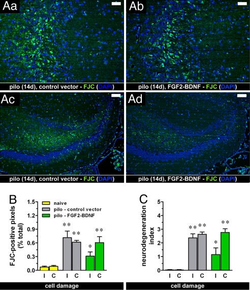
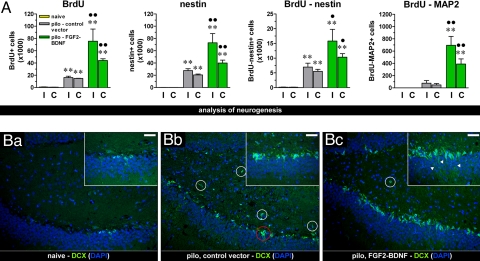
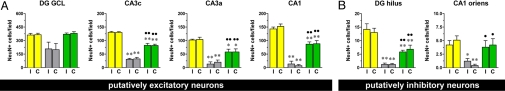
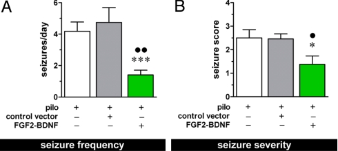
A loss of neurons is observed in the hippocampus of many patients with epilepsies of temporal lobe origin. It has been hypothesized that damage limitation or repair, for example using neurotrophic factors (NTFs), may prevent the transformation of a normal tissue into epileptic (epileptogenesis). Here, we used viral vectors to locally supplement two NTFs, fibroblast growth factor-2 (FGF-2) and brain-derived neurotrophic factor (BDNF), when epileptogenic damage was already in place. These vectors were first characterized in vitro, where they increased proliferation of neural progenitors and favored their differentiation into neurons, and they were then tested in a model of status epilepticus-induced neurodegeneration and epileptogenesis. When injected in a lesioned hippocampus, FGF-2/BDNF expressing vectors increased neuronogenesis, embanked neuronal damage, and reduced epileptogenesis. It is concluded that reduction of damage reduces epileptogenesis and that supplementing specific NTFs in lesion areas represents a new approach to the therapy of neuronal damage and of its consequences.


Conflict of interest statement

The authors declare no conflict of interest.

Figures

Fig. 1.
Fig. 1.
(A) Effect of the treatment with the vector expressing FGF-2 and BDNF together (TH-FGF2/0-BDNF) on SE-induced ongoing damage. Degenerating cells are marked with FJC (green); nuclei marked by DAPI (blue). Fourteen days after pilocarpine (pilo)–induced SE (14d), ongoing hippocampal damage was still detectable in animals injected with the control vector (control), especially in the CA3 area (Aa) and in the dentate (Ac). This pattern was identical to the pattern observed in untreated animals (not shown). In animals treated with TH-FGF2/0-BDNF (FGF2-BDNF), a reduction, but not the abolishment of damage was observed (Ab and Ad). Horizontal bars, 25 μm in (Aa) and (Ab) and 50 μm in (Ac) and (Ad). (B, C) Quantification of neurodegeneration. Shown is the percentage of FJC-positive pixels in the hippocampus (B) and the degeneration index (C), calculated as described in Materials and Methods, in naïve rats (yellow bars) and pilocarpine-treated rats, injected with either the control vector (gray bars) or TH-FGF2/0-BDNF (green bars). I, injected (ipsilateral) hippocampus; C, noninjected (contralateral) hippocampus. Data are means ± SE for seven to eight animals per group. *P < 0.05, **P < 0.01 vs. naïve; Kruskal-Wallis test. No significant difference was observed between pilo – control vector and pilo – FGF2-BDNF.
Fig. 2.
Fig. 2.
Effect of the vector expressing FGF-2 and BDNF together on cell proliferation. (A) Average number of BrdU-positive, nestin-positive, double-labeled BrdU-positive and nestin-positive, double-labeled BrdU-positive and MAP2abc-positive cells in the dorsal hippocampus of naïve rats (yellow bars) and of pilocarpine-treated rats 11 days after inoculation of control vector (pilo – control vector, gray bars) or of the vector expressing FGF-2 and BDNF (pilo – FGF2-BDNF, green bars) in the right, ipsilateral to inoculation (I) and in the left, contralateral (C) hippocampus. Data are the means ± SE of four to five animals per group. ** P < 0.01 vs. naïve; ● P < 0.05, ●● P < 0.01 vs. pilo – control vector; analysis of variance (ANOVA) and post hoc Newman-Keuls test. (B) The vector expressing FGF-2 and BDNF together induced proliferation of DCX-positive cells in vivo. Immunofluorescence (dentate gyrus region) for DCX (green) in control (naïve) rats (Ba), in pilocarpine-treated rats given the control vector (Bb) and in pilocarpine-treated rats given the vector expressing FGF-2 and BDNF (Bc). Nuclei are marked by DAPI (blue). It should be noted that DCX-positive cells in the naïve animal dentate gyrus area are located in the subgranular zone and present detectable elongations projecting across the granular layer (Ba Inset); pilocarpine-induced SE (both in untreated animals and in animals administered the control vector) caused an increase in DCX-positive cells (Bb), which tended to produce relatively fewer elongations (Bb, inset), to group into clusters (example in red circle), and to localize ectopically (white circles). Treatment with the double mutant (Bc) further increased the number of DCX-positive cells while reducing their aberrant features, i.e., lower numbers of ectopic cells and presence of numerous elongations across the granular layer (arrowheads, Bc, inset). Horizontal bar, 25 μm.
Fig. 3.
Fig. 3.
Neuropathological outcome of the treatment with the vector expressing FGF-2 and BDNF (TH-FGF2/0-BDNF), 28 days after pilocarpine-induced status epilepticus. (A) Measure of the density of NeuN-positive, presumably excitatory neurons in selected subareas of the dorsal hippocampus: dentate granule layer (DG GCL), CA3c pyramidal layer (CA3c), CA3a pyramidal layer (CA3a), and CA1 pyramidal layer (CA1). I, injected (ipsilateral) hippocampus; C, noninjected (contralateral) hippocampus. (B) Measure of the density of NeuN-positive, presumably inhibitory neurons in selected subareas of the dorsal hippocampus: hilus of the dentate gyrus (DG hilus) and stratum oriens of CA1 (CA1 oriens). Data are the means ± SE of 10–12 animals per group. *P < 0.05, **P < 0.01 vs. naïve; ● P < 0.05; ●● P < 0.01 vs. pilo – control vector; ANOVA and post hoc Newman-Keuls test.
Fig. 4.
Fig. 4.
EEG and behavioral analysis. Average frequency (A) and severity (B) of spontaneous seizures in the chronic period (14–28 days after pilocarpine-induced SE). Data are the means ± SE for 10–12 animals per group. *P < 0.05 and ***P < 0.001 vs. pilo; ● P < 0.05 and ●● P < 0.01 vs. pilo – control vector; ANOVA and post hoc Newman-Keuls test for (A), Kruskal-Wallis test for (B).

References

    1. Pitkanen A, Sutula TP. Is epilepsy a progressive disorder? Prospects for new therapeutic approaches in temporal-lobe epilepsy. Lancet Neurol. 2002;1:173–181. - PubMed
    1. Lindvall O, Kokaia Z. Stem cells for the treatment of neurological disorders. Nature. 2006;441:1094–1096. - PubMed
    1. Hagg T. Molecular regulation of adult CNS neurogenesis: An integrated view. Trends Neurosci. 2005;28:589–595. - PubMed
    1. Simonato M, Tongiorgi E, Kokaia M. Angels and demons: Neurotrophic factors and epilepsy. Trends Pharmacol Sci. 2006;27:631–638. - PubMed
    1. Kempermann G. They are not too excited: The possible role of adult-born neurons in epilepsy. Neuron. 2006;52:935–937. - PubMed

Publication types

MeSH terms

Substances