. 2009 Jun 5;284(23):15794-804.
doi: 10.1074/jbc.M109.002881. Epub 2009 Apr 14.

Bone morphogenetic protein (BMP) and activin type II receptors balance BMP9 signals mediated by activin receptor-like kinase-1 in human pulmonary artery endothelial cells

Affiliations

Bone morphogenetic protein (BMP) and activin type II receptors balance BMP9 signals mediated by activin receptor-like kinase-1 in human pulmonary artery endothelial cells

Paul D Upton et al. J Biol Chem. .

Abstract

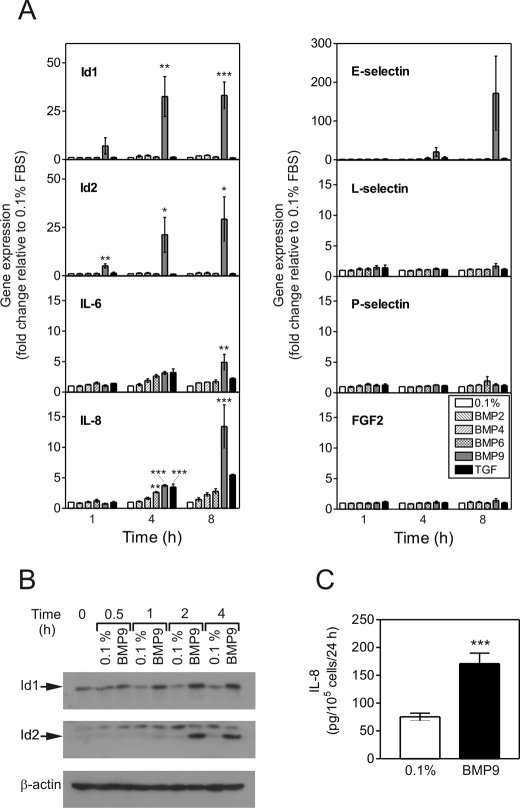
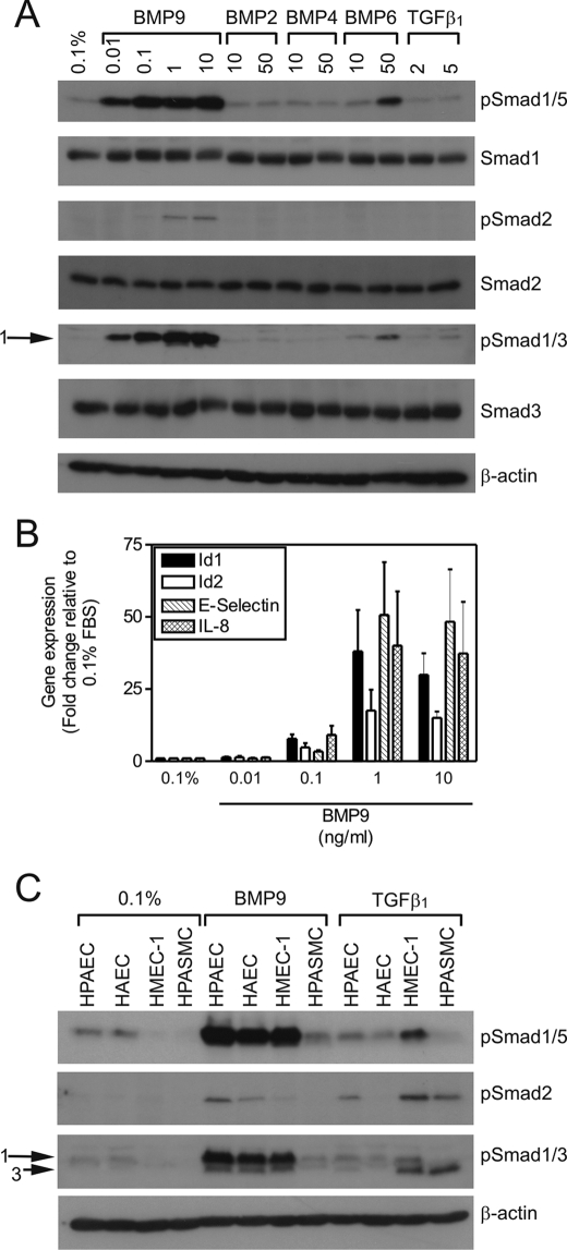
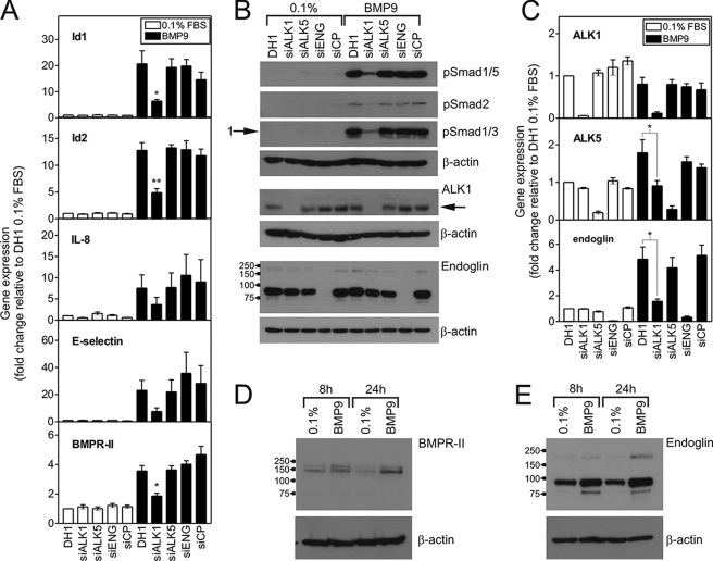
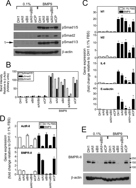
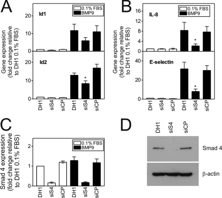
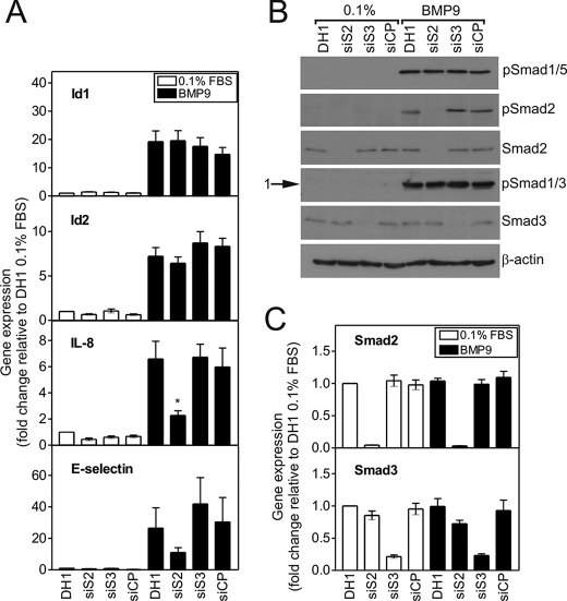
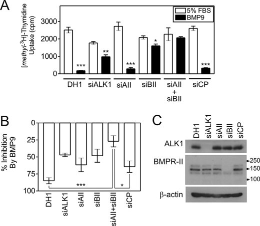
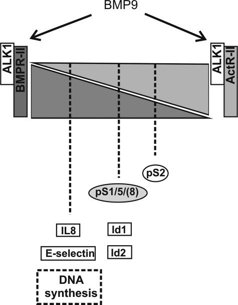
Mutations in transforming growth factor-beta (TGF-beta) receptor superfamily members underlie conditions characterized by vascular dysplasia. Mutations in endoglin and activin-like kinase receptor 1 (ALK1) cause hereditary hemorrhagic telangiectasia, whereas bone morphogenetic protein type II receptor (BMPR-II) mutations underlie familial pulmonary arterial hypertension. To understand the functional roles of these receptors, we examined their relative contributions to BMP signaling in human pulmonary artery endothelial cells (HPAECs). BMP9 potently and selectively induced Smad1/5 phosphorylation and Id gene expression in HPAECs. Contrary to expectations, BMP9 also stimulated Smad2 activation. Furthermore, BMP9 induced the expression of interleukin 8 and E-selectin. Using small interfering RNA, we demonstrate that the type I receptor, ALK1, is essential for these responses. However, small interfering RNA and inhibitor studies showed no involvement of ALK5 or endoglin. We further demonstrate that, of the candidate type II receptors, BMPR-II predominantly mediated IL-8 and E-selectin induction and mitogenic inhibition by BMP9. Conversely, activin receptor type II (ActR-II) contributed more to BMP9-mediated Smad2 activation. Only abolition of both type II receptors significantly reduced the Smad1/5 and Id responses. Both ALK1 and BMPR-II contributed to growth inhibition of HPAECs, whereas ActR-II was not involved. Taken together, our findings demonstrate the critical role of type II receptors in balancing BMP9 signaling via ALK1 and emphasize the essential role for BMPR-II in a subset of BMP9 responses (interleukin 8, E-selectin, and proliferation). This differential signaling may contribute to the contrasting pathologies of hereditary hemorrhagic telangiectasia and pulmonary arterial hypertension.


Figures

FIGURE 1.
FIGURE 1.
BMP9 selectively activates mRNA transcription in HPAECs. A, confluent serum-restricted HPAECs were treated with BMP2, BMP4, BMP6, BMP9 (10 ng/ml), or TGFβ1 (2 ng/ml) in M199, 0.1% FBS (0.1%) for 1, 4, or 8 h. Total RNA was extracted and cDNA prepared. The expression of Id1, Id2, IL-6, IL-8, E-selectin, L-selectin, P-selectin, and fibroblast growth factor 2 (FGF2) were determined by qPCR, with expression being normalized to β-actin and expressed as the fold-change relative to 0.1% at that time point. Data are presented as the mean ± S.E. of three experiments. *, p < 0.05; **, p < 0.01; or ***, p < 0.001 compared with 0.1% FBS (0.1%). B, serum-restricted HPAECs were treated with BMP9 (1 ng/ml) in M199, 0.1% FBS (0.1%) for 0.5, 1, 2, and 4 h. Immunoblotting was performed with antibodies against Id1 and Id2. All blots were reprobed for β-actin to ensure equal loading. C, HPAECs were treated with BMP9 (1 ng/ml) in M199, 0.1% FBS (0.1%) for 24 h. Conditioned medium was assayed for IL-8 using a specific enzyme-linked immunosorbent assay and data are expressed as picograms of IL-8/105 cells. Data are mean ± S.E. (n = 6) from a representative experiment from 3 repeats.
FIGURE 2.
FIGURE 2.
BMP9 induction of Smad phosphorylation and mRNA transcription in HPAECs is concentration-dependent. A, serum-restricted HPAECs were treated with BMP9 (0.01–10 ng/ml), BMP2 (10 or 50 ng/ml), BMP4 (10 or 50 ng/ml), BMP6 (10 or 50 ng/ml), or TGFβ1 (2 or 5 ng/ml) in M199, 0.1% FBS (0.1%) for 1 h. Immunoblotting was performed with antibodies against phospho-Smad1/5, Smad1, phospho-Smad2, Smad2, phospho-Smad1/3, or Smad3. All blots were reprobed for β-actin to ensure equal loading. B, serum-restricted HPAECs were treated with BMP9 (0.01–10 ng/ml) in 0.1% for 8 h. Total RNA was extracted and cDNA prepared. The expression of Id1, Id2, IL-8, and E-selectin were determined by qPCR, with expression being normalized to β-actin and expressed as the fold-change relative to 0.1%. Data are presented as the mean ± S.E. of three experiments. C, serum-restricted HPAECs, HAECs, HMEC-1, and HPASMCs were treated with BMP9 (1 ng/ml) or TGFβ1 (5 ng/ml) in M199,.
FIGURE 3.
FIGURE 3.
siALK1 selectively inhibits BMP9-mediated Smad phosphorylation and mRNA induction in HPAECs. A, HPAECs were transfected with siALK1, siALK5, siEng, or siCP (all 10 nm) using Dharmafect1 (DH1). After 28 h, cells were serum-restricted for 16 h, followed by treatment with BMP9 (1 ng/ml) in 0.1% for 8 h. Total RNA was extracted and cDNA prepared. The expression of Id1, Id2, IL-8, and E-selectin were determined by qPCR, with expression being normalized to β-actin and expressed as the fold-change relative to 0.1%. Data are presented as the mean ± S.E. of four experiments. B, HPAECs were transfected as described above and treated with BMP9 (1 ng/ml) in 0.1% for 1 h. Immunoblotting was performed with antibodies against phospho-Smad1/5, phospho-Smad2, or phospho-Smad1/3. Immunoblotting was also performed to confirm the specific loss of ALK1 (65 kDa) and endoglin with their respective siRNAs. The migration positions of the molecular mass markers (kDa values) are shown on the endoglin blot. C, the expression of ALK1, ALK5, and endoglin were determined in the samples for panel B, confirming each siRNA was selective. Data are presented as the mean ± S.E. of four experiments. D and E, untransfected serum-restricted HPAECs were treated with BMP9 (1 ng/ml) in 0.1% for 8 or 24 h. Immunoblotting was performed with antibodies against (D) BMPR-II or (E) endoglin to confirm the increase in these proteins due to BMP9 treatment. Blots are representative of three separate experiments. *, p < 0.05 or **, p < 0.01 compared with DH1 plus BMP9. All blots were reprobed for β-actin to ensure equal loading.
FIGURE 4.
FIGURE 4.
siActR-II and siBMPR-II differentially alter BMP9-mediated Smad phosphorylation and mRNA induction in HPAECs. A, HPAECs were transfected with siActR-II (siAII), siBMPR-II (siBII), siActR-II + siBMPR-II or siCP (10 nm each siRNA) using Dharmafect1 (DH1). After 28 h, cells were serum-restricted for 16 h, followed by treatment with BMP9 (1 ng/ml) in 0.1% for 1 h. Immunoblotting was performed with antibodies against phospho-Smad1/5, phospho-Smad2, or phospho-Smad1/3. All blots were reprobed for β-actin to ensure equal loading. The blots are representative of three separate experiments. B, graph showing densitometry for the blots in panel A. Band intensities were quantified using Image J and normalized to the β-actin blots corresponding to each Smad blot. C, HPAECs were treated as described above for 8 h. Total RNA was extracted and cDNA prepared. The expression of Id1, Id2, IL-8, and E-selectin were determined by qPCR, with expression being normalized to β-actin and expressed as the fold-change relative to 0.1%. Data are presented as the mean ± S.E. of four experiments. D, the expression of ActR-II and BMPR-II were determined in the samples for panel C, confirming each siRNA was selective. Data are presented as the mean ± S.E. of four experiments. E, cell lysates were immunoblotted for BMPR-II to confirm the specific loss of receptor expression. The migration positions of the molecular mass markers (kDa) are shown. *, p < 0.05 or **, p < 0.01 compared with DH1 plus BMP9.
FIGURE 5.
FIGURE 5.
BMP9 induction of mRNA transcription is Smad4-dependent. A and B, HPAECs were transfected with siSmad4 or siCP (15 nm each) using Dharmafect1 (DH1). After 28 h, cells were serum-restricted for 16 h, followed by treatment with BMP9 (1 ng/ml) in 0.1% for 8 h. Total RNA was extracted and cDNA prepared. The expression of Id1 and Id2 (A) and IL-8 and E-selectin (B) were determined by qPCR, with expression being normalized to β-actin and expressed as the fold-change relative to 0.1%. Data are presented as the mean ± S.E. of five experiments. C, the expression of Smad4 was determined using qPCR, confirming selectivity. Data are presented as the mean ± S.E. of five experiments. D, cell lysates were immunoblotted for Smad4 to confirm the specific loss of receptor expression. *, p < 0.05 compared with DH1 plus BMP9.
FIGURE 6.
FIGURE 6.
The BMP9-mediated induction of IL-8 and E-selectin is regulated by Smad2. A, HPAECs were transfected with siSmad2, siSmad3, or siCP (10 nm each) using Dharmafect1 (DH1). After 28 h, cells were serum-restricted in M199, 0.1% FBS (0.1%) for a further 16 h, followed by treatment with BMP9 (1 ng/ml) in 0.1% for 8 h. Total RNA was extracted and cDNA prepared. The expression of Id1, Id2, IL-8, and E-selectin were determined using qPCR, with expression being normalized to β-actin and expressed as the fold-change relative to 0.1%. Data are presented as the mean ± S.E. of five experiments. B, HPAECs were treated as described above for 1 h. Immunoblotting was performed with antibodies against phospho-Smad1/5, phospho-Smad2, Smad2, phospho-Smad1/3, or Smad3. All blots were reprobed for β-actin to ensure equal loading. The blots are representative of three separate experiments. C, the expression of Smad2 and Smad3 were determined using qPCR, confirming selectivity. Data are presented as the mean ± S.E. of five experiments. *, p < 0.05 compared with DH1 plus BMP9.
FIGURE 7.
FIGURE 7.
BMP9 inhibits DNA synthesis via ALK1 and BMPR-II. A, HPAECs were transfected with siALK1, siActR-II (siAII), siBMPR-II (siBII), siActR-II + siBMPR-II, or siCP (10 nm each siRNA) using Dharmafect1 (DH1). Cells were incubated in EGM-2 overnight and the medium changed to M199, 0.1% FBS for 8 h. At 28 h post-transfection, cells were incubated with M199, 5% FBS (5% FBS) or M199, 5% FBS containing 1 ng/ml BMP9 (BMP9). After a further 18 h, 0.25 μCi of [methyl-3H]thymidine was added to each well for 6 h. The lysates were quantified by liquid scintillation counting. Quantitative PCR analysis confirmed the reduction in mRNA at 28 and 52 h. The graph is representative of five experiments (n = 4 wells/treatment). *, p < 0.05; **, p < 0.01; or ***, p < 0.001 compared with 5% FBS with Student's unpaired t test. B, mean normalized data from four experiments showing the percentage inhibition of [methyl-3H]thymidine uptake by BMP9 in HPAECs transfected with siRNAs as described above. *, p < 0.05; or ***, p < 0.001 compared with DH1 with Student's unpaired t test. C, protein lysates were immunoblotted with antibodies against ALK1 or BMPR-II to confirm selective reduction of these proteins. The migration positions of the molecular mass markers are shown on the BMPR-II blot.
FIGURE 8.
FIGURE 8.
Summary of the relative contributions of BMPR-II and ActR-II to BMP9 signaling through ALK1. Summary figure representing the relative contributions of ALK1, ActR-II, and BMPR-II to BMP9-mediated Smad signaling and transcriptional regulation in HPAECs. The shaded triangles represent the relative contribution and compensation by BMPR-II and ActR-II. The Smad, gene, and functional responses are positioned according to the type II receptor involvement/compensation, such that both receptors compensate for loss of the other with regard to Smad1/5 and Id responses. In contrast, loss of BMPR-II has a dramatic effect upon IL-8 and E-selectin expression, and inhibition of DNA synthesis by BMP9. Conversely, reduction of ActR-II has a greater impact on Smad2 phosphorylation than BMPR-II reduction, but loss of both receptors abolishes the Smad2 response.

References

    1. Tuder R. M., Marecki J. C., Richter A., Fijalkowska I., Flores S. ( 2007) Clin. Chest Med. 28, 23– 42, vii - PMC - PubMed
    1. Lane K. B., Machado R. D., Pauciulo M. W., Thomson J. R., Phillips J. A., 3rd, Loyd J. E., Nichols W. C., Trembath R. C. ( 2000) Nat. Genet. 26, 81– 84 - PubMed
    1. Deng Z., Morse J. H., Slager S. L., Cuervo N., Moore K. J., Venetos G., Kalachikov S., Cayanis E., Fischer S. G., Barst R. J., Hodge S. E., Knowles J. A. ( 2000) Am. J. Hum. Genet. 67, 737– 744 - PMC - PubMed
    1. Machado R. D., Aldred M. A., James V., Harrison R. E., Patel B., Schwalbe E. C., Gruenig E., Janssen B., Koehler R., Seeger W., Eickelberg O., Olschewski H., Elliott C. G., Glissmeyer E., Carlquist J., Kim M., Torbicki A., Fijalkowska A., Szewczyk G., Parma J., Abramowicz M. J., Galie N., Morisaki H., Kyotani S., Nakanishi N., Morisaki T., Humbert M., Simonneau G., Sitbon O., Soubrier F., Coulet F., Morrell N. W., Trembath R. C. ( 2006) Hum. Mutat. 27, 121– 132 - PubMed
    1. Sadick H., Sadick M., Götte K., Naim R., Riedel F., Bran G., Hörmann K. ( 2006) Wien. Klin. Wochenschr. 118, 72– 80 - PubMed

Publication types

MeSH terms