. 2009 Jun 5;284(23):16037-48.
doi: 10.1074/jbc.M109.001123. Epub 2009 Apr 14.

Novel vascular endothelial growth factor D variants with increased biological activity

Affiliations

Novel vascular endothelial growth factor D variants with increased biological activity

Pyry I Toivanen et al. J Biol Chem. .

Abstract

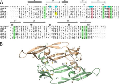
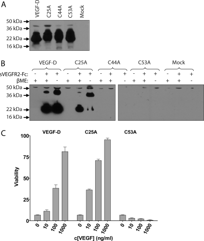
Members of the vascular endothelial growth factor (VEGF) family play a pivotal role in angiogenesis and lymphangiogenesis. They are potential therapeutics to induce blood vessel formation in myocardium and skeletal muscle, when normal blood flow is compromised. Most members of the VEGF/platelet derived growth factor protein superfamily exist as covalently bound antiparallel dimers. However, the mature form of VEGF-D (VEGF-D(DeltaNDeltaC)) is predominantly a non-covalent dimer even though the cysteine residues (Cys-44 and Cys-53) forming the intersubunit disulfide bridges in the other members of the VEGF family are also conserved in VEGF-D. Moreover, VEGF-D bears an additional cysteine residue (Cys-25) at the subunit interface. Guided by our model of VEGF-D(DeltaNDeltaC), the cysteines at the subunit interface were mutated to study the effect of these residues on the structural and functional properties of VEGF-D(DeltaNDeltaC). The conserved cysteines Cys-44 and Cys-53 were found to be essential for the function of VEGF-D(DeltaNDeltaC). More importantly, the substitution of the Cys-25 at the dimer interface by various amino acids improved the activity of the recombinant VEGF-D(DeltaNDeltaC) and increased the dimer to monomer ratio. Specifically, substitutions to hydrophobic amino acids Ile, Leu, and Val, equivalent to those found in other VEGFs, most favorably affected the activity of the recombinant VEGF-D(DeltaNDeltaC). The increased activity of these mutants was mainly due to stabilization of the protein. This study enables us to better understand the structural determinants controlling the biological activity of VEGF-D. The novel variants of VEGF-D(DeltaNDeltaC) described here are potential agents for therapeutic applications, where induction of vascular formation is required.


Figures

FIGURE 1.
FIGURE 1.
The structure of VEGF-DΔNΔC. A, structure-based sequence alignment of the VEGF homology domain regions of the VEGF family members. The crystal structures of VEGF-A (PDB code 1FLT), PlGF (1RV6), VEGF-B (2C7W), VEGF-E (2GNN), and VEGF-F VR-1 (1WQ9) were superimposed as described under “Experimental Procedures.” The sequences of VEGF-D (UniProt accession code O43915) and VEGF-C (UniProt accession code P49767) were aligned to the structure-based alignment. Cysteines of the cystine knot are in green. Cysteines forming the (putative) intermolecular disulfide bond are in pink. The extra cysteine, found in VEGF-D and VEGF-C, is in yellow. Mutated amino acids of the dimer interface are in cyan. Amino acids equivalent to Arg-22 and Cys-25 in VEGF-D are framed. Secondary structure elements (according to VEGF-A structure (22)) are shown in gray. The figure was prepared using Alscript 2.04 (54), Inkscape 0.46, and Gimp 2.6. B, homology model of the VEGF-DΔNΔC dimer. Each monomer (pale green and wheat) consists of a central four-stranded β-sheet (β1, β3, β5, and β6), two additional short β-strands (β4 and β7), and two short α-helices (α1 and α2). Cysteines are shown as sticks. The figure was prepared using Pymol 1.1 (55) and Inkscape 0.46.
FIGURE 2.
FIGURE 2.
Expression, sVEGFR2-Fc binding and dimerization of VEGF-DΔNΔC variants. A, an immunoblot of transiently transfected 293T cell media separated with SDS-PAGE under reducing conditions using an anti-VEGF-D mAb. B, precipitation of receptor binding VEGF-D forms from transiently transfected 293T cell media. The media and sVEGFR2-Fc protein were incubated, and protein complexes were collected using protein A-Sepharose. Samples were separated with SDS-PAGE either with or without β-mercaptoethanol (βME) in the sample buffer and immunoblotted using an anti-VEGF-D mAb to analyze receptor binding and dimerization of the VEGF-D variants. C, Ba/F3-VEGFR-2 cell growth and survival assay of recombinant proteins produced in insect cells. Cells were supplemented with the indicated concentrations of the proteins, and viability was determined after 48 h of incubation.
FIGURE 3.
FIGURE 3.
Amino acids of the dimer interface in homology models of the different VEGF-D variants. A, native VEGF-D. B–E, VEGF-D variants C53A (B), C25L (C), R22I/C25L (D), and G51C (E). Amino acids at the interface are shown as sticks. Mutated amino acids are in pink. Monomers are colored as in Fig. 1B. The figure was prepared using Pymol 1.1 (55) and Inkscape 0.46.
FIGURE 4.
FIGURE 4.
Screening of the biological activity of the VEGF-D mutants on Ba/F3-VEGFR-2 and Ba/F3-VEGFR-3 cell lines. VEGF-DΔNΔC and the mutants were produced in transiently transfected 293T cells. The concentrations of VEGF-DΔNΔC proteins in the media were measured using VEGF-D enzyme-linked immunosorbent assay (R&D Systems), and Ba/F3 cells were supplemented with the indicated concentrations of the proteins. Mock A, B, and C represent similar serial dilutions of media from mock-transfected cells as used for the VEGF-D-containing samples. A and C, assays on the Ba/F3-VEGFR-2 cells. B and D, assays on the Ba/F3-VEGFR-3 cells.
FIGURE 5.
FIGURE 5.
The dimerization of VEGF proteins. A, VEGF-DΔNΔC and the mutants were produced in transiently transfected 293T cells. Samples of the media were separated using SDS-PAGE without reducing agents, and VEGF-D proteins were detected by immunoblotting using an anti-VEGF-D mAb. B and C, purified recombinant VEGF proteins were separated using SDS-PAGE under reducing conditions (B) and non-reducing conditions (C) and stained using Coomassie staining.
FIGURE 6.
FIGURE 6.
Determination of biological activity and receptor binding affinity of the purified recombinant VEGFs. Ba/F3-VEGFR-2 (A) and Ba/F3-VEGFR-3 (B) cell growth and survival assays were performed with purified recombinant VEGF proteins. C, the relative VEGFR-2 binding affinities of recombinant VEGF proteins were measured using a solid phase competition assay. Dilution series of the recombinant VEGF proteins were incubated with sVEGFR2-Fc recombinant protein on VEGF-A121-coated 96-well plates. The amount of bound sVEGFR2-Fc was quantified using an anti-human Fc-AP antibody, and the values are expressed as mean ± S.E. D, the relative VEGFR-3 binding affinities of recombinant VEGF proteins were measured using a competition assay. Experiments were performed as above but using VEGF-C-coated plates and sVEGFR3-Fc recombinant protein. E, the determined IC50 values of each protein in the receptor affinity assays. Sigmoidal dose response curves were fitted to measured data using a non-linear regression in Prism to determine the IC50 values of each protein.
FIGURE 7.
FIGURE 7.
Phosphorylation of VEGFR-2, VEGFR-3, and Akt. Serum-starved cells were incubated with recombinant VEGF proteins, and the phosphorylation status of VEGFRs or Akt was analyzed from cell lysates. The results are representatives of replicate experiments yielding similar results. A, the effect of 500 ng/ml VEGF-DΔNΔC, C25A, and C25L on the phosphorylation of VEGFR-2 in PAE-KDR cells at 15- and 30-min time points. The phosphorylation of Tyr-1175 of VEGFR-2 was quantified from the cell lysates using immunoblotting with a specific antibody for VEGFR-2 phosphorylated at Tyr-1175 (Cell Signaling). Equal loading was confirmed using an antibody against total VEGFR-2 (Cell Signaling). B, the effect of VEGF-DΔNΔC, C25L, and VEGF-A121 on the phosphorylation of VEGFR-2 in PAE-KDR cells at 5-, 15-, and 30-min time points. C, the effect of VEGF-DΔNΔC and C25L on the phosphorylation of VEGFR-3 in PAE-Flt4 cells at 5-, 15-, and 30-min time points. Cell lysates were immunoprecipitated using an anti-VEGFR-3 (Abcam) antibody and analyzed by immunoblotting with an anti phosphotyrosine antibody (Upstate). Equal loading was confirmed using an anti-VEGFR-3 antibody (Abcam). D, the effect of VEGF-DΔNΔC and C25L on the phosphorylation of Akt in PAE-Flt4 cells at 5-, 15-, and 30-min time points. The phosphorylation of Akt was quantified from the cell lysates using immunoblotting with a specific antibody for Akt phosphorylated at Ser-473 (Cell Signaling). Equal loading was confirmed using an antibody against total Akt (Cell Signaling).
FIGURE 8.
FIGURE 8.
Stability of recombinant VEGF-DΔNΔC and its C25L mutant. A, insect cell -derived purified recombinant proteins were incubated both in cell culture medium or Tris-buffered saline from 0 to 48 h. VEGF-D proteins were captured onto anti-FLAG M2 mAb-coated 96-well plates, and the active proteins were detected using sVEGFR2-Fc recombinant protein and an anti-human Fc-AP antibody. A standard curve was generated for both proteins from serial dilutions of the 0-h sample. The amount of remaining active proteins is presented as the percentage of the active protein relative to the concentration in the 0-h sample. The values are expressed as the mean ± S.E. B, immunoblot of the samples from different time points separated under non-reducing conditions and using an anti-VEGF-D mAb. Dimeric (di) and monomer (mo) fractions are indicated. TBS, Tris-buffered saline.
FIGURE 9.
FIGURE 9.
Vascular permeability effects of the recombinant proteins in the skin of New Zealand White rabbits. The recombinant proteins were injected into the rabbit skin, and Evans Blue dye was administered intravenously. The rabbits were sacrificed after 30 min and perfused. The blue color indicates extravasation of proteins from the vasculature.

Similar articles

Cited by

References

    1. Byrne A. M., Bouchier-Hayes D. J., Harmey J. H. ( 2005) J. Cell Mol. Med. 9, 777– 794 - PMC - PubMed
    1. Tammela T., Enholm B., Alitalo K., Paavonen K. ( 2005) Cardiovasc. Res. 65, 550– 563 - PubMed
    1. Lyttle D. J., Fraser K. M., Fleming S. B., Mercer A. A., Robinson A. J. ( 1994) J. Virol. 68, 84– 92 - PMC - PubMed
    1. Yamazaki Y., Takani K., Atoda H., Morita T. ( 2003) J. Biol. Chem. 278, 51985– 51988 - PubMed
    1. Holmes D. I., Zachary I. ( 2005) Genome Biol. 6, 209. - PMC - PubMed

Publication types

MeSH terms

Substances

LinkOut - more resources