. 2009 Jun 12;284(24):16667-16678.
doi: 10.1074/jbc.M109.002816. Epub 2009 Apr 14.

Anterograde transport of surfactant protein C proprotein to distal processing compartments requires PPDY-mediated association with Nedd4 ubiquitin ligases

Affiliations

Anterograde transport of surfactant protein C proprotein to distal processing compartments requires PPDY-mediated association with Nedd4 ubiquitin ligases

Adam Kotorashvili et al. J Biol Chem. .

Abstract

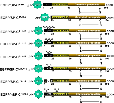
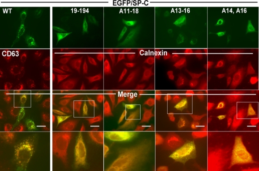
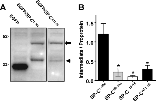
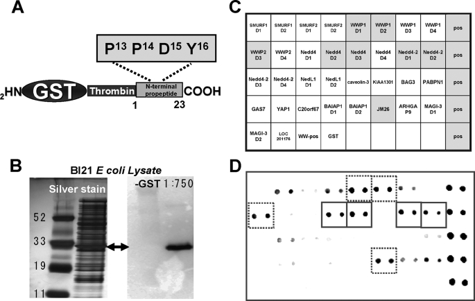
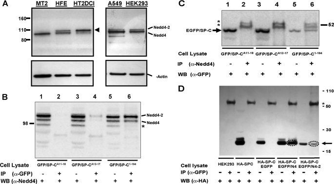
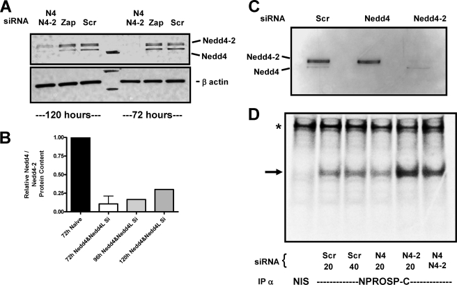
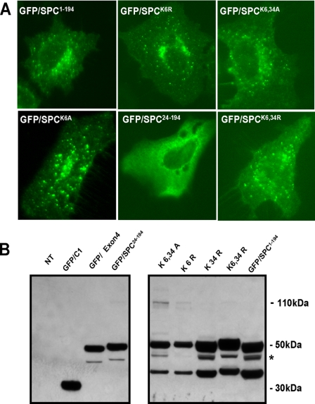
Biosynthesis of surfactant protein C (SP-C) by alveolar type 2 cells requires proteolytic processing of a 21-kDa propeptide (proSP-C21) in post-Golgi compartments to yield a 3.7-kDa mature form. Scanning alanine mutagenesis, binding assays, and co-immunoprecipitation were used to characterize the proSP-C targeting domain. Delivery of proSP-C21 to distal processing organelles is dependent upon the NH2-terminal cytoplasmic SP-C propeptide, which contains a conserved PPDY motif. In A549 cells, transfection of EGFP/proSP-C21 constructs containing polyalanine substitution for Glu11-Thr18, 13PPDY16, or 14P,16Y produced endoplasmic reticulum retention of the fusion proteins. Protein-protein interactions of proSP-C with known WW domains were screened using a solid-phase array that revealed binding of the proSP-C NH2 terminus to several WW domains found in the Nedd4 family of E3 ligases. Specificity of the interaction was confirmed by co-immunoprecipitation of proSP-C and Nedd4 or Nedd4-2 in epithelial cell lines. By Western blotting and reverse transcription-PCR, both forms were detected in primary human type 2 cells. Knockdown of Nedd4-2 by small interference RNA transfection of cultured human type 2 cells blocked processing of 35S-labeled proSP-C21. Mutagenesis of potential acceptor sites for ubiquitination in the cytosolic domain of proSP-C (Lys6, Lys34, or both) failed to inhibit trafficking of EGFP/proSP-C21. These results indicate that PPDY-mediated interaction with Nedd4 E3-ligases is required for trafficking of proSP-C. We speculate that the Nedd4/proSP-C tandem is part of a larger protein complex containing a ubiquitinated component that further directs its transport.


Figures

FIGURE 1.
FIGURE 1.
EGFP/SP-C Constructs. Inserts containing wild-type, mutant rat SP-C, or mutant human SP-C were generated by PCR and subcloned into the EGFPC1 vector as described under “Experimental Procedures.” Amino acid nomenclature is based on published SP-C sequences (10). Top to bottom: EGFP/SP-C1–194 containing wild-type rat SP-C shown with putative intra-chain disulfide bonding between Cys186 and Cys122. “NTP” = N-terminal propeptide; EGFP/SP-C19–194 is a deletional construct produced by removal of amino acids 1–18 of the NTP; EGFP/SP-CA11–18, EGFP/SP-CA12–17, EGFP/SP-CA13–16, and EGFP/SP-CA14A16 each contain polyalanine (A) substitution at the indicated residues in the NH2-flanking propeptide; EGFP/SP-CΔ10–18 contains a deletion of the conserved sequence Met10–Thr18 in the cytoplasmic NTP; EGFP/hSP-CK6K34 represents full-length human SP-C for, which either arginine (R) or alanine (A) is substituted at lysine (K) residues 6 and/or 34.
FIGURE 2.
FIGURE 2.
The PPDY domain within Met10–Thr18 is critical for targeting of proSP-C. The subcellular distribution of wild-type and mutant EGFP/SP-C constructs transiently transfected into A549 cells was determined by fluorescence microscopy. Plasmid DNA encoding EGFP/SP-C1–194 (= WT), the NH2-terminal truncation mutant EGFP/SP-C19–194, polyalanine mutants EGFP/SP-CA11–18, EGFP/SP-CA13–16, or EGFP/SP-CA14A16 were introduced into A549 cells grown on coverslips (70% confluent) using LipofectamineTM as described under “Experimental Procedures.” 48 h after transfection, cells were fixed and stained for the lysosomal-like organelle marker CD63 or the ER marker calnexin using polyclonal antibodies and a Texas Red goat anti-rabbit secondary antiserum. Double label fluorescence images for EGFP (top panels) or Texas Red expression (second row) were acquired, and single images were digitally overlaid using Metamorph imaging software (version 7.5, Molecular Devices, Downingtown, PA). As shown in the merged images (third row), EGFP/SP-C1–194 (= WT) was found to co-localize with CD63 in cytosolic vesicles, whereas EGFP/SP-C19–194, EGFP/SP-CA11–18, EGFP/SP-CA13–16, and EGFP/SP-CA14A16 were each expressed in a diffuse cytosolic reticular pattern that co-localized with calnexin. Bar = 10 μm. Regions delineated by the solid boxes were enlarged to provide additional resolution (bottom row).
FIGURE 3.
FIGURE 3.
Processing of EGFP/SP-C mutants. A549 cells were transiently transfected with EGFP-C1, EGFP/SP-C1–194, EGFP/SP-C19–194, EGFP/SP-CΔ11–18, or EGFP/SP-CA11–18 using CaPO4. Nuclear-free lysates were prepared from cell pellets as described under “Experimental Procedures.” A, 50% of each lysate was subjected to 12% SDS-PAGE and immunoblotted with polyclonal anti-GFP, and bands were visualized using ECL. EGFP-C1 was expressed as a major product with Mr 27,000. EGFP/SP-C1–194 was expressed as a primary translation product with Mr of 48,000 with a lower molecular weight band (Mr = 38,000–40,000) consistent with COOH propeptide processing. EGFP/SP-CA11–18 was expressed predominantly as a primary product of Mr 48,000 with minimal processing intermediate. B, for each condition, densitometry was performed on the primary translation product and processing intermediates, and the ratio of the processed intermediate/proprotein was calculated (n = 3 separate experiments; mean ± S.E.). At steady state, ∼50% of EGFP/SP-C1–194 is in the form of processed intermediates, whereas all NH2-terminal mutants were only ∼10–15% processed (*, p < 0.05 versus EGFP/SP-C1–194 by analysis of variance).
FIGURE 4.
FIGURE 4.
A GST fusion protein containing the SP-C NTP is a ligand for WW domains. A, schematic diagram of the coding insert for expression of a GST-tagged NTP of proSP-C containing the PPDY motif amplified and subcloned into PGEX-2TK as described under “Experimental Procedures” and supplemental Table S1. B, expression of GST-SP-CNTP in BL21 bacterial cells upon induction with isopropyl 1-thio-β-d-galactopyranoside. Lysate was analyzed by SDS-PAGE and silver staining (left) or immunoblotting with α-GST (right). C, schematic diagram of WW Domain Array I (Panomics, Inc.) showing the positions of immobilized WW domain motifs (two spots per motif) used to screen for SP-CNTP/WW domain binding. Pos = positive control. D, dot immunoblot generated by incubation of WHOLE BL21 lysate containing GST/SP-CNTP with the WW Domain I membrane per the manufacturer's guidelines, and binding signals were visualized by secondary incubation with anti-NPROSP-C2–9 and ECL. Representative film shows selective binding to several WW domains contained in Nedd4 and Nedd4-2 E3 ligases (solid boxes) as well as other related proteins (dashed boxes).
FIGURE 5.
FIGURE 5.
Nedd4 and Nedd4-2 are expressed in epithelial cells and interact with proSP-C. A, cell lysates from freshly isolated mouse alveolar type 2 cells (MT2), freshly isolated epithelial cells from second trimester human fetal lung (HFE), human alveolar type 2 cells induced by culture in DCI (HT2DCI), A549 cells and HEK293 cells were analyzed by SDS-PAGE and Western blotting with a panNedd4 antibody. Position of Novex Sharp Standards is shown at left. To control for loading, blots were re-probed for β-actin (lower panel). Human epithelial cell lines express both Nedd4 and Nedd4-2 homologues as indicated. Note the dramatic increase in Nedd4-2 expression in human lung epithelial cells cultured for 96 h in DCI to promote type 2 cell differentiation (arrowhead). B and C, endogenous Nedd4 and Nedd4-2 specifically interact with the NTP of EGFP/proSP-C. A549 cells were transiently transfected with either EGFP/SP-C1–194, or one of two NTP polyalanine mutants EGFP/SP-CA11–18 or EGFP/SP-CA12–17 using CaPO4 as described under “Experimental Procedures.” B, 72 h after transfection, A549 cell lysates expressing EGFP-tagged SP-C isoforms were immunoprecipitated with monoclonal α-GFP and immunoblotted with polyclonal α-Nedd4 (lanes 2, 4, and 6). To control for input, lanes 1, 3, and 5 represent 5% of the indicated total cell lysate that was directly immunoblotted with α-Nedd4. EGFP/SP-C1–194 captures two bands corresponding to Nedd-4 and Nedd4-2, which were not recognized by either EGFP/SP-CA11–18 or EGFP/SP-CA12–17. An additional band (*) present in A549 cell lysates was detected by the panNedd4 antibody by immunoblotting but not by immunoprecipitation. C, the identical lysates were immunoprecipitated with polyclonal α-Nedd4 and immunoblotted with monoclonal α-GFP (lanes 2, 4, and 6). To control for input, lanes 1, 3, and 5 represent 5% of the indicated total cell lysate that was directly immunoblotted with α-GFP. Nedd4 isoforms interacted only with EGFP/SP-C1–194 (arrow). **, nonspecific doublet corresponding to IgG in the immunoprecipitated samples. D, exogenous Nedd4 and Nedd4-2 each interact with the NTP of wild-type EGFP/proSP-C. HEK293 cells were transiently transfected with either HA-SP-C1–194 alone or in combination with the EGFP/Nedd4 (ΔC2 clone) or EGFP/Nedd4-2 plasmid. Cell lysates were immunoprecipitated with monoclonal α-GFP, subjected to SDS-PAGE, and immunoblotted with monoclonal α-HA. To control for input, 5% of the indicated total cell lysate was also directly immunoblotted with α-HA (the position of HA-SP_C is indicated by the arrow). Both EGFP/Nedd4 and EGFP/Nedd4-2 but not EGFP captured HA-SP-C1–194 (circle). “HEK293” = non-transfected controls. *, nonspecific band corresponding to IgG.
FIGURE 6.
FIGURE 6.
Knockdown of Nedd4 homologues inhibits SP-C processing in human type 2 cells. Freshly isolated epithelial cells from second trimester human fetal lung were electroporated with 20 nm Nedd4 siRNA (#120777), Nedd4-2 (#23570), a combination of both Nedd4 and Nedd4-2 (40 nm total), 20 nm to40 nm scrambled control siRNA, or buffer alone and then cultured in DCI for 72–120 h. A, representative Western blot analyses of cell lysates from siRNA treated, scrambled (Scr), and control (Zap) cells immunoblotted with polyclonal α-Nedd4 showing inhibition of expression of both Nedd4 and Nedd4-2 (upper doublet) homologues at 72 and 120 h. B, the time course of the knockdown of Nedd4/Nedd4-2 protein expression was determined by densitometry of immunopositive bands from Western blots. Data were normalized to 72 h untreated cells and expressed as range of mean ± range (n = 2 experiments). C, Western blot analyses with polyclonal α-Nedd4 of lysates from cells subjected to individual Nedd4 siRNA treatments showing specific inhibition of either Nedd4 or Nedd4-2 (doublet) at 120 h. D, cells in culture treated with siRNA for 120 h were continuously labeled with [35S]Met/Cys for 2 h and immunoprecipitated with either non-immune serum (NIS) or NPROSP-C11–23 as described under “Experimental Procedures.” Phosphorimaging acquired autoradiograph (representative of three separate experiments) showing accumulation of proSP-C primary translation product in cells treated with Nedd4-2 siRNA. The arrow depicts the position of proSP-C21 primary translation product; *, nonspecific band shown as a loading control.
FIGURE 7.
FIGURE 7.
Ubiquitination of proSP-C is not required for Nedd4- mediated trafficking. HEK293 cells transiently transfected with either EGFP/hSP-C1–197, or missense mutants containing substitution of cytosolic lysine (K) or arginine (R) at positions 6 and 34 of proSP-C as described under “Experimental Procedures.” Positive controls for ER retention or aggregation, EGFP/SP-C24–194or EGFP/hSP-CΔExon4 were also utilized. A, nuclear-free lysates prepared from cell pellets were immunoblotted with polyclonal α-GFP, and bands were visualized using ECL. EGFP-C1 was expressed as a major product with Mr 27,000. EGFP/SP-C1–194 was expressed as a primary translation product with Mr of 48,000 with a lower molecular weight band (Mr = 33,000) consistent with COOH propeptide processing. All K and R mutants were expressed and exhibited the same relative degree of COOH-terminal processing. In contrast, EGFP/SP-C24–194 and EGFP/hSP-CΔExon4 were expressed predominantly as primary products of Mr 48,000 and 45,000, respectively, with minimal processing. *, nonspecific cleavage product representing cleavage of a portion of EGFP. B, images for EGFP expression were acquired 72 h after transfection by fluorescence microscopy. In contrast to the ER retention mutants, EGFP/SP-C1–194 as well as all ubiquitin-deficient mutants were transported to cytosolic vesicles.

Similar articles

Cited by

References

    1. Beers M. F., Mulugeta S. ( 2005) Annu. Rev. Physiol. 67, 663– 696 - PubMed
    1. Beers M. F., Lomax C. ( 1995) Am. J. Physiol. Lung Cell. Mol. Physiol. 269, L744– 753 - PubMed
    1. Brasch F., ten Brinke A., Johnen G., Ochs M., Kapp N., Müller K. M., Beers M. F., Fehrenbach H., Richter J., Batenburg J. J., Bühling F. ( 2002) Am. J. Respir. Cell Mol. Biol. 26, 659– 670 - PubMed
    1. Beers M. F. ( 1996) J. Biol. Chem. 271, 14361– 14370 - PubMed
    1. Vorbroker D. K., Voorhout W. F., Weaver T. E., Whitsett J. A. ( 1995) Am. J. Physiol. Lung Cell Mol. Physiol. 269, L727– 733 - PubMed

Publication types

MeSH terms

Substances

LinkOut - more resources