. 2009 May 1;122(Pt 9):1390-400.
doi: 10.1242/jcs.043042. Epub 2009 Apr 14.

Recruitment of vimentin to the cell surface by beta3 integrin and plectin mediates adhesion strength

Affiliations

Recruitment of vimentin to the cell surface by beta3 integrin and plectin mediates adhesion strength

Ramona Bhattacharya et al. J Cell Sci. .

Abstract

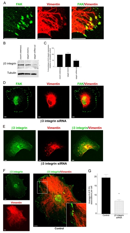
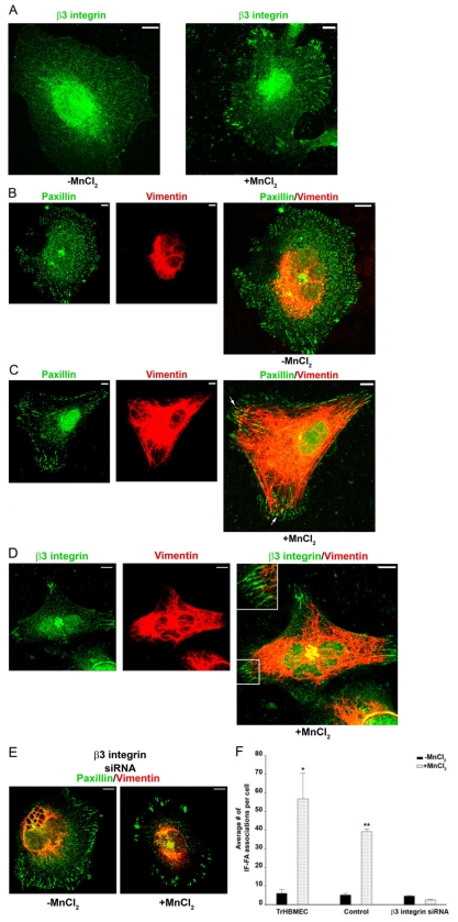
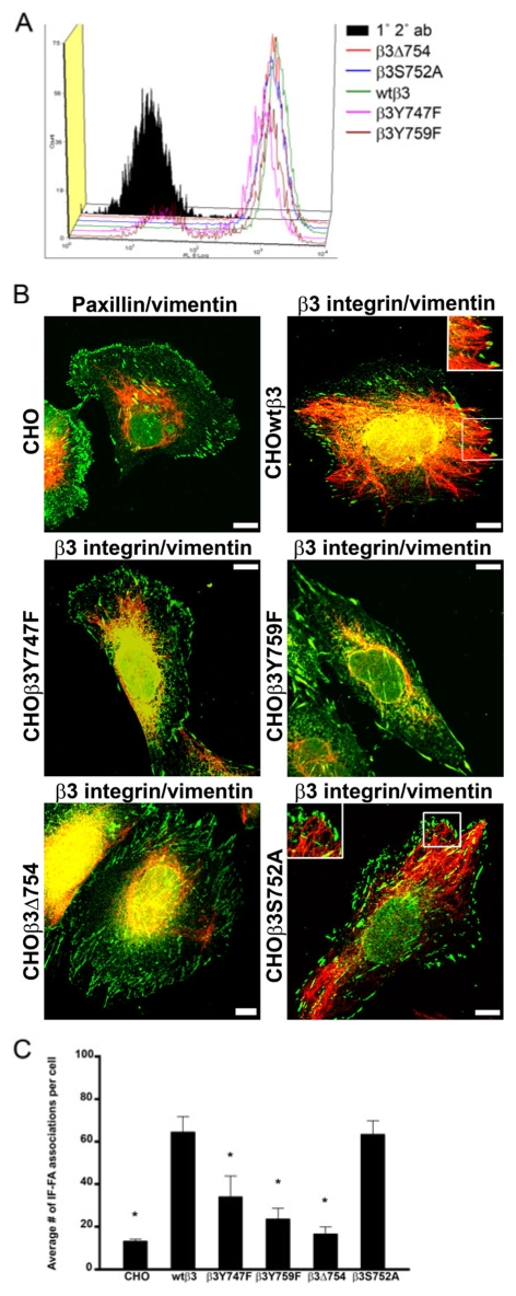
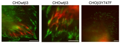
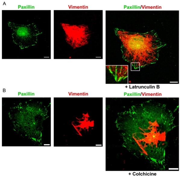
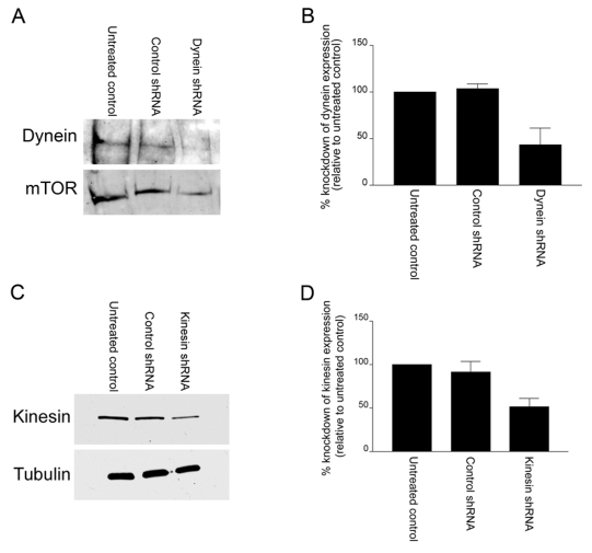
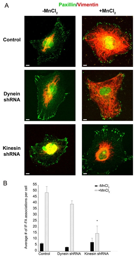
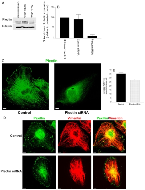
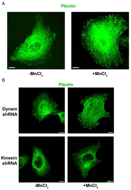
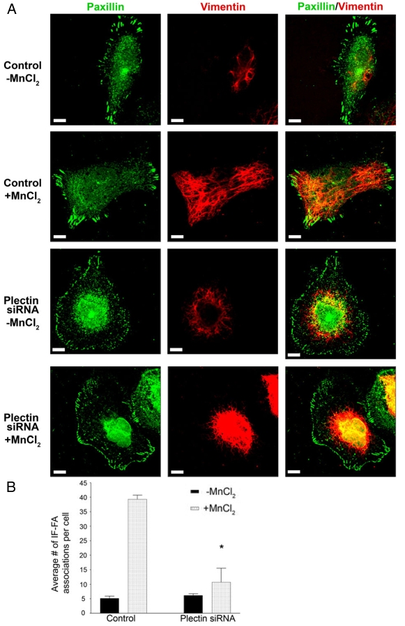
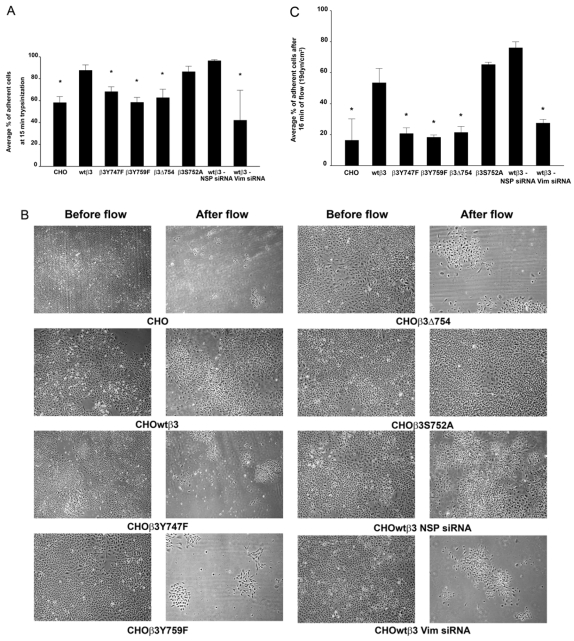
Much effort has been expended on analyzing how microfilament and microtubule cytoskeletons dictate the interaction of cells with matrix at adhesive sites called focal adhesions (FAs). However, vimentin intermediate filaments (IFs) also associate with the cell surface at FAs in endothelial cells. Here, we show that IF recruitment to FAs in endothelial cells requires beta3 integrin, plectin and the microtubule cytoskeleton, and is dependent on microtubule motors. In CHO cells, which lack beta3 integrin but contain vimentin, IFs appear to be collapsed around the nucleus, whereas in CHO cells expressing beta3 integrin (CHOwtbeta3), vimentin IFs extend to FAs at the cell periphery. This recruitment is regulated by tyrosine residues in the beta3 integrin cytoplasmic tail. Moreover, CHOwtbeta3 cells exhibit significantly greater adhesive strength than CHO or CHO cells expressing mutated beta3 integrin proteins. These differences require an intact vimentin network. Therefore, vimentin IF recruitment to the cell surface is tightly regulated and modulates the strength of adhesion of cells to their substrate.


Figures

Fig. 1.
Fig. 1.
Knockdown of β3 integrin in TrHBMECs perturbs interactions between IFs and the cell surface. (A) TrHBMECs were stained for FAK (green) and vimentin (red) as indicated. The panel on the right shows an overlay of the green and red channels. The samples were viewed by confocal microscopy with the focal plane being proximal to the substratum-attached surface of the cells. (B) Extracts of mock-transfected TrHBMECs (untreated control) and TrHBMECs transfected with either control siRNA or β3 integrin siRNA were probed first with a β3 integrin antibody and then reprobed with tubulin. (C) Analysis of densitometric scans of western blots from three different experiments equivalent to those in B. Error bars represent s.e.m. of three experiments. (D) TrHBMECs transfected with β3 integrin siRNA were stained for FAK (green) and vimentin (red) as indicated. (E) TrHBMECs transfected with β3 integrin siRNA were stained for β3 integrin (overexposed to show green) and vimentin (red) as indicated. (F) TrHBMECs transfected with control siRNA were stained for β3 integrin (green) and vimentin (red) as indicated. The inset in the panel on the right is a higher magnification of the boxed area. (G) Quantification of the IF-FA association in TrHBMECs transfected with β3 integrin siRNA compared with the control (*P<0.01). Error bars represent s.e.m. of three experiments, counting a total of 200 cells. Scale bars: 10 μm.
Fig. 2.
Fig. 2.
β3 integrin recruits IFs to FAs. (A-D) TrHBMECs were plated in serum-free medium on PLL-coated coverslips overnight. Untreated cells (left panel in A;B) or cells incubated for 30 minutes in medium supplemented with 0.5 mM MnCl2 (right panel in A;C,D) were processed for immunofluorescence microscopy using antibodies against β3 integrin (green) (A), a combination of antibodies against paxillin (green) and vimentin (red) (B,C), or a combination of antibodies against β3 integrin (green) and vimentin (red) (D). In B-D individual images and overlays of the green and red channels are presented. The arrows in C indicate the IF-FA association. In D, the boxed area is shown at higher magnification in the inset. (E) TrHBMECs were treated with β3 integrin siRNA 72 hours before being plated in serum-free medium on PLL-coated coverslips overnight. Untreated cells (left panel) or cells incubated for 30 minutes in medium supplemented with 0.5 mM MnCl2 (right panel) were processed for immunofluorescence microscopy using a combination of antibodies against paxillin (green) and vimentin (red). (F) Quantification of the IF-FA interaction, as imaged in B,C,E. Error bars represent s.e.m. of three experiments, counting a total of 200 cells. The number of vimentin IF-FA interactions is significantly higher (*P<0.01) in TrHBMECs plated on PLL-coated substrate and treated with MnCl2 compared with that in untreated cells plated on PLL alone. Control cells (cells treated with transfection reagents alone and no siRNA) treated with MnCl2 had significantly more IF-FA associations per cell than control cells plated on PLL alone or TrHBMECs transfected with β3 integrin siRNA and then plated on PLL-coated substrate and treated with MnCl2 (**P<0.01). Indeed, there is no significant difference in IF-FA associations in TrHBMECs transfected with β3 integrin siRNA and then plated on PLL-coated substrate whether they are treated with MnCl2 or not (P>0.1). Scale bars: 10 μm.
Fig. 3.
Fig. 3.
IF recruitment to FAs is regulated by β3 integrin in CHO cells and specifically by tyrosine residues within the tail of the β3 integrin. (A) Constructs encoding wild-type and mutant GFP-tagged β3 integrin were transfected into CHO cells and surface expression of αvβ3 integrin was evaluated by FACS using LM609 antibody (1° 2° ab, nontransfected CHO cells labeled with primary and secondary antibodies). (B) CHO cells (top left) were prepared for immunofluorescence using a combination of paxillin (green) and vimentin (red) antibodies. In addition, CHO cells expressing the indicated GFP-tagged β3 integrin proteins were stained for vimentin (red). The insets (top right, bottom right) are higher magnifications of the boxed areas. (C) Quantification of IF-FA association for all of the CHO cell lines. Over 200 cells were analyzed per cell line in at least three separate experiments. Error bars represent s.e.m. of three experiments. *P<0.03; statistically significant decrease in IF-FA association compared with CHO cells expressing wild-type β3 integrin.
Fig. 4.
Fig. 4.
TIRFM reveals close interaction between vimentin and β3 integrin. CHOwtβ3-GFP cells (left and center panels) and a CHOβ3Y747F-GFP cell (right panel) transfected with vimentin-CFP and imaged by TIRFM. The left and center panels show the close interaction of vimentin IF with FAs at the basal surface of the cell (note regions of yellow in the center panel indicating close association between IFs and β3 integrin). In contrast, vimentin IFs in the CHOβ3Y747F cell are above the level of the plane visualized by TIRFM. Scale bars: 10 μm.
Fig. 5.
Fig. 5.
IF recruitment to FAs is regulated by the microtubule cytoskeleton. (A,B) TrHBMECs were plated in serum-free medium on PLL-coated coverslips overnight. The cells were treated with 0.1 μM latrunculin B (A) or 0.1 μM colchicine (B) for 15 or 30 minutes, respectively, and then incubated in the same drug for an additional 30 minutes with addition of 0.5 mM MnCl2. The treated cells were subsequently processed for double labeling using a combination of antibodies against paxillin (green) and vimentin (red). The inset in A is a higher magnification of the boxed area. Scale bars: 10 μm.
Fig. 6.
Fig. 6.
The use of shRNAs to effectively knockdown dynein and kinesin heavy chain protein expression in TrHBMECs. (A) Extracts of mock electroporated TrHBMECs (untreated control) and cells electroporated with either control (heavy chain kinesin) shRNA or heavy chain dynein shRNA were processed for western immunoblotting, probed first with a dynein heavy chain antibody and then reprobed with an antibody against mTOR. (B) Analysis of densitometric scans of western blots from three different experiments as shown in A. Error bars represent s.e.m. of three experiments. (C) Extracts of mock electroporated TrHBMECs (untreated control) and cells electroporated with either control (heavy chain dynein) shRNA or heavy chain kinesin shRNA were processed for western immunoblotting, probed first with a heavy chain kinesin antibody and then reprobed with an antibody against tubulin. (D) Analysis of densitometric scans of western blots from three different experiments equivalent to that shown in C. Error bars represent s.e.m. of three experiments.
Fig. 7.
Fig. 7.
Kinesin, but not dynein, has a role in IF recruitment to FAs. (A) TrHBMECs electroporated with the control vector, heavy chain dynein shRNA or heavy chain kinesin shRNA were plated in serum-free medium on PLL-coated coverslips overnight. Untreated cells (left panels) or cells incubated for 30 minutes in medium supplemented with 0.5 mM MnCl2 (right panels) were processed for immunofluorescence microscopy using a combination of antibodies against paxillin (green) and vimentin (red). (B) Quantification of the IF-FA association in TrHBMECs electroporated with heavy chain dynein or heavy chain kinesin shRNA compared with cells electroporated with the control vector. Error bars represent s.e.m. of three experiments, counting a total of 200 cells. Upon addition of MnCl2, cells electroporated with heavy chain kinesin shRNA exhibit significantly fewer IF-FA associations per cell than control cells and cells electroporated with heavy chain dynein shRNA (*P<0.01). Scale bars: 10 μm.
Fig. 8.
Fig. 8.
Plectin is not required to maintain IF-FA association. (A) Extracts of mock-transfected TrHBMECs (untreated control) and cells transfected with either control siRNA or plectin siRNA were processed for western blotting, probed first with a plectin antibody and then reprobed with a tubulin antibody. (B) Analysis of densitometric scans of western blots from three different experiments equivalent to that shown in A. Error bars represent s.e.m. of three experiments. (C) Mock-transfected (control) TrHBMECs or TrHBMECs transfected with plectin siRNA were stained for plectin (green). (D) Mock-transfected (control) TrHBMECs or TrHBMECs transfected with plectin siRNA were allowed to spread on glass coverslips before being processed for immunofluorescence microscopy using a combination of antibodies against paxillin (green) and vimentin (red). (E) Quantification of IF-FA association in TrHBMECs transfected with plectin siRNA compared with mock-transfected (control) cells. There is no significant difference in IF-FA association between plectin siRNA-treated cells and control cells (P>0.1). Error bars represent s.e.m. of three experiments. Scale bars: 10 μm.
Fig. 9.
Fig. 9.
Plectin is recruited to FAs in TrHBMECs in a kinesin-dependent manner. (A) TrHBMECs were plated in serum-free medium on PLL-coated coverslips overnight. Untreated cells (left panel) or cells incubated for 30 minutes in medium supplemented with 0.5 mM MnCl2 (right panel) were processed for immunofluorescence microscopy using an antibody against plectin. (B) TrHBMECs electroporated with heavy chain dynein shRNA or heavy chain kinesin shRNA were plated in serum-free medium on PLL-coated coverslips overnight. Untreated cells (left panels) or cells incubated for 30 minutes in medium supplemented with 0.5 mM MnCl2 (right panels) were processed for immunofluorescence microscopy using an antibody against plectin. Scale bars: 10 μm.
Fig. 10.
Fig. 10.
Plectin is required for recruitment of IF to FAs. (A) Mock-transfected (control) TrHBMECs or TrHBMECs transfected with plectin siRNA were plated in serum-free medium on PLL-coated coverslips overnight. In addition, cells in the second and fourth rows were incubated for 30 minutes in medium supplemented with 0.5 mM MnCl2. All cells were processed for immunofluorescence microscopy using a combination of antibodies against paxillin (green) and vimentin (red). Overlays are shown in the panels on the right. (B) Quantification of IF-FA association in TrHBMECs transfected with plectin siRNA compared with mock-transfected (control) cells. Upon addition of MnCl2, cells transfected with plectin siRNA had significantly fewer IF-FA associations per cell than control cells (*P<0.01). Error bars represent s.e.m. of three experiments, counting a total of 200 cells. Scale bars: 10 μm.
Fig. 11.
Fig. 11.
IF-FA interaction enhances cell-substratum adhesion strength of CHO cells. (A) Mock-transfected CHO cells, CHO cells expressing wild-type or mutated β3 integrin, and CHO cells expressing wild-type β3 integrin transfected with control (NSP) or vimentin siRNA were plated into 12-well dishes and allowed to spread for at least 24 hours. They were then treated with trypsin for 15 minutes at room temperature. The percentage of adherent cells was calculated at the end of the treatment protocol. Error bars represent s.e.m. of three experiments. *P<0.03 compared with CHO cells expressing wild-type β3 integrin. (B) The same collection of CHO cells described in A, seeded onto Flexcell glass slides, were subjected to flowing medium (19 dynes/cm2) for 16 minutes. The images show cells before and after flow. (C) The graph represents the percentage of cells, as described in A, remaining adherent to the substrate after they were subjected to flowing medium (19 dynes/cm2) for 16 minutes. Error bars represent s.e.m. of three experiments. *P<0.05 compared with CHO cells expressing wild-type β3 integrin.

References

    1. Arias-Salgado, E. G., Lizano, S., Sarkar, S., Brugge, J. S., Ginsberg, M. H. and Shattil, S. J. (2003). Src kinase activation by direct interaction with the integrin β cytoplasmic domain. Proc. Natl. Acad. Sci. USA 100, 13298-13302. - PMC - PubMed
    1. Axelrod, D. (1989). Total internal reflection fluorescence microscopy. Methods Cell Biol. 30, 245-270. - PubMed
    1. Bershadsky, A. D., Tint, I. S. and Svitkina, T. M. (1987). Association of intermediate filaments with vinculin-containing adhesion plaques of fibroblasts. Cell Motil. Cytoskeleton 8, 274-283. - PubMed
    1. Blystone, S. D. (2004). Integrating an integrin: a direct route to actin. Biochim. Biophys. Acta 1692, 47-54. - PubMed
    1. Blystone, S. D., Lindberg, F. P., Williams, M. P., McHugh, K. P. and Brown, E. J. (1996). Inducible tyrosine phosphorylation of the β3 integrin requires the αv integrin cytoplasmic tail. J. Biol. Chem. 271, 31458-31462. - PubMed

Publication types

MeSH terms