. 2009 May 1;69(9):3955-62.
doi: 10.1158/0008-5472.CAN-08-4450. Epub 2009 Apr 14.

PIK3CA and PIK3CB inhibition produce synthetic lethality when combined with estrogen deprivation in estrogen receptor-positive breast cancer

Affiliations

PIK3CA and PIK3CB inhibition produce synthetic lethality when combined with estrogen deprivation in estrogen receptor-positive breast cancer

Robert J Crowder et al. Cancer Res. .

Abstract

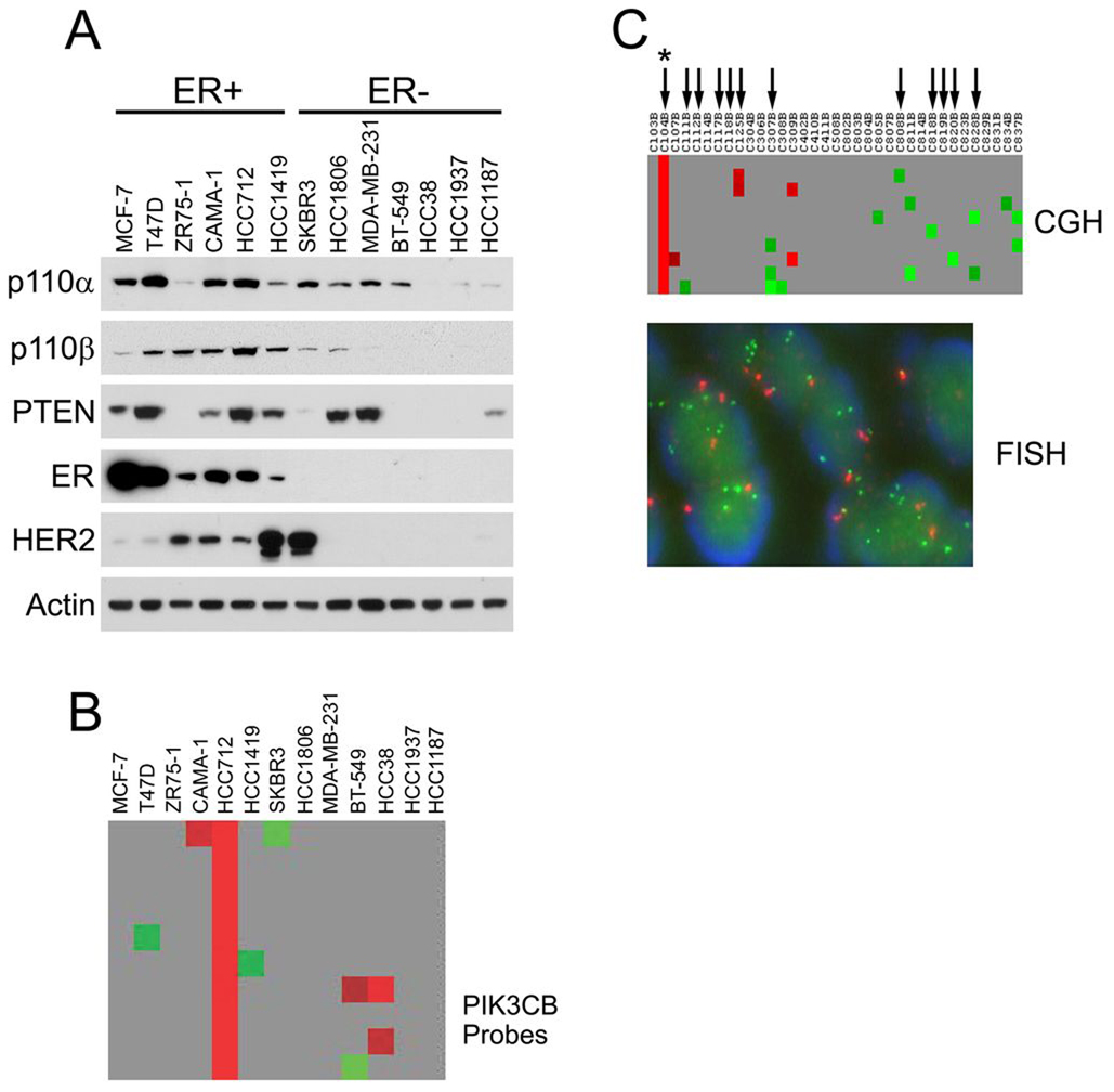
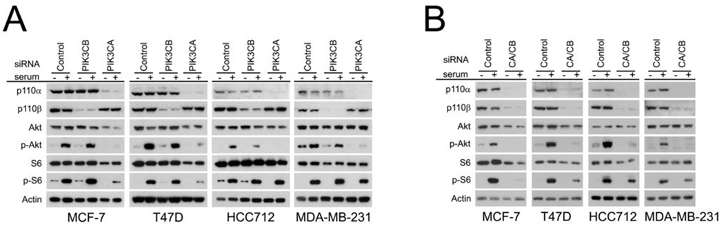
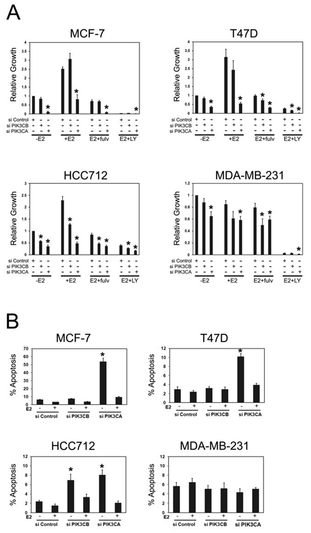
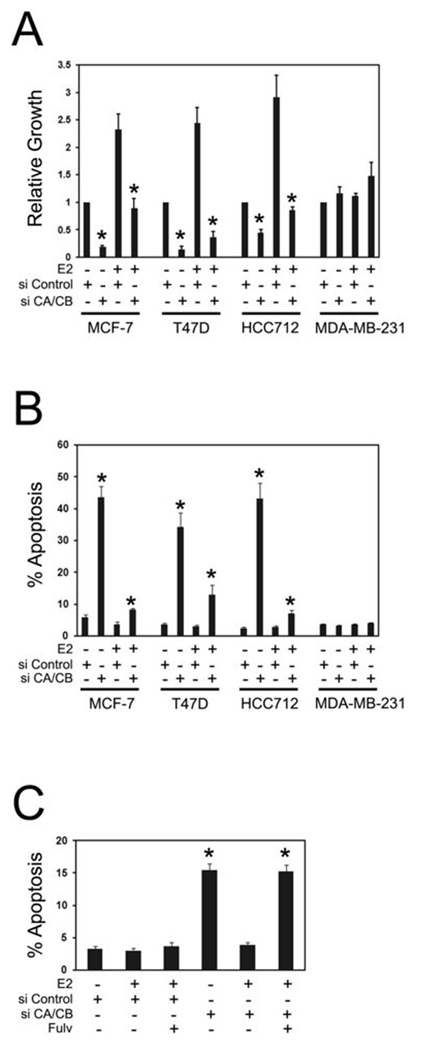
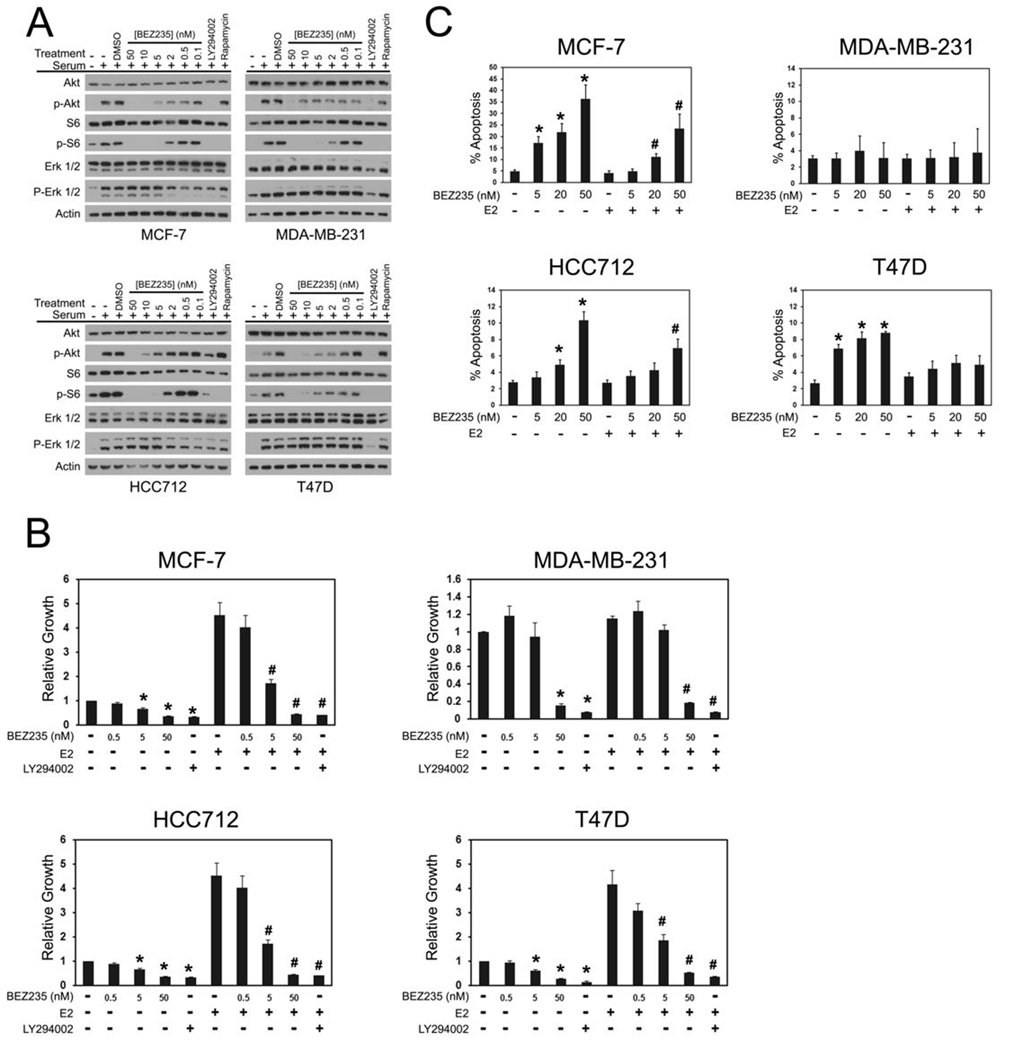
Several phosphoinositide 3-kinase (PI3K) catalytic subunit inhibitors are currently in clinical trial. We therefore sought to examine relationships between pharmacologic inhibition and somatic mutations in PI3K catalytic subunits in estrogen receptor (ER)-positive breast cancer, in which these mutations are particularly common. RNA interference (RNAi) was used to determine the effect of selective inhibition of PI3K catalytic subunits, p110alpha and p110beta, in ER(+) breast cancer cells harboring either mutation (PIK3CA) or gene amplification (PIK3CB). p110alpha RNAi inhibited growth and promoted apoptosis in all tested ER(+) breast cancer cells under estrogen deprived-conditions, whereas p110beta RNAi only affected cells harboring PIK3CB amplification. Moreover, dual p110alpha/p110beta inhibition potentiated these effects. In addition, treatment with the clinical-grade PI3K catalytic subunit inhibitor BEZ235 also promoted apoptosis in ER(+) breast cancer cells. Importantly, estradiol suppressed apoptosis induced by both gene knockdowns and BEZ235 treatment. Our results suggest that PI3K inhibitors should target both p110alpha and p110beta catalytic subunits, whether wild-type or mutant, and be combined with endocrine therapy for maximal efficacy when treating ER(+) breast cancer.


Conflict of interest statement

Conflicts of Interest: No pharmaceutical company funding was received for this research project. Dr Ellis has received honoraria, grants and has served as a consultant for AstraZenica, Novartis and Pfizer.

Figures

Figure 1
Figure 1. PIK3CB is amplified in breast cancer
A, p110α and p110β expression in breast cancer cell lines. Equal amounts (25µg) of protein from each cell line were immunoblotted for the indicated proteins. Longer exposures revealed that both p110α and p110β proteins were expressed in all cell lines. B, PIK3CB aCGH analysis in breast cancer cell lines. Shown is a section of probes on chromosome 3 corresponding to the PIK3CB locus. Individual array probes indicate probable copy number gain (red), loss (green), or no change (gray) relative to female diploid DNA. C, aCGH and PIK3CB FISH in ER+ breast tumors. Top panel, aCGH probes corresponding to the PIK3CB locus. Arrows indicate tumor samples subjected to PIK3CB FISH. One of the FISH-tested tumor samples (*) contained PIK3CB amplification. Bottom panel, FISH results from the PIK3CB-amplified breast tumor above. The CEP3 probe is red; the PIK3CB-specific probe is green.
Figure 2
Figure 2. p110α is the predominant mediator of PI3K signaling in breast cancer cells
A, effect of p110α and p110β knockdown on PI3K signaling. Cells were transfected with control siRNAs (Control) or siRNAs against PIK3CB (PIK3CB) or PIK3CA (PIK3CA). Three days after transfection, serum-deprived cells were stimulated with 20% FBS (final concentration) and lysates were analyzed for effects on PI3K pathway signaling through phospho-Akt (p-Akt) and phospho-S6 (p-S6) immunoblotting. Shown are representative immunoblots obtained from at least three experiments per cell line. B, effect of p110α/p110β dual knockdown on PI3K signaling. Cells were transfected with control siRNAs or a mixture of PIK3CA and PIK3CB siRNAs (CA/CB), treated as above and subjected to immunoblot analysis. Representative results obtained in at least two experiments per cell line are shown.
Figure 3
Figure 3. PIK3CA and PIK3CB RNAi cause synthetic lethality in estrogen-deprived ER+ breast cancer cells
A, PIK3CA and PIK3CB RNAi inhibit growth of ER+ breast cancer cells. Cells in CSS medium were transfected with 10 nM Control (si Control), PIK3CB (si PIK3CB) or PIK3CA (si PIK3CA) siRNAs. Cells were treated without (−E2) or with 10 nM estradiol (+E2) in the absence or presence of 300 nM Fulvestrant (Fulv) or 20 µM LY294002 (LY). Growth was assessed after 10 d of treatment and is expressed relative to untreated (−E2), Control siRNA transfected cells. Results from five experiments per cell line are shown. Significant differences (p< 0.05, *) between treatments in PIK3CB or PIK3CA siRNA transfected cells and identical treatments in Control siRNA transfected cells are indicated. B, PIK3CA and PIK3CB RNAi promote apoptosis in estrogen-deprived ER+ cells. Cells growing in CSS medium were transfected with siRNAs as in A, above and treated without or with 10 nM estradiol for 7d. Apoptosis was assessed by counting TUNEL-positive or pyknotic Hoechst-stained nuclei. Results from four experiments per cell line are shown. Significant differences (p< 0.05. *) between estrogen-deprived Control siRNA and estrogen-deprived PIK3CB or PIK3CA siRNA transfected cells are indicated. Estrogen suppression of PIK3CB RNAi-induced apoptosis in HCC712 cells was not statistically significant (p= 0.06).
Figure 4
Figure 4. Dual PIK3CA/PIK3CB RNAi enhances apoptosis in estrogen-deprived ER+ cells
A, dual PIK3CA/PIK3CB RNAi inhibits growth of ER+ cells. Cells in CSS medium were transfected with 20 nM Control (si Control) or 10 nM each (20 nM final) PIK3CA and PIK3CB siRNAs (si CA/CB). Cells were left untreated or treated with 10 nM estradiol (E2) and growth was assessed after 10d. Growth is expressed relative to untreated, Control siRNA transfected cells. Results are from 4–6 experiments per cell line. Significant differences (p< 0.05, *) between Control siRNA and PIK3CA/PIK3CB siRNA transfected cells in the presence or absence of estradiol are indicated. B, dual p110α/p110β knockdown enhances apoptosis in ER+ cells. Cells were transfected with 20 nM Control or 10 nM each PIK3CA and PIK3CB siRNAs. Cells were left untreated or treated with 10 nM estradiol for 7d and apoptosis was assessed by counting Hoechst-stained nuclei. Results from four experiments per cell line are shown. Significant differences (p< 0.05, *) between Control siRNA and dual PIK3CA/PIK3CA siRNA transfected cells in either the presence or absence of estradiol are indicated. Estradiol significantly suppressed PIK3CA/PIK3CB RNAi-induced apoptosis MCF-7, T47D and HCC712 cells. C, dual p110α/p110β knockdown was performed in T47D cells with alternative PIK3CA and PIK3CB siRNAs. Cells were left untreated, treated with 10 nM estradiol or treated with estradiol + 300 nM Fulvestrant (Fulv) for 7d. Apoptosis was assessed by counting Hoechst-stained nuclei. Results from four experiments are shown. Significant differences (p< 0.05, *) between Control and PIK3CA/PIK3CA siRNA transfected cells in either the presence or absence of estradiol are indicated.
Figure 5
Figure 5. BEZ235 causes synthetic lethality in estrogen-deprived ER+ breast cancer cells
A, BEZ235 treatment inhibits PI3K pathway signaling in breast cancer cells. Serum-starved cells were treated with vehicle (DMSO), the indicated concentrations of BEZ235, LY294002 (20µM), or rapamycin (100 nM) then stimulated with 20% FBS. Cell lysates were immunoblotted to determine effects on PI3K signaling by phospho-Akt (p-Akt) and phospho-S6 (p-S6) antibodies and on MAPK signaling via phospho-ERK (p-ERK 1/2) antibodies. Shown are representative results from at least two experiments per cell line. B, BEZ235 inhibits growth of breast cancer cells. Cells in CSS medium were treated without or with BEZ235 in the absence or presence of 10 nM estradiol (E2). Cell growth was measured after 10 d and is calculated relative to growth in untreated (−E2) cells. Shown are results from five experiments per cell line. Significant differences in growth between estrogen-deprived and estrogen-deprived, drug-treated cells are indicated (p<0.05, *). Significant differences in growth between estrogen-stimulated and estrogen-stimulated cells, drug- treated cells are indicated (p<0.05, #). C, BEZ235 promotes apoptosis in ER+ cells. Cells growing in CSS medium were treated with the indicated concentrations of BEZ235 without or with 10 nM estradiol for 7d. Apoptosis was assessed by counting Hoechst-stained nuclei. Results from 4–6 experiments per cell line are shown. Significant induction of cell death in estrogen-deprived, BEZ235 treated cells (p< 0.05, *) and cells treated with BEZ235 in the presence of estradiol (p< 0.05, #) is indicated.

References

    1. EBCTCG. Effects of chemotherapy and hormonal therapy for early breast cancer on recurrence and 15-year survival: an overview of the randomised trials. Lancet. 2005;365:1687–1717. - PubMed
    1. Ellis MJ, Tao Y, Luo J, et al. Outcome Prediction for Estrogen Receptor-Positive Breast Cancer Based on Postneoadjuvant Endocrine Therapy Tumor Characteristics. JNCI. 2008;100 - PMC - PubMed
    1. Dowsett M, Smith IE, Ebbs SR, et al. Proliferation and apoptosis as markers of benefit in neoadjuvant endocrine therapy of breast cancer. Clin Cancer Res. 2006;12:1024s–1030s. - PubMed
    1. Hennessy BT, Smith DL, Ram PT, Lu Y, Mills GB. Exploiting the PI3K/AKT pathway for cancer drug discovery. Nat Rev Drug Discov. 2005;4:988–1004. - PubMed
    1. Li J, Yen C, Liaw D, et al. PTEN, a putative protein tyrosine phosphatase gene mutated in human brain, breast, and prostate cancer. Science. 1997;275:1943–1947. - PubMed

Publication types

MeSH terms