. 2009 May 1;69(9):3892-900.
doi: 10.1158/0008-5472.CAN-08-4242. Epub 2009 Apr 14.

Computationally guided photothermal tumor therapy using long-circulating gold nanorod antennas

Affiliations

Computationally guided photothermal tumor therapy using long-circulating gold nanorod antennas

Geoffrey von Maltzahn et al. Cancer Res. .

Abstract

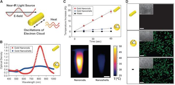
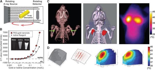
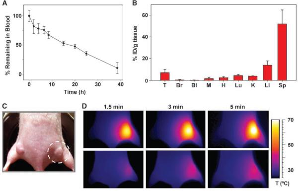
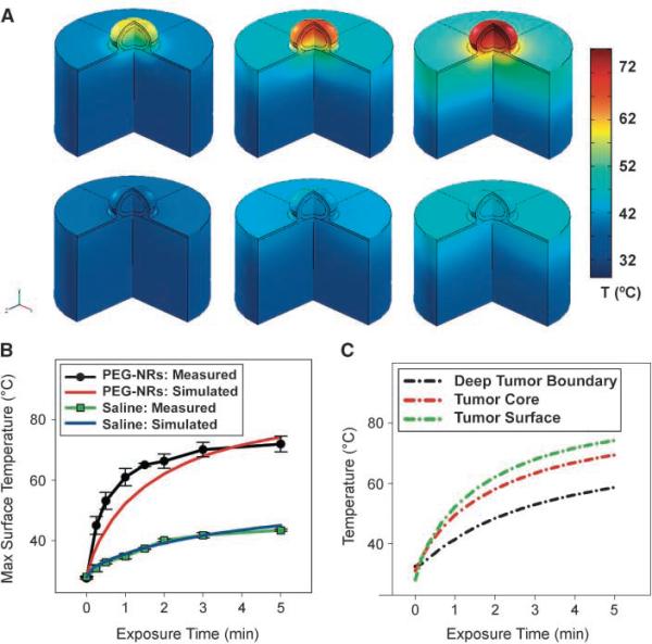
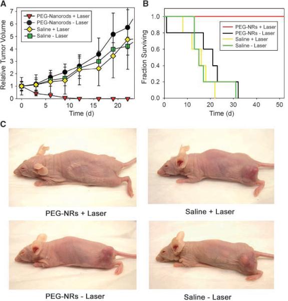
Plasmonic nanomaterials have the opportunity to considerably improve the specificity of cancer ablation by i.v. homing to tumors and acting as antennas for accepting externally applied energy. Here, we describe an integrated approach to improved plasmonic therapy composed of multimodal nanomaterial optimization and computational irradiation protocol development. We synthesized polyethylene glycol (PEG)-protected gold nanorods (NR) that exhibit superior spectral bandwidth, photothermal heat generation per gram of gold, and circulation half-life in vivo (t(1/2), approximately 17 hours) compared with the prototypical tunable plasmonic particles, gold nanoshells, as well as approximately 2-fold higher X-ray absorption than a clinical iodine contrast agent. After intratumoral or i.v. administration, we fuse PEG-NR biodistribution data derived via noninvasive X-ray computed tomography or ex vivo spectrometry, respectively, with four-dimensional computational heat transport modeling to predict photothermal heating during irradiation. In computationally driven pilot therapeutic studies, we show that a single i.v. injection of PEG-NRs enabled destruction of all irradiated human xenograft tumors in mice. These studies highlight the potential of integrating computational therapy design with nanotherapeutic development for ultraselective tumor ablation.


Figures

Figure 1
Figure 1
Structure and synthesis of highly absorbing, PEG-protected gold NRs. A, near-IR absorbing (810nm longitudinal plasmon resonance peak) gold NRs were imaged via transmission electron microscopy. B, schematic of process to drive CTAB-NR conversion to PEG-NRs under dialysis with rendering and molecular schematic of PEG coating on NR surface. C, PEG-NRs show prolonged stability in biological media (>1,000 h), whereas CTAB-coated NRs precipitated over time.
Figure 2
Figure 2
Spectral and photothermal properties of highly absorbing gold NRs compared with gold nanoshells. A, schematic of photothermal heating of gold NRs. The dimensions of gold NRs are tuned to have a near-IR plasmon resonance, at which point nanoparticle electrons resonantly oscillate and dissipate energy as heat. B, spectra for PEG-gold NRs (red) and PEG-gold nanoshells (blue), a benchmark for tunable plasmonic nanomaterials, at equal gold concentrations. C, top, rate of temperature increase for triplicate PEG-NR and PEG-gold nanoshell solutions (7 μg Au/mL, 810nm laser, 2 W/cm2, n = 3 each). Bottom, IR thermographic image of PEG-NRs versus PEG-gold nanoshells after 2 min of irradiation. Scale bar, 5 mm. D, In vitro photothermal toxicity of PEG-NRs over human cancer cells in culture (MDA-MB-435). Tumor cells were incubated with PEG-NRs (14 μg/mL; top), PEG-nanoshells (14 μg/mL; middle), or media alone (bottom) and treated with laser irradiation (2 W/cm2, 810nm, 5 min). Calcein AM staining indicates destruction of cells with PEG-NRs, whereas cells irradiated in the presence of nanoshells or media remained viable. Phase region of calcein staining inset. Scale bar, 10 μm.
Figure 3
Figure 3
X-ray CT, quantitative photothermal modeling, and near-IR photothermal heating of gold NRs In vivo. A, schematic of X-ray absorption by gold NRs in X-ray CT. B, X-ray CT number of PEG-NRs compared with an iodine standard (Isovue-370). C, PEG-NRs were intratumorally given to mice bearing bilateral MDA-MB-435 tumors and imaged using X-ray CT to visualize three-dimensional PEG-NR distribution in tumors (left). A three-dimensional solid model of the complete geometry was rapidly reconstructed by image processing for use with computational photothermal modeling (middle). Red, PEG-NRs. Experimental thermographic surveillance of NIR irradiation after X-ray CT (~0.75 W/cm2, 1 min; right). D, meshed geometry of the left tumor chosen as the computational domain (left). Plot of theoretical heat flux propagation inside the tumor upon irradiation (middle left). Predicted internal temperature distribution at three different planes inside the tumor (middle right) along with surface temperature map (right) matching the left tumor in C.
Figure 4
Figure 4
Long circulation time, passive tumor targeting, and photothermal heating of passively targeted gold NR antennas in tumors. A, PEG-NRs were i.v. given (20mg/kg) to three mice bearing MDA-MB-435 tumors, and blood was withdrawn over time to monitor clearance from circulation. B, PEG-NR biodistribution and targeting to MDA-MB-435 tumors 72 h after i.v. administration, quantified via ICP-MS (three mice). T, tumor; Br, brain; Bl, bladder; M, muscle; H, heart; Lu, lung; K, kidney; Li, liver; SP, spleen. Data are tabulated in Supplementary Table S1. C, PEG-NRs or saline were i.v. given (20mg/kg) to mice bearing MDA-MB-435 tumors on opposing flanks. After NRs had cleared from circulation (72 h after injection), the right flank was irradiated using an 810-nm diode laser (2 W/cm2; beam size indicated by dotted circle). D, thermographic surveillance of photothermal heating in PEG-NR-injected (top) and saline-injected (bottom) mice.
Figure 5
Figure 5
Quantitative photothermal modeling of gold NR tumor heating. A, three-dimensional finite element modeling of PEG-NR heating In vivo. Simulated three-dimensional temperature distributions matching the four-dimensional thermographic time points for PEG-NR (top) and control tumor irradiation (bottom). B, thermographically measured and simulated tumor surface temperatures over time for irradiation of PEG-NR or saline mice. C, simulated temperature increases various depths for PEG-NR-injected and saline-injected mice. By 5 min after the onset of irradiation, the entire PEG-NR tumor is predicted to have reached ablative temperatures of >60°, motivating the choice of this irradiation regimen for subsequent therapeutic trials.
Figure 6
Figure 6
Photothermal destruction of human tumors in mice using long-circulating gold NRs. A, mice harboring two MDA-MB-435 human tumors on opposite flanks were injected with either saline or PEG-NRs. After PEG-NRs had cleared from circulation (72 h after injection), the right flank of each mouse was exposed to the computationally designed irradiation regimen (810nm, 2 W/cm2, 5 min). Volumetric changes in tumor sizes are plotted over time after irradiation. B, mice harboring one MDA-MB-435 human tumor were injected with either saline or PEG-NRs and irradiated as in A. Survival of mice after irradiation is plotted versus time after irradiation. C, at 20d after irradiation, NIR-irradiated, all PEG-NR-injected mice showed only a minor scar and no evidence of tumor regrowth whereas all other treatment groups harbored thriving tumors.

References

    1. Elghanian R, Storhoff JJ, Mucic RC, Letsinger RL, Mirkin CA. Selective colorimetric detection of polynucleotides based on the distance-dependent optical properties of gold nanoparticles. Science. 1997;277:1078–81. - PubMed
    1. Grubisha DS, Lipert RJ, Park HY, Driskell J, Porter MD. Femtomolar detection of prostate-specific antigen: an immunoassay based on surface-enhanced Raman scattering and immunogold labels. Anal Chem. 2003;75:5936–43. - PubMed
    1. Jackson JB, Westcott SL, Hirsch LR, West JL, Halas NJ. Controlling the surface enhanced Raman effect via the nanoshell geometry. Appl Phys Lett. 2003;82:257–9.
    1. Qian XM, Peng XH, Ansari DO, et al. In vivo tumor targeting and spectroscopic detection with surfaceenhanced Raman nanoparticle tags. Nat Biotechnol. 2008;26:83–90. - PubMed
    1. Hirsch LR, Stafford RJ, Bankson JA, et al. Nanoshellmediated near-infrared thermal therapy of tumors under magnetic resonance guidance. P Natl Acad Sci USA. 2003;100:13549–54. - PMC - PubMed

Publication types

MeSH terms