. 2009 May 1;69(9):3856-65.
doi: 10.1158/0008-5472.CAN-08-2940. Epub 2009 Apr 14.

A double hit to kill tumor and endothelial cells by TRAIL and antiangiogenic 3TSR

Affiliations

A double hit to kill tumor and endothelial cells by TRAIL and antiangiogenic 3TSR

Bin Ren et al. Cancer Res. .

Abstract

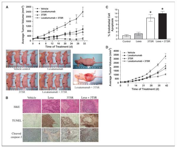
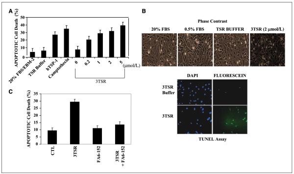
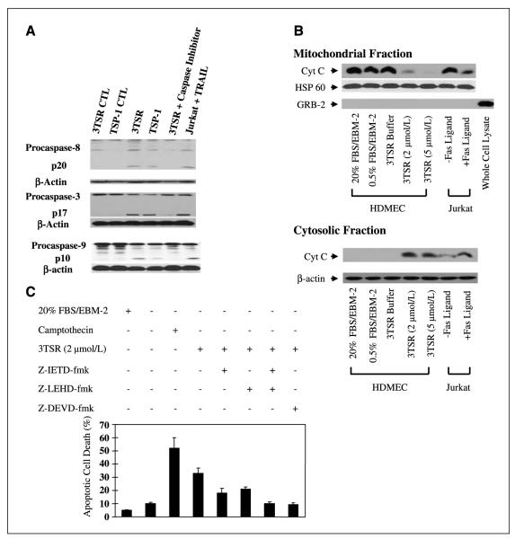
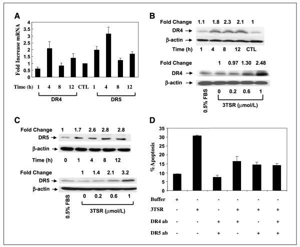
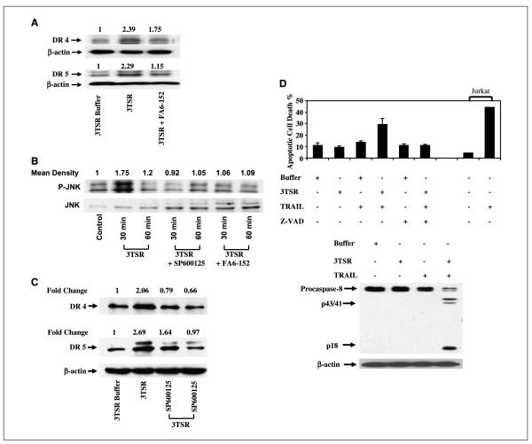
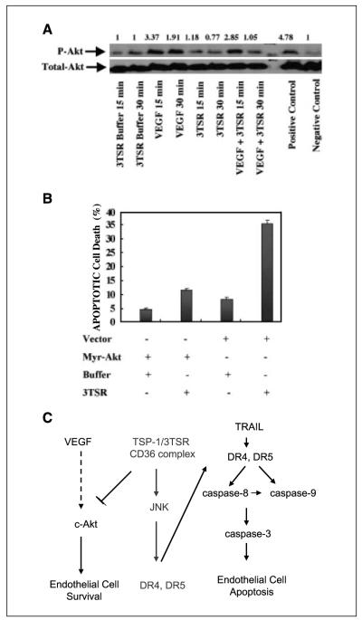
As tumor development relies on a coordination of angiogenesis and tumor growth, an efficient antitumor strategy should target both the tumor and its associated vessels. Tumor necrosis factor-related apoptosis-inducing ligand (TRAIL) induces apoptosis in a tumor-selective manner. Additionally, thrombospondin-1, a naturally occurring inhibitor of angiogenesis, and a recombinant protein containing functional domains of thrombospondin-1, 3TSR, have been shown to be necessary and sufficient to inhibit tumor angiogenesis. Here, we show that a combination of a TRAIL receptor 2 agonist antibody, Lexatumumab, and 3TSR results in a significantly enhanced and durable tumor inhibition. We further observed that 3TSR induces apoptosis in primary endothelial cells by up-regulating the expression of TRAIL receptors 1 and 2 in a CD36 and Jun NH(2)-terminal kinase-dependent manner leading to the activation of both intrinsic and extrinsic apoptotic machineries. The modulation of these pathways is critical for 3TSR-induced apoptosis as disrupting either via specific inhibitors reduced apoptosis. Moreover, 3TSR attenuates the Akt survival pathway. These studies indicate that 3TSR plays a critical role in regulating the proapoptotic signaling pathways that control growth and death in endothelial cells and that a combination of TRAIL and 3TSR acts as a double hit against tumor and tumor-associated vessels.


Figures

Figure 1
Figure 1
3TSR and TRAIL cooperate in inhibiting colon cancer. A, human colon cancer SW480 cells were implanted into the right flankof nu/nu mice. Seven days post-implantation, mice were injected daily with Lexatumumab (3 mg/kg intravenously) and 3TSR (1 mg/kg intraperitoneally) or 0.9% saline and TSR buffer. Mean from 10 mice in each group (n = 10). By treatment day 31, ANOVA indicates P < 0.01 for Lexatumumab versus vehicle, P < 0.001 for 3TSR versus vehicle, P < 0.001 for Lexatumumab + 3TSR versus vehicle, and P < 0.05 for Lexatumumab + 3TSR versus Lexatumumab. By day 31, TGI = 60% for Lexatumumab, TGI = 69% for 3TSR, and TGI = 93% for Lexatumumab + 3TSR. B, immunohistochemical staining on SW480 tumor sections for TUNEL and cleaved caspase-3. SW480 xenograft tumors were collected from three mice from each treatment group on day 28 after tumor cell inoculation. Similar results were observed from tumor samples of three mice from each treatment group. Original photos were taken at ×200 magnification. C, analysis of tumor vasculature endothelial cell apoptosis. The apoptosis of tumor-associated vascular endothelial cells was evaluated via double labeling of CD31 and TUNEL. 3TSR significantly induced apoptosis of tumor vascular endothelial cells (P = 0.001 versus vehicle control), whereas Lexatumumab (Lexa) treatment showed little effect on tumor vascular endothelial cell apoptosis. The combination of Lexatumumab and 3TSR resulted in a similar level of endothelial cell apoptosis as with 3TSR alone (*, P < 0.001 versus control). D, growth of HCT116 colon cancer xenograft in nude mice. Tumor implantation and treatments were same as A, except 1 × 10⁶ cells were used for implantation. Points, mean from 12 mice in each group (n = 12). By treatment day 41, statistical analysis by ANOVA indicates P < 0.01 for ×Lexatumumab versus vehicle, P < 0.05 for 3TSR versus vehicle, P < 0.001 for Lexatumumab + 3TSR versus vehicle, and P < 0.05 for Lexatumumab + 3TSR versus 3TSR. By treatment day 41, TGI = 46% for Lexatumumab, TGI = 36% for 3TSR, and TGI = 68% for Lexatumumab + 3TSR.
Figure 2
Figure 2
3TSR induces apoptosis of HDMEC. A, primary HDMEC were treated with 3TSR, TSP-1, and camptothecin at the indicated concentrations for 24 h. The apoptotic cell population was assayed by fluorescence-activated cell sorting analysis and indicated that 3TSR-mediated apoptosis increased in a dose-dependent manner. B, phase-contrast microscopy showed that 3TSR-treated cells have a rounded/apoptotic morphology and are positive for TUNEL staining. C, HDMEC were preincubated with anti-CD36 antibody FA6-152 for 1 h before adding 3TSR. Apoptotic cell population was measured as in A and showed that 3TSR-mediated apoptosis is dependent on the activity of the CD36 receptor.
Figure 3
Figure 3
3TSR stimulates caspase-8, caspase-3, and cytochrome c release from mitochondria and promotes activation of caspase-9 in HDMEC. A, HDMEC cells were treated with buffer (CTL), 3TSR, hTSP-1, and caspase-8 (z-IEDT-fmk), caspase-3 (z-DEVD-fmk), or caspase-9 (z-LEHD-fmk) inhibitor. Caspase cleavage was detected by Western blotting with specific caspase-8. Caspase-3 or caspase-9 antibodies. Jurkat cells treated with TRAIL in the presence or absence of z-IETD-fmkwere used as controls. B, cytochrome c (Cyt C) levels in mitochondrial and cytosolic fractions were determined by Western blotting after treatment of cells with 2 or 5 μmol/L 3TSR or buffer. Jurkat cells treated with or without Fas ligand for 6 h were used as controls. Western blot analysis indicated that the mitochondrial preparations were free of GRB2, a protein that is normally excluded from mitochondria. C, HDMEC were treated with 3TSR in the absence or presence of the caspase-8 inhibitor z-IETD-fmk, the caspase-9 inhibitor z-LRHD-fmk, either alone or in combination, and the caspase-3 inhibitor z-DEVD-fmk. Camptothecin was used as a positive control for induction of apoptosis in endothelial cells. Mean ± SD of three separate experiments.
Figure 4
Figure 4
3TSR regulates expression of the proapoptotic death receptors DR4 and DR5. A, 3TSR treatment of HDMEC for 1 to 12 h significantly increased expression of DR4 and DR5 at the transcriptional level. B and C, 3TSR treatment of HDMEC for 1 to 12 h increased both DR4 and DR5 protein expression, whereas 3TSR treatment of HDMEC for 8 h increased both DR4 and DR5 expression in a concentration-dependent manner. D, pretreatment of HDMEC cells with DR4- and/or DR5-blocking antibody for 30 min inhibited 3TSR-mediated apoptosis.
Figure 5
Figure 5
3TSR-induced DR4 and DR5 expression is mediated by JNK activation. A, 3TSR and IgG treatment of HDMEC for 4 h up-regulated DR4 and DR5 expression and this increase was suppressed with the CD36-blocking antibody FA6-152. B, both 3TSR-stimulated phosphorylation of SAPK/JNK and JNK activity in HDMEC were inhibited by treatment with SP600125, a selective inhibitor of JNK and FA6-152. C, 3TSR treatment of HDMEC for 4 h up-regulated DR4 and DR5 expression. Treatment of these cells with SP600125, however, similarly decreased DR4 and DR5 levels. D, 3TSR sensitizes HDMEC to TRAIL-induced apoptosis. HDMEC were treated with control buffer, 3TSR, and TRAIL alone or in combination. 3TSR and TRAIL combination enhances apoptosis induced in HDMEC. Inhibition of caspase activity by z-VAD suppresses this cooperation. Additionally, combination of 3TSR and TRAIL enhances caspase-8 activity in HDMEC. Processing of caspase-8 was evaluated by Western blot analysis. Arrow, processed p43/p41 signature fragments of caspase-8 processing. Jurkat cells are used as a control to show the activity of TRAIL.
Figure 6
Figure 6
VEGF induces Akt activation in HDMEC and 3TSR inhibits basal and VEGF-induced Akt activation of HDMEC. A, HDMEC were treated with control buffer, 3TSR (2 μmol/L), VEGF (100 ng/mL), or a combination of 3TSR and VEGF for the indicated times. B, exogenous expression of Myr-Akt inhibits 3TSR-induced apoptosis in HDMEC. Fluorescence-activated cell sorting analysis indicated that exogenous expression of myr-Akt inhibited 3TSR-mediated apoptosis in microvascular endothelial cells. C, 3TSR acts as a double-edged sword promoting apoptosis via up-regulation of DR4 and DR5 and inhibiting cell survival via attenuation of the Akt pathway.

Similar articles

Cited by

References

    1. Armstrong LC, Bornstein P. Thrombospondins 1 and 2 function as inhibitors of angiogenesis. Matrix Biol. 2003;22:63–71. - PubMed
    1. Nor JE, Mitra RS, Sutorik MM, Mooney DJ, Castle VP, Polverini PJ. Thrombospondin-1 induces endothelial cell apoptosis and inhibits angiogenesis by activating the caspase death pathway. J Vasc Res. 2000;37:209–18. - PubMed
    1. Stupack DG, Cheresh DA. Apoptotic cues from the extracellular matrix: regulators of angiogenesis. Oncogene. 2003;22:9022–9. - PubMed
    1. Jimenez B, Volpert OV, Crawford SE, Febbraio M, Silverstein RL, Bouck N. Signals leading to apoptosis-dependent inhibition of neovascularization by thrombospondin 1. Nat Med. 2000;6:41–8. - PubMed
    1. Danial NN, Korsmeyer SJ. Cell death: critical control points. Cell. 2004;116:205–19. - PubMed

Publication types

MeSH terms