

The role of Robo3 in the development of cortical interneurons
- PMID: 19366869
- PMCID: PMC2693537
- DOI: 10.1093/cercor/bhp041
The role of Robo3 in the development of cortical interneurons
Abstract
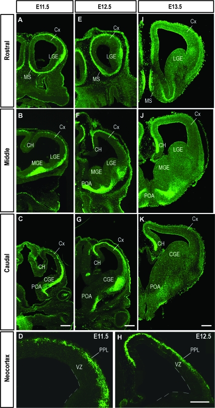
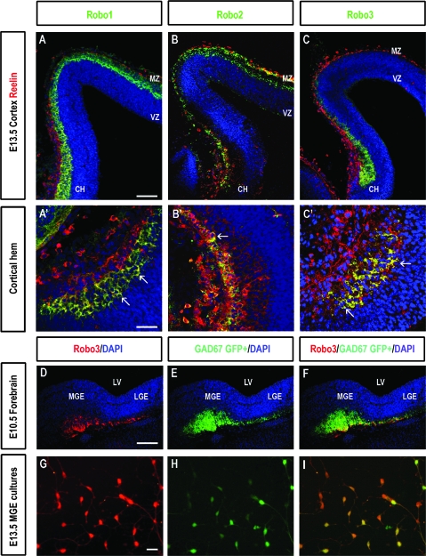
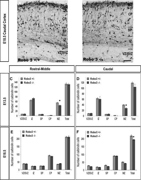
A number of studies in recent years have shown that members of the Roundabout (Robo) receptor family, Robo1 and Robo2, play significant roles in the formation of axonal tracks in the developing forebrain and in the migration and morphological differentiation of cortical interneurons. Here, we investigated the expression and function of Robo3 in the developing cortex. We found that this receptor is strongly expressed in the preplate layer and cortical hem of the early cortex where it colocalizes with markers of Cajal-Retzius cells and interneurons. Analysis of Robo3 mutant mice at early (embryonic day [E] 13.5) and late (E18.5) stages of corticogenesis revealed no significant change in the number of interneurons, but a change in their morphology at E13.5. However, preliminary analysis on a small number of mice that lacked all 3 Robo receptors indicated a marked reduction in the number of cortical interneurons, but only a limited effect on their morphology. These observations and the results of other recent studies suggest a complex interplay between the 3 Robo receptors in regulating the number, migration and morphological differentiation of cortical interneurons.
Figures
Similar articles
-
The role of Slit-Robo signaling in the generation, migration and morphological differentiation of cortical interneurons.Dev Biol. 2008 Jan 15;313(2):648-58. doi: 10.1016/j.ydbio.2007.10.052. Epub 2007 Nov 13. Dev Biol. 2008. PMID: 18054781
-
Robo1 regulates semaphorin signaling to guide the migration of cortical interneurons through the ventral forebrain.J Neurosci. 2011 Apr 20;31(16):6174-87. doi: 10.1523/JNEUROSCI.5464-10.2011. J Neurosci. 2011. PMID: 21508241 Free PMC article.
-
Robo1 regulates the development of major axon tracts and interneuron migration in the forebrain.Development. 2006 Jun;133(11):2243-52. doi: 10.1242/dev.02379. Development. 2006. PMID: 16690755
-
Slit-Robo interactions during cortical development.J Anat. 2007 Aug;211(2):188-98. doi: 10.1111/j.1469-7580.2007.00750.x. Epub 2007 Jun 6. J Anat. 2007. PMID: 17553100 Free PMC article. Review.
-
The origin and specification of cortical interneurons.Nat Rev Neurosci. 2006 Sep;7(9):687-96. doi: 10.1038/nrn1954. Epub 2006 Aug 2. Nat Rev Neurosci. 2006. PMID: 16883309 Review.
Cited by
-
BCL11A Haploinsufficiency Causes an Intellectual Disability Syndrome and Dysregulates Transcription.Am J Hum Genet. 2016 Aug 4;99(2):253-74. doi: 10.1016/j.ajhg.2016.05.030. Epub 2016 Jul 21. Am J Hum Genet. 2016. PMID: 27453576 Free PMC article.
-
Bilateral Whisker Representations in the Primary Somatosensory Cortex in Robo3cKO Mice Are Reflected in the Primary Motor Cortex.Neuroscience. 2024 Apr 19;544:128-137. doi: 10.1016/j.neuroscience.2024.02.031. Epub 2024 Mar 5. Neuroscience. 2024. PMID: 38447690 Free PMC article.
-
Molecules and mechanisms involved in the generation and migration of cortical interneurons.ASN Neuro. 2010 Mar 31;2(2):e00031. doi: 10.1042/AN20090053. ASN Neuro. 2010. PMID: 20360946 Free PMC article. Review.
-
A global gene regulatory program and its region-specific regulator partition neurons into commissural and ipsilateral projection types.Sci Adv. 2024 May 24;10(21):eadk2149. doi: 10.1126/sciadv.adk2149. Epub 2024 May 23. Sci Adv. 2024. PMID: 38781326 Free PMC article.
-
Peripheral nerve injury is accompanied by chronic transcriptome-wide changes in the mouse prefrontal cortex.Mol Pain. 2013 Apr 18;9:21. doi: 10.1186/1744-8069-9-21. Mol Pain. 2013. PMID: 23597049 Free PMC article.
References
-
- Anderson SA, Eisenstat DD, Shi L, Rubenstein JL. Interneuron migration from basal forebrain to neocortex: dependence on Dlx genes. Science. 1997;278:474–476. - PubMed
-
- Andrews W, Liapi A, Plachez C, Camurri L, Zhang J, Mori S, Murakami F, Parnavelas JG, Sundaresan V, Richards LJ. Robo1 regulates the development of major axon tracts and interneuron migration in the forebrain. Development. 2006;133:2243–2252. - PubMed
-
- Andrews W, Barber M, Hernandez-Miranda LR, Xian J, Rakic S, Sundaresan V, Rabbitts TH, Pannell R, Rabbitts P, Thompson H, et al. The role of Slit-Robo signalling in the generation, migration and morphological differentiation of cortical interneruons. Dev Biol. 2008;313:648–658. - PubMed
Publication types
MeSH terms
Substances
Grants and funding
LinkOut - more resources
Full Text Sources
Molecular Biology Databases
