. 2009 Jul;17(7):1187-96.
doi: 10.1038/mt.2009.71. Epub 2009 Apr 14.

Intravenous administration of self-complementary AAV9 enables transgene delivery to adult motor neurons

Affiliations

Intravenous administration of self-complementary AAV9 enables transgene delivery to adult motor neurons

Sandra Duque et al. Mol Ther. 2009 Jul.

Abstract

Therapeutic gene delivery to the whole spinal cord is a major challenge for the treatment of motor neuron (MN) diseases. Systemic administration of viral gene vectors would provide an optimal means for the long-term delivery of therapeutic molecules from blood to the spinal cord but this approach is hindered by the presence of the blood-brain barrier (BBB). Here, we describe the first successful study of MN transduction in adult animals following intravenous (i.v.) delivery of self-complementary (sc) AAV9 vectors (up to 28% in mice). Intravenous MN transduction was achieved in adults without pharmacological disruption of the BBB and transgene expression lasted at least 5 months. Importantly, this finding was successfully translated to large animals, with the demonstration of an efficient systemic scAAV9 gene delivery to the neonate and adult cat spinal cord. This new and noninvasive procedure raises the hope of whole spinal cord correction of MN diseases and may lead to the development of new gene therapy protocols in patients.


Figures
<b>Figure 1</b>
Figure 1
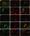
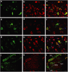
Intraperitoneal injection of GFP-encoding scAAV9 mediates CNS transduction in neonatal mice. (a–i) Representative transverse brain and (j–l) spinal cord sections treated for GFP/NeuN double-immunofluorescence, 7 days following injection of scAAV9 vectors. (a,d,g,j) GFP-positive cells and fibers (green), (b,e,h,k) NeuN-positive neurons (red), (c,f,i,l) merge (arrows: neuronal cells, arrowhead: GFP-positive glial cell profile) in (a–c) the hippocampus, (d–f) the entorhinal cortex, (g–i) the motor cortex, and (j–l) the spinal cord (DRf, dorsal root fibers; VRf, ventral root fibers; SAf, somatic afferent fibers; MNs, motor neurons). In (c,f,i) the nuclei were stained with DAPI (blue). Bars = (a–i) 40 µm and (j–l) 60 µm.
<b>Figure 2</b>
Figure 2
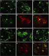
Comparison of i.p., i.m., and i.v. scAAV9-GFP delivery for spinal cord neurons transduction in neonatal mice. Representative transverse sections of the (a–b,d–l) cervical spinal cord and (c,m–o) lumbar spinal cord treated for GFP/NeuN double-immunofluorescence 7 days after (a,d–f) i.p., (b,g–i) i.m., or (c,j–o) i.v. scAAV9 injection. (d,g,j,m) GFP-positive cells (green) (e,h,k,n) NeuN-expressing neuronal cells (red). (a–c,f,i,l,o) Merge (yellow), transduced spinal cord neurons (arrows). Bars = (a–c) 40 µm and (d–o) 80 µm.
<b>Figure 3</b>
Figure 3
Intravenous injected scAAV9 vectors mediate transgene expression in the adult mouse spinal cord. Representative sections of (a–h,j–l) the spinal cord and (i) the dorsal root ganglion (DRG) from adult mice 4 weeks after i.v. delivery of 2 × 10¹² vg GFP-encoding scAAV9. (a–c,g–i) Sections treated for GFP-immunofluorescence revealed transduction of cells with (a–c) a clear MN phenotype and location (arrows), (g,h) a glial phenotype (arrowheads), (i) GFP-positive cells in the DRG. (d–f) Colabeling using GFP native fluorescence and ChAT immunofluorescence, (d) GFP-positive cells (green), (e) ChAT-expressing MNs (red), (f) merge (yellow) transduced spinal cord MNs. (j–l) Colabeling using GFP native fluorescence and GFAP immunofluorescence (j) GFP-positive cells (green) (k) GFAP-expressing astrocytes (red) (l) Merge (yellow) transduced spinal cord astrocytes. Bar = 20 µm.
<b>Figure 4</b>
Figure 4
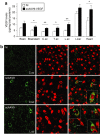
Intravenous scAAV9 injection mediates long-term VEGF delivery in mice. (a) ELISA analysis of VEGF levels in the CNS, the liver and the heart of scAAV9-VEGF injected (n = 4) and noninjected (n = 3) mice 21 weeks following i.v. injection. Values are expressed in pg/mg of total proteins. *P < 0.05; **P < 0.01. C.sc, cervical spinal cord; T.sc, thoracic spinal cord; L.sc, lumbar spinal cord; NI, noninjected. (b) Representative brain sections from an adult mouse treated for c-Myc (green, left panels)/NeuN (red, middle panels) double-immunofluorescence 19 weeks following i.v. scAAV9-VEGF-c-Myc injection; right panels: merge (arrows: transduced neurons). Bar = 20 µm.
<b>Figure 5</b>
Figure 5
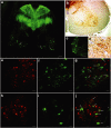
Intravenous delivery of scAAV9-GFP in neonatal LIX1 cats mediates transgene expression throughout the spinal cord. (a–d) Representative transverse sections of spinal cord from a LIX1 heterozygous cat observed by (a,c) laser scanning confocal microscopy (arrows: GFP-positive MNs; star: dorsal sensory tracts) or (b,d) treated for GFP immunohistochemistry (arrows: GFP-immunopositive MNs) 10 days after the injection of scAAV9-GFP into the jugular vein. (e–j) Double-labeled transverse sections of the spinal cord of both affected (e–g) and nonaffected (h–j) kitten, treated for ChAT immunofluorescence (e,h, red) and GFP native fluorescence (f,i, green). (g,j) Merge (arrows: transduced MNs). Bars = (a) 200 µm; (b,c) 100 µm; (d–j) 50 µm.
<b>Figure 6</b>
Figure 6
Intravenous scAAV9 delivery mediates GFP expression in the lower MNs and in non-nervous tissues of adult LIX1 cats. Representative transverse sections of (a–d) the cervical spinal cord and (e–h) non-nervous tissues 12 days after scAAV9-GFP injection in an adult cat. (a–c) GFP native fluorescence was detected in spinal cord cells with a MN-like morphology (arrows: GFP-positive cells with a MN morphology; arrowheads: GFP-positive MN axons in the ventral root) (d) GFP/ChAT colabeling showing GFP expression in ChAT-immunopositive MNs (arrows indicate double-labeled MNs). GFP expression was also detected in (e) the heart (f) the diaphragm (g) the liver and (h) the adrenal gland. Bars = (a) 135 µm; (b–f) 50 µm; (g–h) 65 µm.

References

    1. Monani UR. Spinal muscular atrophy: a deficiency in a ubiquitous protein; a motor neuron-specific disease. Neuron. 2005;48:885–896. - PubMed
    1. Pasinelli P., and , Brown RH. Molecular biology of amyotrophic lateral sclerosis: insights from genetics. Nat Rev Neurosci. 2006;7:710–723. - PubMed
    1. Miller RG, Mitchell JD, Lyon M., and , Moore DH. Riluzole for amyotrophic lateral sclerosis (ALS)/motor neuron disease (MND) Cochrane Database Syst Rev. 2007;24:CD001447. - PubMed
    1. Azzouz M, Hottinger A, Paterna JC, Zurn AD, Aebischer P., and , Büeler H. Increased motoneuron survival and improved neuromuscular function in transgenic ALS mice after intraspinal injection of an adeno-associated virus encoding Bcl-2. Hum Mol Genet. 2000;9:803–811. - PubMed
    1. Kaspar BK, Lladó J, Sherkat N, Rothstein JD., and , Gage FH. Retrograde viral delivery of IGF-1 prolongs survival in a mouse ALS model. Science. 2003;301:839–842. - PubMed

Publication types

Substances