. 2009;4(4):e5191.
doi: 10.1371/journal.pone.0005191. Epub 2009 Apr 15.

Analysis of pmpD expression and PmpD post-translational processing during the life cycle of Chlamydia trachomatis serovars A, D, and L2

Affiliations

Analysis of pmpD expression and PmpD post-translational processing during the life cycle of Chlamydia trachomatis serovars A, D, and L2

Andrey O Kiselev et al. PLoS One. 2009.

Abstract

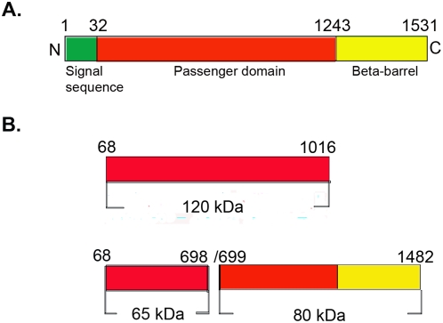
Background: The polymorphic membrane protein D (PmpD) in Chlamydia is structurally similar to autotransporter proteins described in other bacteria and may be involved in cellular and humoral protective immunity against Chlamydia. The mechanism of PmpD post-translational processing and the role of its protein products in the pathogenesis of chlamydial infection have not been very well elucidated to date.

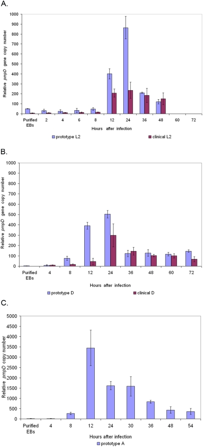
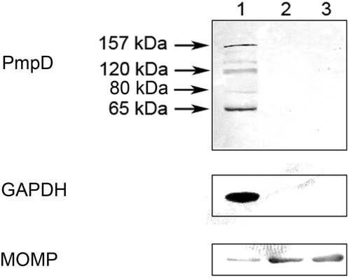
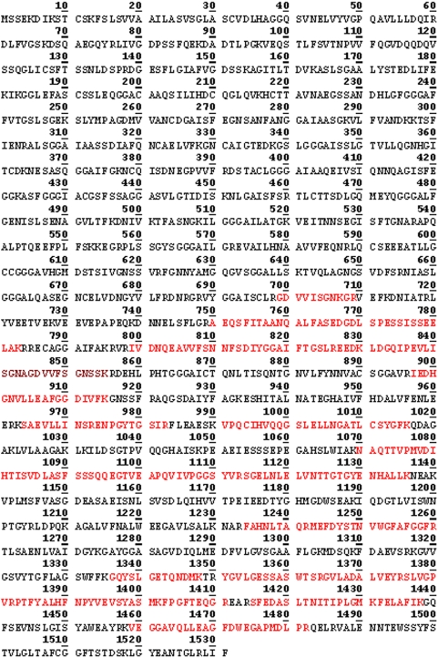
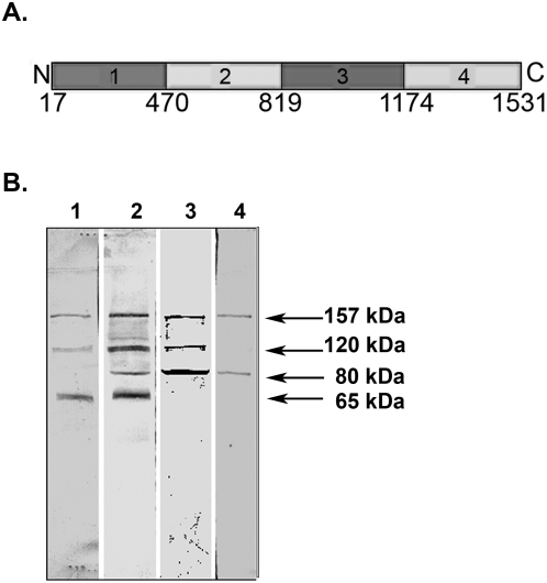
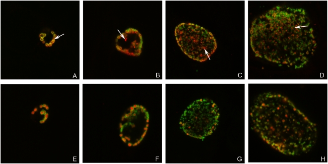
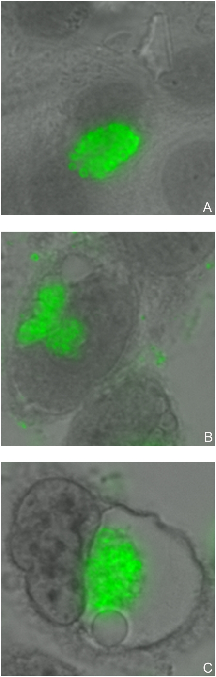
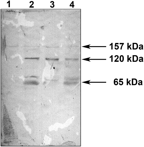
Methodology/principal findings: Here we examined the expression and post-translational processing of the protein product of the pmpD gene during the life cycle of C. trachomatis serovars A, D, and L2. Each of these three serovars targets different human organs and tissues and encodes a different pmpD gene nucleotide sequence. Our quantitative real-time reverse transcription polymerase chain reaction results demonstrate that the pmpD gene is up-regulated at 12-24 hours after infection regardless of the Chlamydia serovar. This up-regulation is coincidental with the period of exponential growth and replication of reticulate bodies (RB) of Chlamydia and indicates a probable similarity in function of pmpD in serovars A, D, and L2 of Chlamydia. Using mass spectrometry analysis, we identified the protein products of post-translational processing of PmpD of C. trachomatis serovar L2 and propose a double pathway model for PmpD processing, with one cleavage site between the passenger and autotransporter domains and the other site in the middle of the passenger domain. Notably, when Chlamydia infected culture cells were subjected to low (28 degrees C) temperature, PmpD post-translational processing and secretion was found to be uninhibited in the resulting persistent infection. In addition, confocal microscopy of cells infected with Chlamydia confirms our earlier hypothesis that PmpD is secreted outside Chlamydia and its secretion increases with growth of the chlamydial inclusion.

Conclusion/significance: The results of this current study involving multiple Chlamydia serovars support the general consensus that the pmpD gene is maximally expressed at mid infection and provide new information about PmpD as an autotransporter protein which is post-translationally processed and secreted outside Chlamydia during normal and low temperature induced persistent chlamydial infection.


Conflict of interest statement

Competing Interests: The authors have declared that no competing interests exist.

Figures

Figure 1
Figure 1. Relative pmpD transcription in prototype and clinical isolate strains of C. trachomatis serovar L2 (A), D (B), and A (C).
Total RNA was isolated from Chlamydia infected McCoy cells at 2 (L2), 4, 6 (L2), 8, 12, 24, 30 (A), 36, 48, 54 (A), 60 (D), and 72 (D) h p.i., and from purified EBs, random primed, and assayed by real-time RT-PCR for 16S rRNA and pmpD copy numbers. Relative pmpD expression at each time point was calculated by dividing the average pmpD copy number by the average 16S RNA copy number at the same time point and multiplying by 1×10⁶ . Error bars were based on the standard error of the mean . Note that serovar A pmpD expression levels are much higher than levels in serovars D and L2.
Figure 2
Figure 2. PmpD processing and secretion in C. trachomatis serovar A.
McCoy cells infected with C. trachomatis serovar A were harvested at 24, 48, and 72 h p.i., and soluble and insoluble protein fractions prepared and loaded as described in METHODS. The proteins in the soluble fractions were reacted in an immunoblot with antibodies against fragment 2 of PmpD. Data not shown for PmpD in the insoluble fractions. Antibodies against MOMP and GAPDH were reacted with both the soluble and insoluble fractions to assess the efficiency of their separation. Soluble fractions: Lanes 1–3; Insoluble fractions: Lanes 4–6. Lanes 1 and 4, 24 h p.i. Lanes 2 and 5, 48 h p.i. Lanes 3 and 6, 72 h p.i.; Note that the PmpD post-translational products and the GAPDH protein are found exclusively in the soluble fractions while chlamydial MOMP is found in the insoluble fractions which contain chlamydial organisms.
Figure 3
Figure 3. Differential extraction of proteins from culture cells based on their subcellular localization.
McCoy cells infected with C. trachomatis serovar L2 were subsequently treated at 48 h p.i. with different Extraction buffers (Calbiochem®). The proteins in each extract were loaded in equal volumes and reacted in an immunoblot with antibodies against fragment 2 of PmpD, MOMP, and GAPDH. Lane 1, Cytosolic fraction (Extract I). Lane 2, Membrane and membrane organelles (Extract II). Lane 3, Nuclear fraction (Extract III). Note that the PmpD post-translational products and the GAPDH protein are found exclusively in the cytosolic fraction and almost all of the chlamydial MOMP is found in the subsequent fractions which contain chlamydial organisms.
Figure 4
Figure 4. Peptides identified by MS in the ≈65 kDa protein spot (raw data).
The peptides identified by MS are highlighted in red.
Figure 5
Figure 5. Peptides identified by MS in the ≈120 kDa protein spot (raw data).
The peptides identified by MS are highlighted in red.
Figure 6
Figure 6. Peptides identified by MS in the ≈80 kDa protein spot (raw data).
The peptides identified by MS are highlighted in red.
Figure 7
Figure 7. Position of PmpD post-translational products identified by MS in the PmpD aa sequence.
A.The full length PmpD of C. trachomatis serovar L2. B. Position of the PmpD post-translational products in the PmpD sequence.
Figure 8
Figure 8. The amino acid sequence of the PmpD fragments used to raise antibodies and antibody reaction with proteins present in the soluble fraction.
A. The amino acid sequence of the PmpD fragments used to raise antibodies (Fragment 1, aa 17–517; fragment 2, aa 470–818; fragment 3, aa 819–1180; fragment 4, aa 1174–1531). B. Reaction of antibodies raised against PmpD fragments with proteins present in the soluble fraction. McCoy cells infected with C. trachomatis serovar L2 were harvested at 48 h p.i. and the soluble fraction was prepared as described in METHODS. Lane 1, pAb against fragment 1. Lane 2, pAb against fragment 2. Lane 3, pAb against fragment 3. Lane 4, pAb against fragment 4.
Figure 9
Figure 9. The passenger domain of PmpD is secreted outside Chlamydia and accumulated in the inclusion lumen during the life cycle of C. trachomatis.
McCoy cells were infected with C. trachomatis serovar L2 and fixed with methanol at 16 (A and E), 24 (B and F), 36 (C and G), and 48 (D and H) h p.i. and reacted with mAb against chlamydial MOMP (green) in combination with pAb against fragment 2 of PmpD (red) (A–D) or pAb against chlamydial HSP60 protein (red) (E–H) (internal control). Arrow indicates localization of PmpD during the chlamydial life cycle on the surface of Chlamydia (A) or in the inclusion lumen outside Chlamydia (red spots) (B–D). When reacted with anti-HSP60 pAb, no material was visible outside Chlamydia. The photographs were made using a Leica SL confocal microscope.
Figure 10
Figure 10. The beta-barrel of PmpD is associated with the interior of the chlamydial cell wall during the life cycle of C. trachomatis.
McCoy cells were infected with C. trachomatis serovar L2, fixed with methanol at 24 (A, C, and E) and 48 (B, D, and F) h p.i., and reacted with mAb against chlamydial MOMP (green) in combination with pAb against fragment 4 of PmpD (A and B), pAb against chlamydial PmpA (C and D), or mAb against chlamydial IncA (E and F) (red). PmpA is localized in the cytoplasm while IncA is secreted outside Chlamydia and localized to the inclusion membrane. The beta-barrel (transporter domain) alone and included in the ≈80 kDa protein, remains associated with the interior of the cell wall of Chlamydia resulting in staining of chlamydial RBs with pAb against fragment 4 of PmpD in a doughnut-like pattern. Note, that contrary to pAb against fragments 1, 2, and 3, pAb against fragment 4 do not stain unfixed chlamydial RBs (not shown). The photographs were made using a Leica SL confocal microscope.
Figure 11
Figure 11. Chlamydial inclusions in culture cells produced by low (28°C) temperature induced persistent infection.
McCoy cells were infected with C. trachomatis serovar L2, fixed with methanol at 24 (A) h p.i. at 37°C and 48 (B) and 120 (C) h p.i. at 28°C and reacted with pAb against fragment 2 of PmpD followed by staining with anti-rabbit IgG conjugated with FITC (Sigma-Aldrich) and counterstaining with Evans Blue. Multiple small non-fusogenic (B) or single “empty” looking giant (C) inclusions stained green are produced by persistent infection. A single inclusion filled with typical RBs produced by normal (37°C) infection (A) is shown for comparison. The photographs were made using Nikon Eclipse E600 upright microscope with a QImaging Retigia EX CCD Camera.
Figure 12
Figure 12. Low (28°C) temperature induced persistent infection of culture cells with C. trachomatis does not block PmpD post-translational processing.
McCoy cells were infected with C. trachomatis serovar L2, fixed with methanol at 24 (A and D) h p.i. at 37°C and 48 (B and E) and 120 (C and F) h p.i. at 28°C and reacted with mAb against chlamydial MOMP (green) in combination with pAb against fragment 2 of PmpD (red) (A–C) or pAb against chlamydial HSP60 protein (red) (D–F) (internal control). RBs produced during persistent infection are abnormally large with thin cell wall (B–C and E–F) compared with uniformly sized and round shaped RBs (A and D) produced during normal infection. Arrow indicates localization of PmpD during the chlamydial life cycle on the surface of aberrant RBs (A) or in the inclusion lumen outside Chlamydia (red spots) (B). When reacted with anti-HSP60 pAb, no material was visible outside Chlamydia. Note that the rate of PmpD secretion outside Chlamydia is much lower and slower when compared with normal (37°C) infection. The photographs were made using a Leica SL confocal microscope.
Figure 13
Figure 13. PmpD processing and secretion at 28°C.
McCoy cells infected with C. trachomatis serovar L2 were incubated at 28°C, harvested every 48 h, and soluble fractions prepared as described in METHODS. The proteins in each soluble fraction were reacted in an immunoblot with antibodies against fragment 2 of PmpD. Lane 1, 48 h p.i. Lane 2, 96 h p.i. Lane 3, 144 h p.i. Lane 4, 192 h p.i.

Similar articles

Cited by

References

    1. Stephens RS, Kalman S, Lammel C, Fan J, Marathe R, et al. Genome sequence of an obligate intracellular pathogen of humans: Chlamydia trachomatis. Science. 1998;282(5389):754–9. - PubMed
    1. Carlson JH, Porcella SF, McClarty G, Caldwell HD. Comparative genomic analysis of Chlamydia trachomatis oculotropic and genitotropic strains. Infect Immun. 2005;73(10):6407–18. - PMC - PubMed
    1. Thomson NR, Holden MT, Carder C, Lennard N, Lockey SJ, et al. Chlamydia trachomatis: genome sequence analysis of lymphogranuloma venereum isolates. Genome Res. 2008;18(1):161–71. Epub 2007 Nov 21. - PMC - PubMed
    1. Read TD, Brunham RC, Shen C, Gill SR, Heidelberg JF, et al. Genome sequences of Chlamydia trachomatis MoPn and Chlamydia pneumoniae AR39. Nuc Acids Res. 2000;28(6):1397–1406. - PMC - PubMed
    1. Gomes JP, Bruno WJ, Borrego MJ, Dean D. Recombination in the genome of Chlamydia trachomatis involving the polymorphic membrane protein C gene relative to ompA and evidence for horizontal gene transfer. J Bacteriol. 2004;186(13):4295–306. - PMC - PubMed

Publication types

MeSH terms

Substances