. 2008 Dec;7(12):5157-66.
doi: 10.1021/pr8004887.

Characterization and comprehensive proteome profiling of exosomes secreted by hepatocytes

Affiliations

Characterization and comprehensive proteome profiling of exosomes secreted by hepatocytes

Javier Conde-Vancells et al. J Proteome Res. 2008 Dec.

Abstract

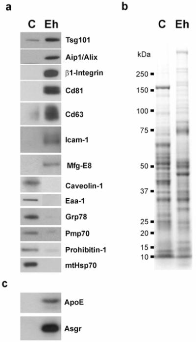
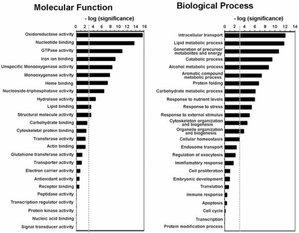
Exosomes represent a discrete population of vesicles that are secreted from various cell types to the extracellular media. Their protein and lipid composition are a consequence of sorting events at the level of the multivesicular body, a central organelle which integrates endocytic and secretory pathways. Characterization of exosomes from different biological samples has shown the presence of common as well as cell-type specific proteins. Remarkably, the protein content of the exosomes is modified upon pathological or stress conditions. Hepatocytes play a central role in the body response to stress metabolizing potentially harmful endogenous substances as well as xenobiotics. In the present study, we described and characterized for the first time exosome secretion in nontumoral hepatocytes, and with the use of a systematic proteomic approach, we establish the first extensive proteome of a hepatocyte-derived exosome population which should be useful in furthering our understanding of the hepatic function and in the identification of components that may serve as biomarkers for hepatic alterations. Our analysis identifies a significant number of proteins previously described among exosomes derived from others cell types as well as proteins involved in metabolizing lipoproteins, endogenous compounds and xenobiotics, not previously described in exosomes. Furthermore, we demonstrated that exosomal membrane proteins can constitute an interesting tool to express nonexosomal proteins into exosomes with therapeutic purposes.


Figures

Figure 1
Figure 1
Characterization of exosome-like vesicles from murine hepatocytes. (a) Cryo-electron micrograph of extracellular vesicles secreted by MLP-29 cells. Bar, 100 nm. (b) Western blot analysis of protein extracts prepared from MLP-29 cells (“C”) or from MLP-29-secreted vesicles (“Ex”). Proteins (10 μg) were analyzed by western blotting using antibodies against Tsg101 (exosome marker) or Grp78 (endoplasmic reticulum marker) proteins.
Figure 2
Figure 2
Targeting of Enhanced Green Fluorescent Protein (EGFP) to exosomes mediated by fusion to CD63 protein. (a) EGFP- or (b) CD63-EGFP-expressing MLP-29 cells were grown on glass coverlips, fixed using 2% (w/v) formaldehyde, washed two times with PBS and mounted on glass slides using Fluoromount-G (Southern Biotech, Birmingham, AL) containing 0.7 mg/liter of DAPI. Fluorescent samples were examined on a Leica TCS SP multiphoton confocal microscope. Images from GFP (green) and DAPI (blue) fluorescence were acquired using a 40X objetive and the Leica Confocal Software. Note the cytosolic distribution of EGFP versus the vesicular localization around the nucleus of CD63-EGFP protein. Bars, 40 μm. (c) MLP-29 cellular clones expressing EGFP or CD63-EGFP proteins were cultured in 150-mm dishes, trypsinized and washed two times with ice-cold PBS. GFP fluorescence was acquired using BD FACSDiva flow cytometry software version 5.0 in a FACScanto II flow cytometer. A threshold for GFP fluorescence was established based on the fluorescence observed for the parental cell line MLP-29. Between parentheses are indicated both the percentage of cells with GFP fluorescence above the threshold and the median GFP fluorescence intensity determined in this percentage of cells. (d) Protein extracts prepared from cells (“C”, 12μg protein) and exosomes (“Ex”, 3 μg protein) of MLP29 cellular clones expressing EGFP- or CD63-EGFP proteins were electrophoretically separated on 4-12% SDS-PAGE gel and analyzed by western blotting using antibodies against the following proteins: EGFP tag, Tsg101 (exosomal marker), Grp78 (endoplasmic reticulum marker) and Pallidin (“Pldn”, cytosolic and early-endosomal protein).
Figure 3
Figure 3
Ultrastructural analysis of vesicles secreted by rat hepatocytes. Representative (a) negative stainning- and (b) cryo- electron micrographs of vesicles secreted by primary culture of rat hepatocytes. Bars, 200 nm. (c) Quantitative analysis of vesicle diameter was performed in cryo-electron micrographs acquired and processed by using Digital Micrograph Software. Distributions of percentage of cells, number of vesicles analyzed (n), and mean and standard deviation of two independent preparations are indicated.
Figure 4
Figure 4
Western blot analysis and Coomassie Blue staining of exosomes secreted by rat hepatocytes. (a) Protein extracts (10 μg) prepared from rat hepatocytes (“C”) or from exosomes-derived from those cells (“Eh”) were analyzed by western blotting using antibodies against either exosomal markers (Tsg101, Aip1/Alix, β1-Integrin, Cd81, Cd63, Icam-1, Mfg-E8) or protein markers from other subcellular compartments; caveolae (Caveolin1), early endosome (Eaa1), endoplasmic reticulum (Grp78), peroxisome (Pmp70), mitochondria (Prohibitin-1, mtHsp70). Note the enrichment of exosomal markers in the exosome preparation compared to the cellular lysate. (b) Coomassie staining pattern of protein extracts (10 μg) prepared from rat hepatocytes (“C”) or from exosomes-derived from those cells (“Eh”). Notice that the pattern of bands presents in both samples is different. (c) Protein extracts (10 μg) prepared from rat hepatocytes or from exosomes-derived from those cells were loaded on a 4-12% SDS gel followed by western blotting using antibodies against ApoE and Asgr proteins.
Figure 5
Figure 5
Genome Ontology (GO) Analysis. 244 exosomal proteins identified in this study were classified in molecular function and biological process categories defined by GO consortium. GO categories with a higher or lower number of proteins assigned than expected by random were identified by stadistical analysis. The enrichment and depletion statistics was calculated according to the equation (2), described in Experimental Procedures, using all entries in Swiss-Prot rat protein database as reference set. We represented the [-log(P)], where P is the significance calculated according to the mentioned equation, for the categories indicated. A threshold (discontinued line) was established as P=0.001. Categories with significance above the threshold have significantly greater number of identified protein than expected by random.

References

    1. Fevrier B, Raposo G. Exosomes: endosomal-derived vesicles shipping extracellular messages. Current Opinion in Cell Biology. 2004;16(4):415–421. - PubMed
    1. Johnstone RM. Revisiting the road to the discovery of exosomes. Blood Cells Mol Dis. 2005;34(3):214–9. - PubMed
    1. van Niel G, Porto-Carreiro I, Simoes S, Raposo G. Exosomes: a common pathway for a specialized function. J Biochem (Tokyo) 2006;140(1):13–21. - PubMed
    1. Trajkovic K, Hsu C, Chiantia S, Rajendran L, Wenzel D, Wieland F, Schwille P, Brugger B, Simons M. Ceramide triggers budding of exosome vesicles into multivesicular endosomes. Science. 2008;319(5867):1244–7. - PubMed
    1. Novikoff AB, Essner E, Quintana N. Golgi Apparatus and Lysosomes. Fed Proc. 1964;23:1010–22. - PubMed

Publication types