. 2009 Apr 28;106(17):6992-7.
doi: 10.1073/pnas.0812620106. Epub 2009 Apr 14.

Single molecule mechanics of the kinesin neck

Affiliations

Single molecule mechanics of the kinesin neck

Thomas Bornschlögl et al. Proc Natl Acad Sci U S A. .

Abstract

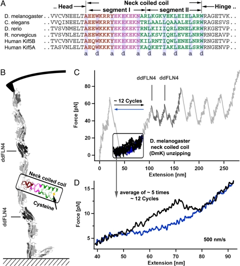
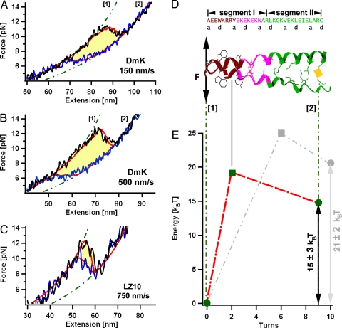
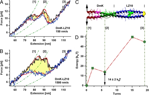
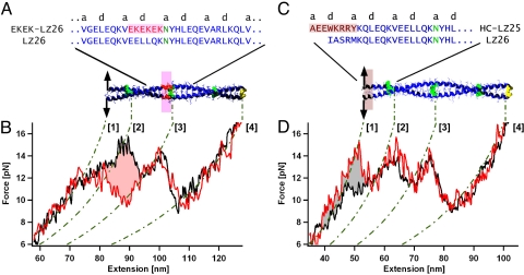
Structural integrity as well as mechanical stability of the parts of a molecular motor are crucial for its function. In this study, we used high-resolution force spectroscopy by atomic force microscopy to investigate the force-dependent opening kinetics of the neck coiled coil of Kinesin-1 from Drosophila melanogaster. We find that even though the overall thermodynamic stability of the neck is low, the average opening force of the coiled coil is >11 pN when stretched with pulling velocities >150 nm/s. These high unzipping forces ensure structural integrity during motor motion. The high mechanical stability is achieved through a very narrow N-terminal unfolding barrier if compared with a conventional leucine zipper. The experimentally mapped mechanical unzipping profile allows direct assignment of distinct mechanical stabilities to the different coiled-coil subunits. The coiled-coil sequence seems to be tuned in an optimal way to ensure both mechanical stability as well as motor regulation through charged residues.


Conflict of interest statement

The authors declare no conflict of interest.

Figures

Fig. 1.
Fig. 1.
Experimental method for the mechanical coiled-coil unzipping. (A) Alignment of different Kinesin-1 neck sequences. (B) Schematic view of the experimental setup. The coiled coil (in this case the D. melanogaster neck coiled coil) is inserted between globular ddFLN1–5 domains, which act as handles for the proper attachment to the AFM. (C) Representative force trace of the whole protein construct. The saw-tooth pattern at forces of 50 pN corresponds to the unfolding of single ddFLN1–5 domains, whereas coiled-coil unzipping occurs in the low force regime (framed). (D) Averaged forward (black) and backward (blue) force trace obtained by averaging traces from 5 independent experiments where every experiment contains ≈12 forward and backward cycles.
Fig. 2.
Fig. 2.
Mechanical unzipping profiles and mapped energy landscapes of the kinesin neck (DmK) and a leucine zipper. (A and B) Averaged forward (black) and backward (blue) force traces pulled at 150 nm/s (A) and 500 nm/s (B) measured on the DmK coiled coil. Red lines are the results of the kinetic Monte Carlo simulation. Green dotted lines represent the WLC elasticity (19) for polypeptides with fixed contour length. (C) For comparison, the averaged force traces from a canonical coiled coil (LZ10) at velocities of 750 nm/s are shown. (D) Sequence of the DmK-neck coiled coil aligned to the structure model of the comparable neck coiled coil from Rattus norwegicus (13). The C-terminal half (segment II) shows the canonical knob-into-holes scheme, whereas within segment I noncanonical amino acids are found. (E) Energy landscape of the DmK coiled coil (red) and the LZ10 coiled coil (gray) used for the Monte Carlo simulation.
Fig. 3.
Fig. 3.
Mechanical unzipping profiles and mapped energy landscapes of an elongated coiled coil containing the kinesin neck. (A) Averaged forward (black) and backward (blue) force traces of the DmK-LZ10 coiled coil obtained at a pulling velocity of 150 nm/s. Red lines represent the results of the Monte Carlo simulation. Between totally closed state [1] and totally opened state [3], an intermediate state with 6 ± 1 opened turns [2] is resolvable. (B) Averaged force traces at 1,500 nm/s. (C) Schematic structure of the DmK-LZ10 coiled-coil construct. The colors characterize the hydrophobic collar region (brown), the EKEK-motif (pink), the canonical segment II (green), and the canonical LZ10 sequence (blue). (D) Underlying energy landscape used for Monte Carlo simulations. We find, in agreement with the data for the DmK construct, a narrow N-terminal energy barrier at 2 ± 1 opened turns. The position of the intermediate state lays with 6 opened turns close to the boundary between segments I and II of the DmK coiled coil.
Fig. 4.
Fig. 4.
Energetic contributions of single subfragments from the kinesin neck within a canonical coiled coil. (A) Amino acid sequence and schematic structure of the LZ26 coiled coil with inserted EKEK-sequence (red). (B) Averaged forward force trace of the resulting EKEK-LZ26 coiled coil (red), compared to averaged forward force trace of the unmodified LZ26 coiled coil (black). The shaded area indicates a local decrease in equilibrium energy of 10.6 ±1.5 kBT. (C) Amino acid sequence and schematic structure of the LZ26 coiled coil, where the N terminus is replaced by the hydrophobic collar sequence from DmK (brown). (D) Average forward force trace of the resulting HC-LZ25 coiled coil (red) compared with the unmodified LZ26 forward trace (black). The positive free energy of the hydrophobic collar sequence was calculated from the gray shaded area as 10 ± 1.5 kBT.

References

    1. Hackney DD, Stock MF, Moore J, Patterson RA. Modulation of kinesin half-site ADP release and kinetic processivity by a spacer between the head groups. Biochemistry. 2003;42:12011–12018. - PubMed
    1. Rosenfeld SS, Fordyce PM, Jefferson GM, King PH, Block SM. Stepping and stretching. How kinesin uses internal strain to walk processively. J Biol Chem. 2003;278:18550–18556. - PMC - PubMed
    1. Crevel IM, et al. What kinesin does at roadblocks: The coordination mechanism for molecular walking. EMBO J. 2004;23:23–32. - PMC - PubMed
    1. Hahlen K, et al. Feedback of the kinesin-1 neck-linker position on the catalytic site. J Biol Chem. 2006;281:18868–18877. - PubMed
    1. Yildiz A, Tomishige M, Gennerich A, Vale RD. Intramolecular strain coordinates kinesin stepping behavior along microtubules. Cell. 2008;134:1030–1041. - PMC - PubMed

Publication types

LinkOut - more resources