

Flow-induced clustering and alignment of vesicles and red blood cells in microcapillaries
- PMID: 19369212
- PMCID: PMC2669370
- DOI: 10.1073/pnas.0811484106
Flow-induced clustering and alignment of vesicles and red blood cells in microcapillaries
Abstract
The recent development of microfluidic devices allows the investigation and manipulation of individual liquid microdroplets, capsules, and cells. The collective behavior of several red blood cells (RBCs) or microcapsules in narrow capillaries determines their flow-induced morphology, arrangement, and effective viscosity. Of fundamental interest here is the relation between the flow behavior and the elasticity and deformability of these objects, their long-range hydrodynamic interactions in microchannels, and thermal membrane undulations. We study these mechanisms in an in silico model, which combines a particle-based mesoscale simulation technique for the fluid hydrodynamics with a triangulated-membrane model. The 2 essential control parameters are the volume fraction of RBCs (the tube hematocrit, H(T)), and the flow velocity. Our simulations show that already at very low H(T), the deformability of RBCs implies a flow-induced cluster formation above a threshold flow velocity. At higher H(T) values, we predict 3 distinct phases: one consisting of disordered biconcave-disk-shaped RBCs, another with parachute-shaped RBCs aligned in a single file, and a third with slipper-shaped RBCs arranged as 2 parallel interdigitated rows. The deformation-mediated clustering and the arrangements of RBCs and microcapsules are relevant for many potential applications in physics, biology, and medicine, such as blood diagnosis and cell sorting in microfluidic devices.
Conflict of interest statement
The authors declare no conflict of interest.
Figures

 α
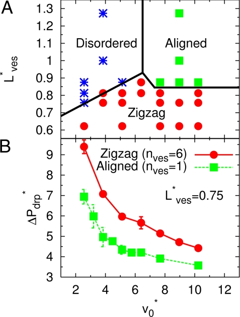
α , which measures the deviation from a spherical shape, as a function of the mean flow velocity v*0. Here, the asphericity is given by α = [ (λ1 − λ2)2 + (λ2 − λ3)2 + (λ3 − λ1)2]/2Rg4, with the eigenvalues λ1 ≤ λ2 ≤ λ3 of the gyration tensor and the squared radius of gyration Rg2 = λ1 + λ2 + λ3. RBCs transits from the discocyte (with α ≃ 0.15) to the parachute shape (with α ≲ 0.05). Simulation snapshots show the parachute (Lower Left) and bowl (Upper Right) shapes for v*0 = 7.7 with L*ves = 2.25 and L*ves = 1, respectively. (B) Average inclination angle θ for a discocyte vesicle for low flow velocities v*0 < v*c, as a function of the vesicle distance Lves. The inclination angle θ measures the deviation of the vesicle symmetry axis (determined by the eigenvector associated with the minimum eigenvalue of the gyration tensor) with the flow direction (z axis).

Similar articles
-
Dynamical clustering of red blood cells in capillary vessels.J Mol Model. 2003 Feb;9(1):16-33. doi: 10.1007/s00894-002-0105-x. Epub 2003 Jan 16. J Mol Model. 2003. PMID: 12638008
-
SPH-DEM approach to numerically simulate the deformation of three-dimensional RBCs in non-uniform capillaries.Biomed Eng Online. 2016 Dec 28;15(Suppl 2):161. doi: 10.1186/s12938-016-0256-0. Biomed Eng Online. 2016. PMID: 28155717 Free PMC article.
-
Microconfined flow behavior of red blood cells.Med Eng Phys. 2016 Jan;38(1):11-6. doi: 10.1016/j.medengphy.2015.05.007. Epub 2015 Jun 10. Med Eng Phys. 2016. PMID: 26071649
-
Hydrodynamic lift of vesicles and red blood cells in flow--from Fåhræus & Lindqvist to microfluidic cell sorting.Adv Colloid Interface Sci. 2014 Jun;208:161-76. doi: 10.1016/j.cis.2014.03.002. Epub 2014 Mar 12. Adv Colloid Interface Sci. 2014. PMID: 24674656 Review.
-
Blood flow structure related to red cell flow: determinant of blood fluidity in narrow microvessels.Jpn J Physiol. 2001 Feb;51(1):19-30. doi: 10.2170/jjphysiol.51.19. Jpn J Physiol. 2001. PMID: 11281993 Review.
Cited by
-
Optical-force-controlled red-blood-cell microlenses for subwavelength trapping and imaging.Biomed Opt Express. 2022 Apr 25;13(5):2995-3004. doi: 10.1364/BOE.457700. eCollection 2022 May 1. Biomed Opt Express. 2022. PMID: 35774333 Free PMC article.
-
Surface-active microrobots can propel through blood faster than inert microrobots.PNAS Nexus. 2024 Oct 15;3(10):pgae463. doi: 10.1093/pnasnexus/pgae463. eCollection 2024 Oct. PNAS Nexus. 2024. PMID: 39474503 Free PMC article.
-
Single-molecule imaging of NGF axonal transport in microfluidic devices.Lab Chip. 2010 Oct 7;10(19):2566-73. doi: 10.1039/c003385e. Epub 2010 Jul 9. Lab Chip. 2010. PMID: 20623041 Free PMC article.
-
Weak Shape Anisotropy Leads to a Nonmonotonic Contribution to Crowding, Impacting Protein Dynamics under Physiologically Relevant Conditions.J Phys Chem B. 2018 Dec 27;122(51):12396-12402. doi: 10.1021/acs.jpcb.8b07901. Epub 2018 Dec 13. J Phys Chem B. 2018. PMID: 30499666 Free PMC article.
-
Dynamic and rheological properties of soft biological cell suspensions.Rheol Acta. 2016 Jun;55(6):433-449. doi: 10.1007/s00397-015-0869-4. Epub 2015 Sep 3. Rheol Acta. 2016. PMID: 27540271 Free PMC article.
References
-
- Gompper G, Kroll DM. In: Mechanics of Membranes and Surfaces. 2nd Ed. Nelson DR, Piran T, Weinberg S, editors. Singapore: World Scientific; 2004. pp. 359–426.
-
- Seifert U. Configurations of fluid membranes and vesicles. Adv Phys. 1997;46:13–137.
-
- Vaziri A, Gopinath A. Cell and biomolecular mechanics in silico. Nat Mater. 2008;7:15–23. - PubMed
Publication types
MeSH terms
Substances
LinkOut - more resources
Full Text Sources
Other Literature Sources
