. 2009 Apr 14;106(15):6152-7.
doi: 10.1073/pnas.0813061106. Epub 2009 Apr 6.

VEGF-B is dispensable for blood vessel growth but critical for their survival, and VEGF-B targeting inhibits pathological angiogenesis

Affiliations

VEGF-B is dispensable for blood vessel growth but critical for their survival, and VEGF-B targeting inhibits pathological angiogenesis

Fan Zhang et al. Proc Natl Acad Sci U S A. .

Abstract

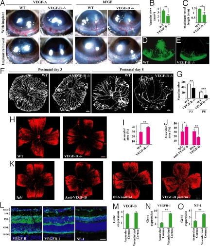
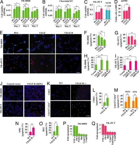
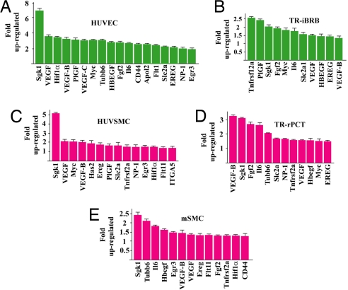
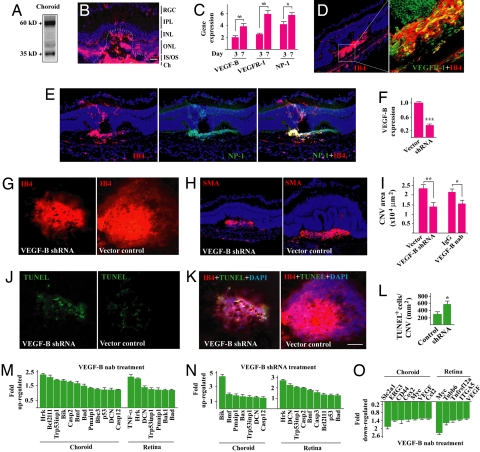
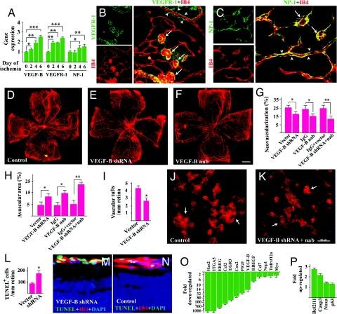
VEGF-B, a homolog of VEGF discovered a long time ago, has not been considered an important target in antiangiogenic therapy. Instead, it has received little attention from the field. In this study, using different animal models and multiple types of vascular cells, we revealed that although VEGF-B is dispensable for blood vessel growth, it is critical for their survival. Importantly, the survival effect of VEGF-B is not only on vascular endothelial cells, but also on pericytes, smooth muscle cells, and vascular stem/progenitor cells. In vivo, VEGF-B targeting inhibited both choroidal and retinal neovascularization. Mechanistically, we found that the vascular survival effect of VEGF-B is achieved by regulating the expression of many vascular prosurvival genes via both NP-1 and VEGFR-1. Our work thus indicates that the function of VEGF-B in the vascular system is to act as a "survival," rather than an "angiogenic" factor and that VEGF-B inhibition may offer new therapeutic opportunities to treat neovascular diseases.


Conflict of interest statement

Conflict of interest statement: A.N. and P.S. are employees of CSL Limited and A.N. holds stock options. R.J.W. and A.W.K. are employees of Genentech, Inc.

Figures

Fig. 1.
Fig. 1.
VEGF-B is required for blood vessel survival in pathological conditions. (A) VEGF-A- and bFGF-induced blood vessel growth in wild-type (WT) and VEGF-B deficient (VEGF-B −/−) mouse cornea to a similar extent (Upper). After removing the growth factors, blood vessels degenerated faster in the VEGF-B −/− cornea (Lower). (B–E) Three weeks after removing the bFGF implants, fewer vessels were left in VEGF-B deficient cornea. (F and G) VEGF-B −/− hyaloid blood vessels (HBVs) displayed poorer survival at postnatal day 3 and 8 (P3, P8). (Scale bar: 200 μm.) (H and I) VEGF-B deficiency led to more blood vessel regression (bigger avascular areas) in the hyperoxia-induced retinal blood vessel degeneration model measured by Alexa568-isolectin GS-IB4 (IB4) staining. (Scale bar: 300 μm.) (J and K) VEGF-B neutralizing antibody intravitreal injection exacerbated blood vessel regression; VEGF-B167 protein intravitreal treatment inhibited blood vessel regression measured by IB4 staining. (Scale bar: 300 μm.) (L) VEGF-B, VEGFR-1, and NP-1 (green color) were expressed in P8 retina. RGC: retinal ganglion cell layer; IPL: inner plexiform layer; INL/ONL: inner/outer nuclear layer; IS/OS: inner/outer segment. (Scale bar: 50 μm.) (M–O) VEGF-B, VEGFR-1, and NP-1 were expressed in avascular and vascularized cornea measured by real-time PCR (arbitrary unit normalized against beta-actin), with the later 2 up-regulated in vascularized cornea. *, P < 0.05, **, P < 0.01, ***, P < 0.001.
Fig. 2.
Fig. 2.
VEGF-B is a survival factor for multiple types of vascular cells and their progenitors. (A–D) VEGF-B167 protein treatment increased survival of rat retina-derived ECs (A) TR-iBRB, mouse choroidal ECs (B) choroidal EC, rat retina-derived vascular pericytes (C) TR-rPCT, and mouse aortic SMCs (D) mSMC cultured in serum-free medium. In TR-iBRB and choroidal ECs, the survival effect of VEGF-B167 was as potent as VEGF. In TR-rPCT pericytes and mSMC cells, VEGF-B167, but not VEGF, promoted their survival (C and D). VEGF-B167 did not increase survival of T47D human tumor cells (C), which expressed minimum level of VEGFR-1 and NP-1 (data not shown). (E–G) VEGF-B167 treatment inhibited serum starvation-induced apoptosis in TR-iBRB and TR-rPCT cells [TUNEL staining at day 6 after serum starvation, (E Upper and Lower) green and red fluorescein-labeled dUTP incorporations, respectively]. VEGF had an effect only on the TR-iBRB cells. (Scale bar: 20 μm.) (H–J) Two different VEGF-B shRNA treatment increased apoptosis in both TR-iBRB and TR-rPCT cells cultured in serum free medium (day 6 after serum starvation). (Scale bar: 20 μm.) (K–M) VEGF-B deficient CHEC and adult bone marrow derived CD133+CD34+ vascular progenitor/stem cells displayed an increased apoptosis when cultured in serum-free medium (day 6 after serum starvation) or under H2O2-inducedoxidative challenge. (Scale bar: 20 μm.) (N and O) VEGF-B deficient aortic SMCs and retinal ECREC displayed an increased apoptosis when cultured in serum-free medium. (P and Q) VEGF-B shRNA treatment up-regulated the expression of many apoptotic/cell death-related genes in TR-iBRB and TR-rPCT cells measured by real-time PCR. *, P < 0.05, **, P < 0.01, ***, P < 0.001.
Fig. 3.
Fig. 3.
VEGF-B up-regulates the expression of many prosurvival genes in vascular cells. VEGF-B167 protein treatment up-regulated the expression of many prosurvival genes in vascular ECs, (A) human umbilical vein EC (HUVEC) and (B) TR-iBRB; vascular pericytes, (D) TR-rPCT; and SMCs, (C) human umbilical vein SMC (HUVSMC) and (E) (mSMC) in hypoxia (1% oxygen) measured by real-time PCR.
Fig. 4.
Fig. 4.
VEGF-B targeting inhibits CNV. (A) Western blot showed that VEGF-B was highly expressed in adult mouse choroid. (B) Immunofluorescent staining showed that VEGF-B was highly expressed in the choroidal neovascularization area (lined). RGC: retinal ganglion cell layer, IPL: inner plexiform layer, INL/ONL: inner/outer nuclear layer, IS/OS: inner/outer segment; Ch: choroid. (Scale bar: 20 μm.) (C) Real-time PCR (arbitrary unit normalized against beta-actin) showed that VEGF-B, VEGFR-1, and NP-1 expression increased at day 7 after laser treatment in choroid. (D and E) Immunofluorescent staining showed that VEGFR-1 (green, D) and NP-1 (green, E) were expressed in the CNV area and colocalized with IB4 staining (red). (F) VEGF-B shRNA intravitreal treatment decreased VEGF-B transcript level in adult retina measured by the real-time PCR. (G and I) VEGF-B shRNA and neutralizing antibody (nab) intravitreal treatment reduced CNV area. (H) VEGF-B shRNA treated CNV displayed less SMA staining. (J and L) VEGF-B shRNA treatment led to more apoptotic cells (TUNEL staining) within the CNV area. (K) The TUNEL positive cells within the CNV area were colocalized with IB4 staining. (Scale bar: 50 μm.) (M–O) VEGF-B inhibition by neutralizing antibody (nab) or shRNA up- and down-regulated the expression of many apoptotic/cell death-related (M and N) and prosurvival (O) genes respectively in choroids and retinae measured by real-time PCR. *, P < 0.05, **, P < 0.01, ***, P < 0.001.
Fig. 5.
Fig. 5.
VEGF-B targeting inhibits retinal neovascularization. (A) VEGF-B, VEGFR-1, and NP-1 expression was up-regulated in retinal ischemia measured by real-time PCR (arbitrary unit normalized against beta-actin). (B and C) VEGFR-1 and NP-1 were expressed (immunofluorescent staining) in both normal retinal vasculature (arrowheads) and in retinal neovascular tufts (arrows) with a higher expression level in the later (arrows). (Scale bar: 50 μm.) (D–H) VEGF-B shRNA and neutralizing antibody (nab) intravitreal injection reduced retinal neovascularization and led to greater avascular areas in the retinae. VEGF-B nab + shRNA treatment inhibited retinal neovascularization to a greater extent. (Scale bar: 300 μm.) (I–K) VEGF-B shRNA and nab treatment reduced neovascular tuft numbers in the retina. (Scale bar: 50 μm.) (L–N) VEGF-B shRNA treatment led to more TUNEL+ cells colocalized with IB4 staining in the tuft area. (O and P) Real-time PCR showed that VEGF-B inhibition (nab + shRNA) suppressed and increased the expression of many prosurvival (O) and cell death-related/apoptotic (P) genes respectively in the ischemic retinae. *, P < 0.05, **, P < 0.01, ***, P < 0.001.

References

    1. Carmeliet P. Angiogenesis in life, disease and medicine. Nature. 2005;438:932–936. - PubMed
    1. Ferrara N, Kerbel RS. Angiogenesis as a therapeutic target. Nature. 2005;438:967–974. - PubMed
    1. Benjamin LE, Hemo I, Keshet E. A plasticity window for blood vessel remodelling is defined by pericyte coverage of the preformed endothelial network and is regulated by PDGF-B and VEGF. Development (Cambridge, UK)t. 1998;125:1591–1598. - PubMed
    1. Olofsson B, et al. Vascular endothelial growth factor B (VEGF-B) binds to VEGF receptor-1 and regulates plasminogen activator activity in endothelial cells. Proc Natl Acad Sci USA. 1998;95:11709–11714. - PMC - PubMed
    1. Makinen T, et al. Differential binding of vascular endothelial growth factor B splice and proteolytic isoforms to neuropilin-1. J Biol Chem. 1999;274:21217–21222. - PubMed

Publication types

MeSH terms

Substances

LinkOut - more resources