. 2009 Jun;37(11):3670-9.
doi: 10.1093/nar/gkp156. Epub 2009 Apr 15.

Constructing RNA dynamical ensembles by combining MD and motionally decoupled NMR RDCs: new insights into RNA dynamics and adaptive ligand recognition

Affiliations

Constructing RNA dynamical ensembles by combining MD and motionally decoupled NMR RDCs: new insights into RNA dynamics and adaptive ligand recognition

Aaron T Frank et al. Nucleic Acids Res. 2009 Jun.

Abstract

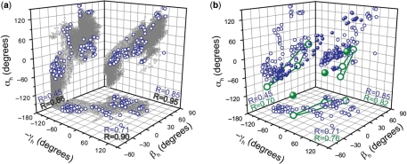
We describe a strategy for constructing atomic resolution dynamical ensembles of RNA molecules, spanning up to millisecond timescales, that combines molecular dynamics (MD) simulations with NMR residual dipolar couplings (RDC) measured in elongated RNA. The ensembles are generated via a Monte Carlo procedure by selecting snap-shot from an MD trajectory that reproduce experimentally measured RDCs. Using this approach, we construct ensembles for two variants of the transactivation response element (TAR) containing three (HIV-1) and two (HIV-2) nucleotide bulges. The HIV-1 TAR ensemble reveals significant mobility in bulge residues C24 and U25 and to a lesser extent U23 and neighboring helical residue A22 that give rise to large amplitude spatially correlated twisting and bending helical motions. Omission of bulge residue C24 in HIV-2 TAR leads to a significant reduction in both the local mobility in and around the bulge and amplitude of inter-helical bending motions. In contrast, twisting motions of the helices remain comparable in amplitude to HIV-1 TAR and spatial correlations between them increase significantly. Comparison of the HIV-1 TAR dynamical ensemble and ligand bound TAR conformations reveals that several features of the binding pocket and global conformation are dynamically preformed, providing support for adaptive recognition via a 'conformational selection' type mechanism.


Figures

Figure 1.
Figure 1.
SAS analysis of HIV-1 E-TAR RDCs. (a) Secondary structure of HIV-1 TAR with helix I highlighted in red, helix II in green and tri-nucleotide bulge in orange. HIV-2 TAR lacks bulge residue C24. (b and c) Plots of experimental RDCs versus values computed from the 80 ns MD trajectory for (b) EI-TAR and (c) EII-TAR. Data for helix I, helix II, and bulge, are shown in red, green and orange, respectively. Also shown is the root-mean-square-deviation (rmsd) and correlation coefficient (R). (d) RMSD (Hz) between calculated and experimental RDCs as a function of N, following a SAS analysis using both EI and EII-TAR HIV-1 RDCs. (e–g) Plots of experimental RDCs versus values calculated from the N = 20 SAS ensemble using (e) EI-TAR, (f) EII-TAR and (g) EI-TAR+EII-TAR RDCs.
Figure 2.
Figure 2.
Local motions in the HIV-1 TAR dynamical ensemble. (a) Shown in black are the mean values for the base opening (σ), buckle (κ), propeller twist (ω) and twist (Ω) angles and their SD calculated over 121 HIV-1 TAR conformers obtained from multiple N = 20 SAS runs. For comparison, shown in red are corresponding values for an idealized A-form helix as obtained from a statistical survey of high resolution X-ray structures (48). (b) Average conformation of the HIV-1 TAR bulge and neighboring base-pairs calculated from the 121-membered SAS ensemble. The bases of the bulge and flanking base pairs are color-coded based on the root mean square fluctuations (r.m.s.f.) calculated over the ensemble.
Figure 3.
Figure 3.
Global inter-helical dynamics in the HIV-1 TAR dynamical ensemble. (a) Shown in gray are the inter-helical twist (αh and γh) and bend (βh) angles for 80 000 HIV-1 TAR conformers derived from an 80 ns MD trajectory (conformer selected every 1 ps). In blue are the corresponding SAS selected conformers. The correlation coefficient (R) is shown on individual planes. (b) Comparison of the SAS selected inter-helical angles and those derived previously (26) based on a three-state rigid body ensemble analysis of E-TAR RDCs.
Figure 4.
Figure 4.
HIV-2 TAR dynamical ensemble. Plot of experimental HIV-2 EI-TAR RDCs versus values calculated from (a) an 80 ns MD trajectory of HIV-2 TAR and (b) following SAS selection of N = 20 conformers from the MD trajectory based on HIV2 EI-TAR RDCs. Coloring scheme is same as in Figure 1. (c) Shown in black are the mean base-pair angles and their SD calculated over a 279-membered HIV-2 TAR SAS ensemble. For comparison shown in green are corresponding angles for an idealized A-form helix as obtained from a statistical survey of high-resolution X-ray structures (48). (d) Average conformation of the bulge of HIV-2 TAR calculated from the 279-membered HIV-2 TAR SAS ensemble. Bases of the bulge and flanking base pairs are color-coded based on the root mean square fluctuations (r.m.s.f.) calculated over the ensemble. (e) Comparison of SAS selected HIV-1 (blue) and HIV-2 (red) inter-helical conformations. The correlation coefficient (R) is shown on individual planes.
Figure 5.
Figure 5.
Comparison of the SAS derived HIV-1 TAR dynamical ensemble and ligand bound TAR conformations. (a) Comparison of the global inter-helical angles. Shown in blue are the SAS selected angles and in gray seven distinct ligand bound TAR structures. (b and c) Comparison of the b, global and c, local structure of SAS TAR conformers and seven distinct ligand bound TAR conformations (1QD3, 1UUI, 1UTS, 1UUD, 1ARJ, 1LVJ, and 397D). Shown are the pairs yielding the lowest RMSD fit when superimposing b, all heavy atoms excluding terminal base-pairs G17-C45 and the apical loop and c, all heavy atoms in the bulge and immediately adjacent base-pairs. Every model in the ligand bound NMR ensembles was used in the superposition. The corresponding ligand is colored yellow.

Similar articles

Cited by

References

    1. Perez-Canadillas JM, Varani G. Recent advances in RNA-protein recognition. Curr. Opin. Struct. Biol. 2001;11:53–58. - PubMed
    1. Tucker BJ, Breaker RR. Riboswitches as versatile gene control elements. Curr. Opin. Struct. Biol. 2005;15:342–348. - PubMed
    1. Nudler E. Flipping riboswitches. Cell. 2006;126:19–22. - PubMed
    1. Al-Hashimi HM, Walter NG. RNA dynamics: it is about time. Curr. Opin. Struct. Biol. 2008;18:321–329. - PMC - PubMed
    1. Hall KB. RNA in motion. Curr. Opin. Chem. Biol. 2008;12:612–618. - PMC - PubMed

Publication types