Comparative Study
. 2009 Jun 18;113(25):6465-76.
doi: 10.1182/blood-2009-02-203307. Epub 2009 Apr 15.

Impact of donor CMV status on viral infection and reconstitution of multifunction CMV-specific T cells in CMV-positive transplant recipients

Affiliations
Comparative Study

Impact of donor CMV status on viral infection and reconstitution of multifunction CMV-specific T cells in CMV-positive transplant recipients

Wendi Zhou et al. Blood. .

Abstract

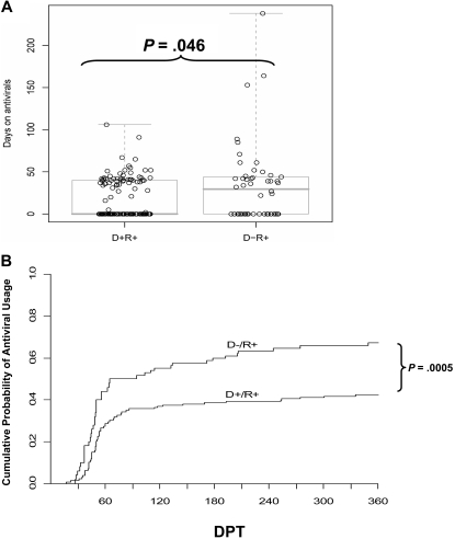
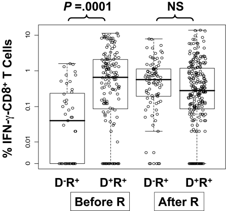
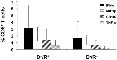
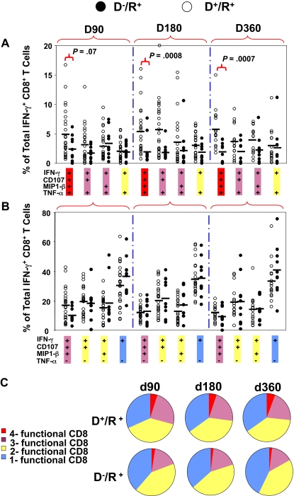
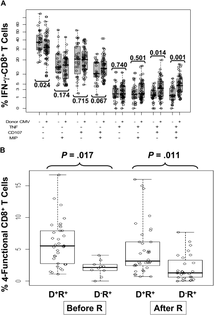
Reconstitution of cytomegalovirus (CMV)-specific CD8(+) T cells is essential to the control of CMV infection in CMV-positive recipients (R(+)) after allogeneic hematopoietic stem cell transplantation (HCT). Six-color flow cytometry was used to assess the functional profile of CMV-specific CD8(+) T cells in 62 of 178 R(+) HCT recipients followed virologically for CMV reactivation. R(+) recipients receiving grafts from CMV-negative donors (D(-); D(-)/R(+)) reconstituted fewer multifunctional CD8(+) T cells expressing tumor necrosis factor-alpha (TNF-alpha), macrophage inflammatory protein-1beta (MIP-1beta), and CD107 in addition to interferon-gamma (IFN-gamma), compared with D(+)/R(+) recipients. Unlike monofunctional CD8(+) T cells secreting IFN-gamma, which were abundantly generated during CMV reactivation in D(-)/R(+) recipients, the relative lack of multifunctional CD8(+) T cells persisted until at least 1 year post-HCT. D(-)/R(+) recipients were more likely to require recurrent and prolonged use of antivirals. These findings were robust to statistical adjustment for pretransplant factors, as well as for posttransplant factors including graft-versus-host disease (GVHD) and its treatment by steroids. These analyses suggest that D(+)/R(+) transplants, on average, generate higher levels of multifunctional CMV-specific T cells and require less antiviral therapy compared with D(-)/R(+) HCT recipients. These results highlight the benefit of D(+) donors in improving outcomes of R(+) HCT recipients by reducing the duration and recurrent need of antiviral treatment, aided by increased levels of multifunctional CMV-specific T cells.


Figures

Figure 1
Figure 1
Impact of donor CMV status on antiviral use. (A) The number of days that each D+/R+ or D/R+ recipient received antiviral drug therapy (GCV or foscarnet). The box plot overlays show the median and the central 50% of the data. The median is zero for the D+/R+ group, as the majority did not require antivirals. P value determined by rank-sum test. (B) The cumulative incidence of antiviral use, estimated by the method of Kalbfleisch and Prentice, incorporates recurrent events. Most events occur between 30 to 90 days posttransplant, but the separation of the curves continues to increase, as late events occur more frequently in the D group. The excess risk in the D group was statistically significant, adjusted for pre-HCT covariates (P < .001) and for GVHD (P = .009, Table 3). DPT, days posttransplant.
Figure 2
Figure 2
Levels of CMV pp65-specific IFN-γ–CD8+ in PBMC samples from R+ HCT recipients. The circles are average percentages of IFN-γ–CD8+ within PBMC samples from R+ HCT recipients in response to stimulation with CMV pp65 peptide library. All measurements from day 90 to day 360 were pooled together and divided into 4 groups according to donor CMV status and CMV reactivation post-HCT assessed by quantitative PCR (“Prospective study subjects”). The D/R+ prereactivation group had significantly lower values compared with the other groups (P < .001, see Table 4). Within the prereactivation subset, D has significantly lower levels than D+ (P = .002, adjusted as in Table 4, P < .001 unadjusted). The effect of donor status was significantly different in prereactivation versus postreactivation samples (P < .001 interaction test, adjusted as in Table 4). Each dot represents a single IFN-γ measurement. The lower and upper horizontal border of the box drawn for each group marks the 25th and 75th percentiles, with a solid bar at the median and whiskers covering the range of the data. Horizontal scatter within boxes is introduced to avoid overlap. Pre-R and post-R refers to CMV reactivation.
Figure 3
Figure 3
CD8+ T cells in D+/R+ and D/R+ HCT recipients expressing each of 4 functional markers. The average percentage of CD8+ T cells in samples from D+/R+ (n = 41) and D/R+ (n = 21) HCT recipients (Table 1) that were positive for each of the markers IFN-γ, MIP-1β, CD107, or TNF-α are shown as separate bars. The data bars represent an average of measurements from all 3 time points in all HCT recipients in group 3 (Table 1). Error bars illustrate the range of measurements for each marker.
Figure 4
Figure 4
Comparison of levels of multifunctional CD8+ T cells in D+/R+ and D/R+ HCT recipients. The percentages of 8 combinations of single-, double-, triple-, and quadruple-functional subsets within the total population of IFN-γ–CD8+ were compared between D+/R+ and D/R+ recipients at 3 time points after HCT. Each dot represents an individual measurement. Average percentages are shown as horizontal black bars. Four combinations of TNF-α+ subsets are shown in (A) and 4 combinations of TNF-α subsets are shown in panel B. (C) All 8 possible combinations of response categories are summarized and shown in the pie chart, in which each slice of the pie represents the average response across all individual samples. The fraction of the total IFN-γ–CD8+ response for 8 different functional marker combinations are shown sequentially in different colors from single-functional cells (blue) through double-functional (yellow), triple-functional (pink), and quadruple-functional (red).
Figure 5
Figure 5
Relationship between multifunctional CMV-specific CD8+ T cell levels, CMV reactivation, and donor CMV status. (A) The 8 combinations of T cells positive or negative for TNF-α, CD107, and MIP are shown as a percentage of IFN-γ–CD8+ T cells and subdivided according to donor CMV status. Each circle represents a single measurement, and samples from all 3 time points are pooled for display. The vertical scale uses square-root spacing, labeled with percentages. Boxes cover the central 50% of the observations, with a central bar at the median. Significance probabilities (P), shown for each subset are from a 2-sided rank-sum test after reducing the data to a single mean per subject. (B) The percentages of 4-functional CD8+ T cells from D+/R+ and D/R+ recipients were further divided according to CMV reactivation status as defined by PCR. There was no statistically significant time trend, so samples from 3 time points were pooled for display from day 90, day 180, and day 360 observations. Each dot represents an individual measurement. Boxes cover the central 50% of the observations, with a central bar at the median. Nomenclature for patients and CMV reactivation defined in Figure 2 legend. The P values are from rank-sum tests on the pre- or postreactivation subsets, using one mean value for each subject, and are not adjusted. Using all data and adjusting for covariates, recipients with D donors have significantly lower average 4-function T cell levels (P = .01, Table 5).

References

    1. Nichols WG, Corey L, Gooley T, Davis C, Boeckh M. High risk of death due to bacterial and fungal infection among cytomegalovirus (CMV)-seronegative recipients of stem cell transplants from seropositive donors: evidence for indirect effects of primary CMV infection. J Infect Dis. 2002;185:273–282. - PubMed
    1. Schmidt GM, Horak DA, Niland JC, et al. A randomized, controlled trial of prophylactic ganciclovir for cytomegalovirus pulmonary infection in recipients of allogeneic bone marrow transplants; The City of Hope-Stanford-Syntex CMV Study Group. N Engl J Med. 1991;324:1005–1011. - PubMed
    1. Chakrabarti S, Mackinnon S, Chopra R, et al. High incidence of cytomegalovirus infection after nonmyeloablative stem cell transplantation: potential role of Campath-1H in delaying immune reconstitution. Blood. 2002;99:4357–4363. - PubMed
    1. Boeckh M, Nichols WG. The impact of cytomegalovirus serostatus of donor and recipient before hematopoietic stem cell transplantation in the era of antiviral prophylaxis and preemptive therapy. Blood. 2004;103:2003–2008. - PubMed
    1. Goodrich JM, Bowden RA, Fisher L, et al. Ganciclovir prophylaxis to prevent cytomegalovirus disease after allogeneic marrow transplant. Ann Intern Med. 1993;118:173–178. - PubMed

Publication types

MeSH terms