. 2009 Jun 19;284(25):17013-17020.
doi: 10.1074/jbc.M901618200. Epub 2009 Apr 15.

Structure of the thiostrepton resistance methyltransferase.S-adenosyl-L-methionine complex and its interaction with ribosomal RNA

Affiliations

Structure of the thiostrepton resistance methyltransferase.S-adenosyl-L-methionine complex and its interaction with ribosomal RNA

Mark S Dunstan et al. J Biol Chem. .

Abstract

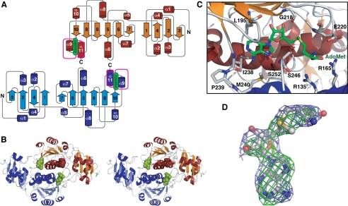
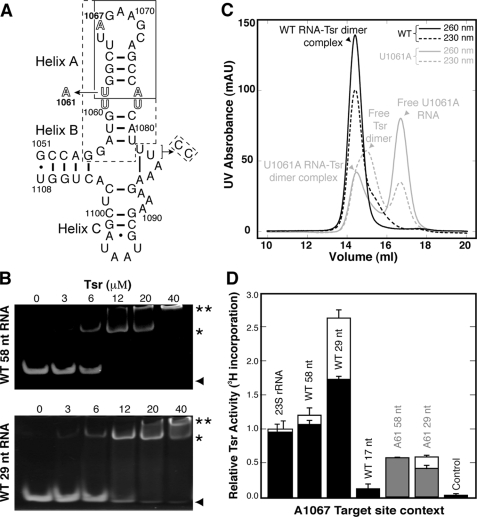
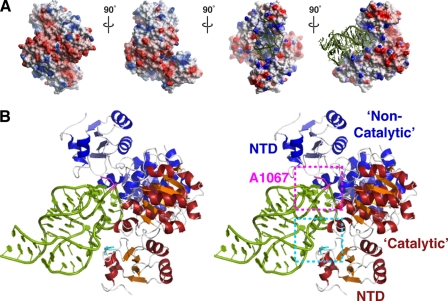
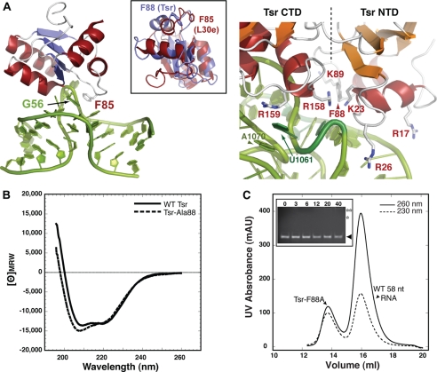
The x-ray crystal structure of the thiostrepton resistance RNA methyltransferase (Tsr).S-adenosyl-L-methionine (AdoMet) complex was determined at 2.45-A resolution. Tsr is definitively confirmed as a Class IV methyltransferase of the SpoU family with an N-terminal "L30-like" putative target recognition domain. The structure and our in vitro analysis of the interaction of Tsr with its target domain from 23 S ribosomal RNA (rRNA) demonstrate that the active biological unit is a Tsr homodimer. In vitro methylation assays show that Tsr activity is optimal against a 29-nucleotide hairpin rRNA though the full 58-nucleotide L11-binding domain and intact 23 S rRNA are also effective substrates. Molecular docking experiments predict that Tsr.rRNA binding is dictated entirely by the sequence and structure of the rRNA hairpin containing the A1067 target nucleotide and is most likely driven primarily by large complementary electrostatic surfaces. One L30-like domain is predicted to bind the target loop and the other is near an internal loop more distant from the target site where a nucleotide change (U1061 to A) also decreases methylation by Tsr. Furthermore, a predicted interaction with this internal loop by Tsr amino acid Phe-88 was confirmed by mutagenesis and RNA binding experiments. We therefore propose that Tsr achieves its absolute target specificity using the N-terminal domains of each monomer in combination to recognize the two distinct structural elements of the target rRNA hairpin such that both Tsr subunits contribute directly to the positioning of the target nucleotide on the enzyme.


Figures

FIGURE 1.
FIGURE 1.
X-ray crystal structure of the Tsr-AdoMet complex. A, Tsr dimer protein secondary structure topology diagram. The knotted sequence is colored magenta. B, stereo view schematic of the Tsr dimer with bound AdoMet (green). C, the AdoMet binding pocket with key Tsr amino acids indicated. D, AdoMet in an extended conformation shown with omit FoFcc (green) and omit 2Fo − 1Fc (light blue) maps contoured at 3.5 and 1.0 σ, respectively.
FIGURE 2.
FIGURE 2.
rRNA binding and methylation. A, the 58-nucleotide L11 rRNA binding domain containing the Tsr target nucleotide A1067. Boxed regions correspond to smaller RNA hairpins: 17 nucleotides (solid line) and 29 nucleotides (dashed). A modification of the 3′-UU end for the latter RNA to generate Watson-Crick pairing is indicated. The mutation U1061 to A is indicated, and the internal loop within Helix A is shown in outline type. B, gel electrophoresis shift assays with wild-type 58-nucleotide (upper panel) and 29-nucleotide (lower panel) RNAs at a constant concentration of 6 μm RNA per assay and Tsr input at the concentrations indicated above each gel. Free RNA (▴), RNA-Tsr dimer 1:1 complex (*) and higher molecular weight complexes (**) are indicated on the right hand side. C, gel filtration chromatography of 1:1 mixtures of wild-type (black) and U1061A mutant (gray) 58-nucleotide RNAs (3 μm) and Tsr dimer (3 μm). Elution from the column was monitored at 260 (solid line) and 230 nm (dashed line). The content of each peak is identified as indicated on the basis of apparent molecular weight and relative intensity of absorbance at each wavelength. D, methylation activity was measured for 23 S rRNA and three wild-type (58, 29, and 17 nucleotides) and two U1061A mutant (58 and 29 nucleotides) L11-binding domain RNAs by 3H incorporation. Solid bars represent data at the 10-min time point, and where present open bars represent the 30-min time point (both were measured for all RNAs but for some maximum methylation was reached by the earlier time point). Control experiments used an unrelated 54-nucleotide RNA (see supplemental Fig. S4). Error bars are the standard deviation from at least three independent experiments.
FIGURE 3.
FIGURE 3.
Molecular modeling of Tsr-rRNA interactions. A, four orthogonal views around the vertical axis of the Tsr dimer with electrostatic surface potential indicated in red (negative) and blue (positive). Docked RNA is shown in the two orientations on the right only. B, stereo view schematic of the docked Tsr·rRNA complex. Regions encompassing the A1067 target loop (magenta) recognized by the non-catalytic Tsr and the internal loop (cyan) recognized by the catalytic Tsr, including Phe-88 (also see Fig. 4), are shown in dashed boxes.
FIGURE 4.
FIGURE 4.
Tsr Phe-88 is a critical determinant of recognition. A, interaction of L30e Phe-85 with the L30e mRNA kink turn (44, 45) (left) and close-up view of the modeled Tsr-rRNA interaction in the region of Phe-88 (right). The inset shows an alignment of the Tsr L30-like NTD and L30e. B, CD spectra of wild-type and Ala-88 Tsr proteins. C, the Tsr-Ala-88 protein is defective in RNA binding as monitored by gel filtration chromatography and gel mobility shift assay (inset; compare with data of Fig. 2, B and C). Protein concentrations in the gel shift assay are indicated above each lane, and bands corresponding to free RNA (▴) and approximate positions of absent shifted bands (asterisks) are indicated.

References

    1. Gustilo E. M., Vendeix F. A., Agris P. F. (2008) Curr. Opin. Microbiol. 11, 134–140 - PMC - PubMed
    1. Clouet-d'Orval B., Gaspin C., Mougin A. (2005) Biochimie (Paris) 87, 889–895 - PubMed
    1. Ferré-D'Amaré A. R. (2003) Curr. Opin. Struct. Biol. 13, 49–55 - PubMed
    1. Rozenski J., Crain P. F., McCloskey J. A. (1999) Nucleic Acids Res. 27, 196–197 - PMC - PubMed
    1. Xu Z., O'Farrell H. C., Rife J. P., Culver G. M. (2008) Nat. Struct. Mol. Biol. 15, 534–536 - PubMed

Publication types

MeSH terms

LinkOut - more resources