. 2009 Jun;296(6):F1514-20.
doi: 10.1152/ajprenal.00068.2009. Epub 2009 Apr 15.

Caveolin-1 directly interacts with UT-A1 urea transporter: the role of caveolae/lipid rafts in UT-A1 regulation at the cell membrane

Affiliations

Caveolin-1 directly interacts with UT-A1 urea transporter: the role of caveolae/lipid rafts in UT-A1 regulation at the cell membrane

Xiuyan Feng et al. Am J Physiol Renal Physiol. 2009 Jun.

Abstract

The cell plasma membrane contains specialized microdomains called lipid rafts which contain high amounts of sphingolipids and cholesterol. Lipid rafts are involved in a number of membrane protein functions. The urea transporter UT-A1, located in the kidney inner medullary collecting duct (IMCD), is important for urine concentrating ability. In this study, we investigated the possible role of lipid rafts in UT-A1 membrane regulation. Using sucrose gradient cell fractionation, we demonstrated that UT-A1 is concentrated in the caveolae-rich fraction both in stably expressing UT-A1 HEK293 cells and in freshly isolated kidney IMCD suspensions. In these gradients, UT-A1 at the cell plasma membrane is codistributed with caveolin-1, a major component of caveolae. The colocalization of UT-A1 in lipid rafts/caveolae was further confirmed in isolated caveolae from UT-A1-HEK293 cells. The direct association of UT-A1 and caveolin-1 was identified by immunoprecipitation and GST pull-down assay. Examination of internalized UT-A1 in pEGFP-UT-A1 transfected HEK293 cells fluorescent overlap with labeled cholera toxin subunit B, a marker of the caveolae-mediated endocytosis pathway. Disruption of lipid rafts by methyl-beta-cyclodextrin or knocking down caveolin-1 by small-interference RNA resulted in UT-A1 cell membrane accumulation. Functionally, overexpression of caveolin-1 in oocytes decreased UT-A1 urea transport activity and UT-A1 cell surface expression. Our results indicate that lipid rafts/caveolae participate in UT-A1 membrane regulation and this effect is mediated via a direct interaction of caveolin-1 with UT-A1.


Figures
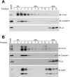
Fig. 1.
Fig. 1.
Cell membrane UT-A1 is concentrated in lipid raft microdomains and codistributed with caveolin-1. UT-A1-HEK293 cells (A) or freshly prepared rat kidney inner medullary collecting duct (IMCD) suspension (B) were lysed with 1% Triton X-100 and loaded on 5–40% sucrose gradients for lipid raft isolation. After 20-h centrifugation, equal volume fractions were collected from the top to bottom and Western blotted for UT-A1. Caveolin-1 was used as a positive control for lipid raft fractions. Clathrin adaptor protein μ2 was used as the indicator for nonlipid raft fractions. The same membrane (B) was stripped and immunoblotted for aquaporin-2 (AQP2). The experiments of A were replicated in 10 separate experiments. B was repeated in 4 separate cohorts of animals with similar results.
Fig. 2.
Fig. 2.
UT-A1 associates with caveolin and colocalizes with lipid rafts. A: caveolae were isolated from UT-A1-HEK293 cells and blotted with UT-A1 antibody. UT-A1 was detected in the isolated caveolae. B: UT-A1 was immunoprecipitated from the total lysate and the isolated caveolae. The immunoprecipites were blotted for caveolin-1. *Unknown nonspecific band. C: GST pull-down assay. GST-caveolin-1 was incubated with rat IM tissue lysates (2 rats). The pulled-down samples were analyzed with UT-A1 antibody. D: HEK293 cells were transfected with pEGFP-UT-A1 for 48 h. Cells were fixed in 4% paraformaldehyde, incubated with Alexa-594-conjugated cholera toxin B (CTB), and visualized with a confocal microscope. A, B, and D were repeated twice. C was repeated 3 times.
Fig. 3.
Fig. 3.
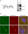
UT-A1 endocytosis mediated by caveolae. A, left: schematic diagram shows the FLAG tag inserted in the first large extracellular loop. Right: Western blot analysis of FLAG-UT-A1 expression in HEK293 cells. FLAG-UT-A1 in pcDNA3 was transiently transfected into cells for 2 days. Cell lysates were immunoblotted with UT-A1 COOH-terminus antibody or FLAG. The pcDNA3-UT-A1 and FLAG HSP70 were used as controls. B: FLAG-UT-A1-transfected HEK293 cells were incubated with FLAG antibody and Alexa-CTB for 45 min on ice. Cell endocytosis was induced by incubation with prewarmed complete cell medium at 37°C for 5 min and analyzed under confocal microscope as described in materials and methods. Arrows indicate sites of colocalization as determined by yellow color. The results are representative of 2 separate experiments.
Fig. 4.
Fig. 4.
Knock-down of caveolin-1 by RNA interference increases UT-A1 membrane expression. UT-A1-HEK293 cells were transfected with pSuppressor/scramble, dynamin, or caveolin-1 for 48 h. Cells were processed for biotinylation. Total cell lysates and biotinylated samples were separated on SDS-PAGE and probed with antibodies against the indicated proteins. The experiments were repeated 3 times with similar results.
Fig. 5.
Fig. 5.
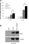
Lipid raft disruption by methyl-β-cyclodextrin (MβCD) increases UT-A1 expression at the cell surface. Three days after cRNA microinjection, the oocytes were incubated with 5 mM MβCD for 2 h at room temperature and then processed for [14C]urea uptake assay (n = 5∼6 oocytes/time point; A) or cell surface UT-A1 expression by biotinylation (B). This experiment was repeated 2 additional times with similar results. **P < 0.01.
Fig. 6.
Fig. 6.
Overexpression of caveolin-1 reduces UT-A1 cell surface protein expression and urea transport activity. Oocytes were coinjected with cRNAs encoding UT-A1 (2 ng/cell), plus dynamin or caveolin-1 (5 ng/cell) for 3 days. A: urea transport activity was measured by [14C]urea flux (n = 5∼6 oocytes/time point). *P < 0.05; **P < 0.01. B: cell surface UT-A1 protein expression was measured by biotinylation. These results were reproduced 2 additional times.

References

    1. Blount MA, Mistry AC, Froehlich O, Price RS, Chen G, Sands JM, Klein JD. Phosphorylation of UT-A1 urea transporter at serines 486 and 499 is important for vasopressin-regulated activity and membrane accumulation. Am J Physiol Renal Physiol 295: F295–F299, 2008. - PMC - PubMed
    1. Bradford AD, Terris JM, Ecelbarger CA, Klein JD, Sands JM, Chou CL, Knepper MA. 97- And 117-kDa forms of collecting duct urea transporter UT-A1 are due to different states of glycosylation. Am J Physiol Renal Physiol 281: F133–F143, 2001. - PubMed
    1. Cha SK, Wu T, Huang CL. Protein kinase C inhibits caveolae-mediated endocytosis of TRPV5. Am J Physiol Renal Physiol 294: F1212–F1221, 2008. - PubMed
    1. Chen G, Fröhlich O, Yang Y, Klein JD, Sands JM. Loss of N-linked glycosylation reduces urea transporter UT-A1 response to vasopressin. J Biol Chem 281: 27436–27442, 2006. - PubMed
    1. Chen G, Huang H, Fröhlich O, Yang Y, Klein JD, Price SR, Sands JM. MDM2 E3 ubiquitin ligase mediates UT-A1 urea transporter ubiquitination and degradation. Am J Physiol Renal Physiol 295: F1528–F1534, 2008. - PMC - PubMed

Publication types