. 2009 Jul 1;18(13):2431-42.
doi: 10.1093/hmg/ddp181. Epub 2009 Apr 15.

Mouse models of MeCP2 disorders share gene expression changes in the cerebellum and hypothalamus

Affiliations

Mouse models of MeCP2 disorders share gene expression changes in the cerebellum and hypothalamus

Shay Ben-Shachar et al. Hum Mol Genet. .

Abstract

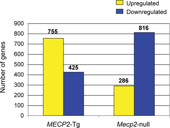
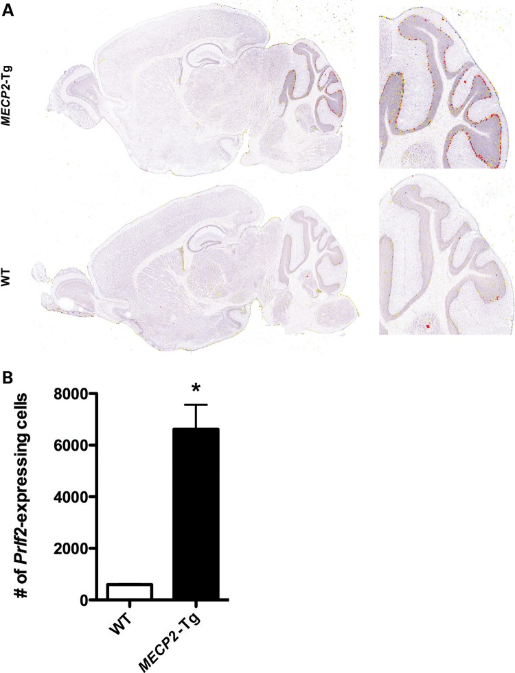
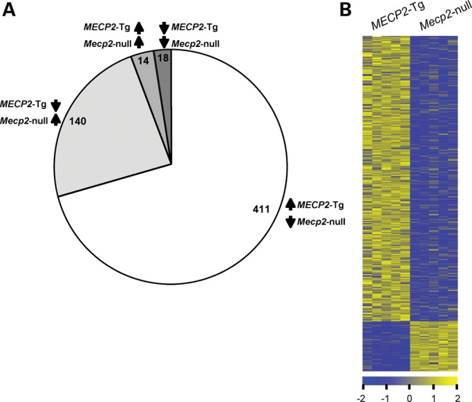
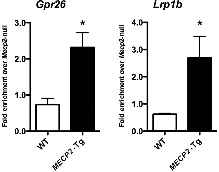
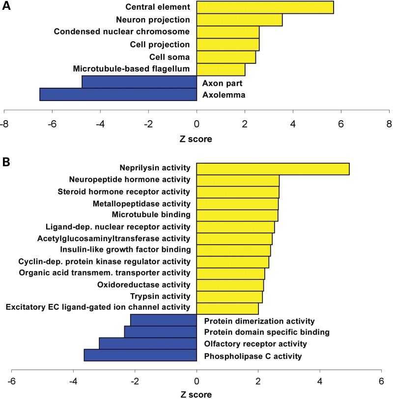
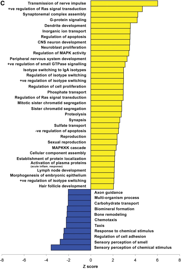
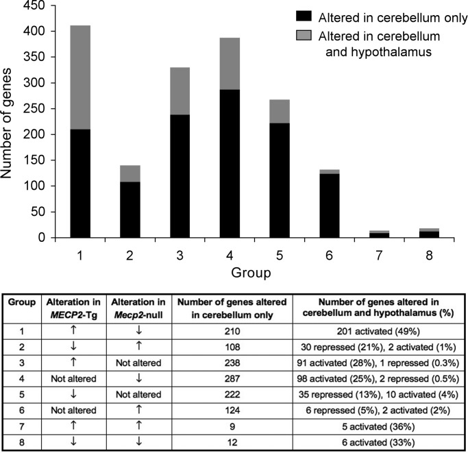
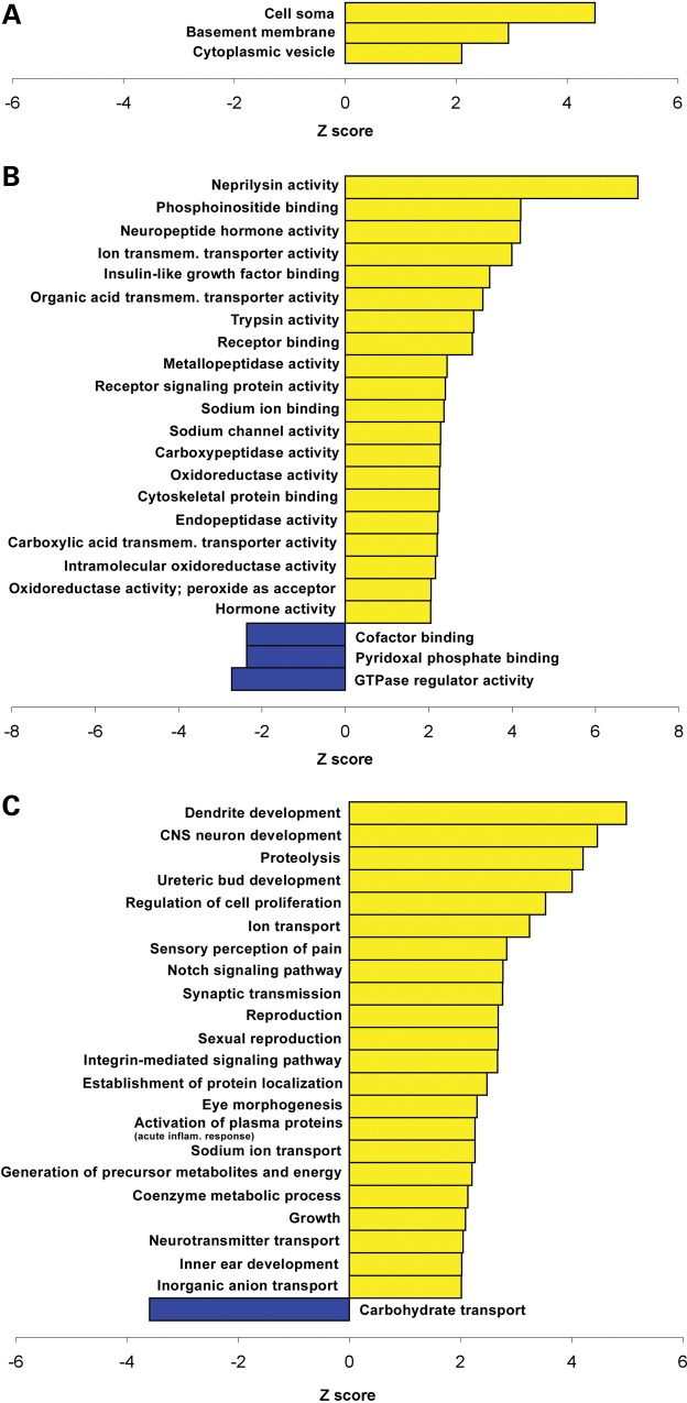
A group of post-natal neurodevelopmental disorders collectively referred to as MeCP2 disorders are caused by aberrations in the gene encoding methyl-CpG-binding protein 2 (MECP2). Loss of MeCP2 function causes Rett syndrome (RTT), whereas increased copy number of the gene causes MECP2 duplication or triplication syndromes. MeCP2 acts as a transcriptional repressor, however the gene expression changes observed in the hypothalamus of MeCP2 disorder mouse models suggest that MeCP2 can also upregulate gene expression, given that the majority of genes are downregulated upon loss of MeCP2 and upregulated in its presence. To determine if this dual role of MeCP2 extends beyond the hypothalamus, we studied gene expression patterns in the cerebellum of Mecp2-null and MECP2-Tg mice, modeling RTT and MECP2 duplication syndrome, respectively. We found that abnormal MeCP2 dosage causes alterations in the expression of hundreds of genes in the cerebellum. The majority of genes were upregulated in MECP2-Tg mice and downregulated in Mecp2-null mice, consistent with a role for MeCP2 as a modulator that can both increase and decrease gene expression. Interestingly, many of the genes altered in the cerebellum, particularly those increased by the presence of MeCP2 and decreased in its absence, were similarly altered in the hypothalamus. Our data suggest that either gain or loss of MeCP2 results in gene expression changes in multiple brain regions and that some of these changes are global. Further delineation of the expression pattern of MeCP2 target genes throughout the brain might identify subsets of genes that are more amenable to manipulation, and can thus be used to modulate some of the disease phenotypes.


Figures

Figure 1.
Figure 1.
Studies of cerebellar gene expression changes in MECP2-Tg and Mecp2-null mice. Most of the genes altered in both mouse models are upregulated in the presence of MeCP2 and downregulated in its absence.
Figure 2.
Figure 2.
RNA ISH demonstrates that Prlf2 is highly upregulated in MECP2-Tg cerebellum compared with WT (A). Prlf2-expressing cells were counted on sections throughout the cerebellum (B) (*P < 0.004).
Figure 3.
Figure 3.
Cerebellar gene expression changes common to both MECP2-Tg and Mecp2-null mice. (A) The majority of genes altered in both mouse models (95%) show opposite direction of change, usually activation. (B) Heatmap showing cerebellar gene expression profiles in MECP2-Tg and Mecp2-null mice. Yellow and blue colors indicate increased and decreased expression, respectively, relative to WT. Each column represents one RNA sample from each genotype, and each row represents one gene. Expression levels are depicted according to the color scale at the bottom (FDR-corrected P < 0.05).
Figure 4.
Figure 4.
MeCP2 binds to the promoter regions of an activated (Gpr26) and a repressed (Lrp1b) target gene. ChIP with antibody to MeCP2 detects MeCP2 binding to the promoter region of two novel targets. QPCR values were normalized to the input and plotted as relative enrichment over Mecp2-null (n = 3, *P < 0.05).
Figure 5.
Figure 5.
Gene ontology analysis performed on genes that are differentially expressed in cerebella of MECP2-Tg and Mecp2-null mice. Cellular component (A), molecular function (B) and biological process (C) categories that differ significantly between the genes activated by MeCP2 (yellow) and those repressed by MeCP2 (blue) are plotted. The standardized scores (Z values) are presented on the x-axis, and significance was determined by a Z-value of more than ± 2 and a count of at least two genes. +ve, positive; −ve, negative; CNS, central nervous system; inflam., inflammatory; dep., dependent; transmem., transmembrane; EC, extracellular.
Figure 5.
Figure 5.
Gene ontology analysis performed on genes that are differentially expressed in cerebella of MECP2-Tg and Mecp2-null mice. Cellular component (A), molecular function (B) and biological process (C) categories that differ significantly between the genes activated by MeCP2 (yellow) and those repressed by MeCP2 (blue) are plotted. The standardized scores (Z values) are presented on the x-axis, and significance was determined by a Z-value of more than ± 2 and a count of at least two genes. +ve, positive; −ve, negative; CNS, central nervous system; inflam., inflammatory; dep., dependent; transmem., transmembrane; EC, extracellular.
Figure 6.
Figure 6.
Gene expression changes common to the cerebellum and hypothalamus. Groups 1–8 in the top panel are described in the lower panel. For example, genes in Group 1 are upregulated in MECP2-Tg and downregulated in Mecp2-null mice, 201 such genes are altered in both the cerebellum and hypothalamus, and activated in the hypothalamus.
Figure 7.
Figure 7.
Gene ontology analysis performed on genes that are differentially expressed in cerebella and hypothalami of MECP2-Tg and Mecp2-null mice. Cellular component (A), molecular function (B) and biological process (C) categories that differ significantly between the genes activated by MeCP2 (yellow) and those repressed by MeCP2 (blue) are plotted. The standardized scores (Z-values) are presented on the x-axis, and significance was determined by a Z-value of more than ± 2 and a count of at least two genes. CNS, central nervous system; inflam., inflammatory; transmem., transmembrane.

References

    1. Amir R.E., Van den Veyver I.B., Wan M., Tran C.Q., Francke U., Zoghbi H.Y. Rett syndrome is caused by mutations in X-linked MECP2, encoding methyl-CpG-binding protein 2. Nat. Genet. 1999;23:185–188. - PubMed
    1. Chahrour M., Zoghbi H.Y. The story of Rett syndrome: from clinic to neurobiology. Neuron. 2007;56:422–437. - PubMed
    1. Friez M.J., Jones J.R., Clarkson K., Lubs H., Abuelo D., Bier J.A., Pai S., Simensen R., Williams C., Giampietro P.F., et al. Recurrent infections, hypotonia, and mental retardation caused by duplication of MECP2 and adjacent region in Xq28. Pediatrics. 2006;118:e1687–e1695. - PubMed
    1. Lugtenberg D., de Brouwer A.P., Kleefstra T., Oudakker A.R., Frints S.G., Schrander-Stumpel C.T., Fryns J.P., Jensen L.R., Chelly J., Moraine C., et al. Chromosomal copy number changes in patients with non-syndromic X linked mental retardation detected by array CGH. J. Med. Genet. 2006;43:362–370. - PMC - PubMed
    1. Van Esch H., Bauters M., Ignatius J., Jansen M., Raynaud M., Hollanders K., Lugtenberg D., Bienvenu T., Jensen L.R., Gecz J., et al. Duplication of the MECP2 region is a frequent cause of severe mental retardation and progressive neurological symptoms in males. Am. J. Hum. Genet. 2005;77:442–453. - PMC - PubMed

Publication types

Substances

Associated data