. 2009 Jul;83(13):6825-36.
doi: 10.1128/JVI.00301-09. Epub 2009 Apr 15.

Herpes simplex virus glycoprotein B associates with target membranes via its fusion loops

Affiliations

Herpes simplex virus glycoprotein B associates with target membranes via its fusion loops

Brian P Hannah et al. J Virol. 2009 Jul.

Abstract

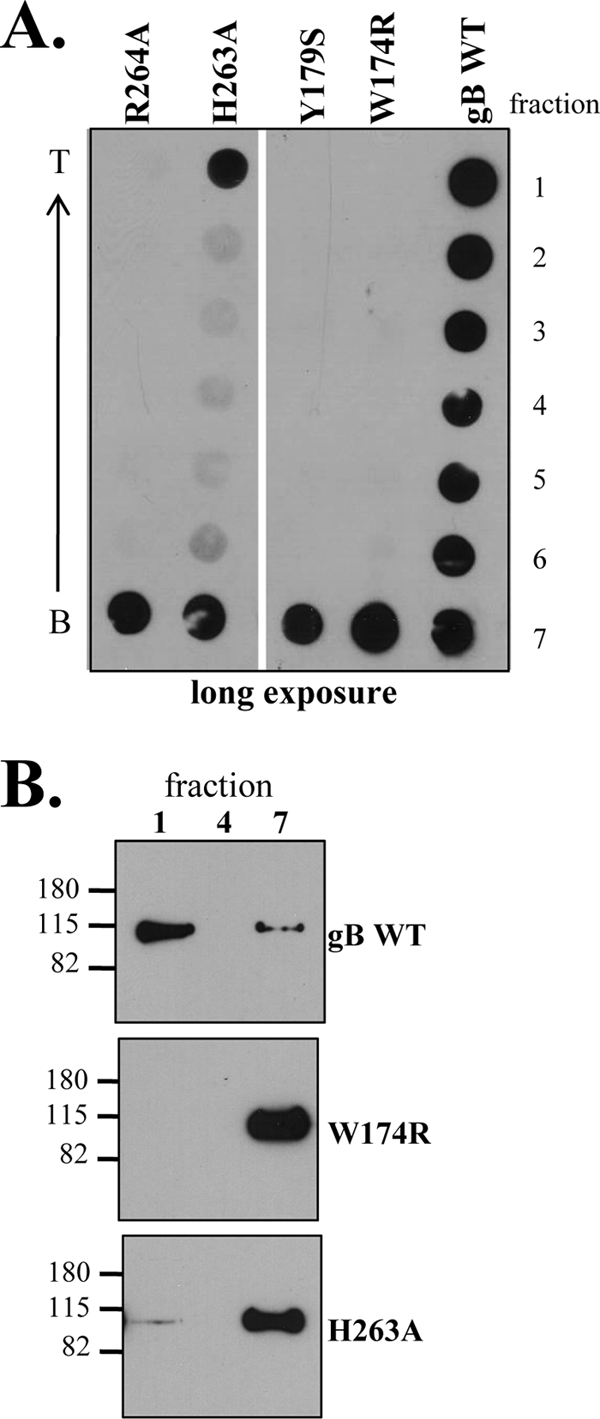
Herpes simplex virus (HSV) glycoproteins gB, gD, and gH/gL are necessary and sufficient for virus entry into cells. Structural features of gB are similar to those of vesicular stomatitis virus G and baculovirus gp64, and together they define the new class III group of fusion proteins. Previously, we used mutagenesis to show that three hydrophobic residues (W174, Y179, and A261) within the putative gB fusion loops are integral to gB function. Here we expanded our analysis, using site-directed mutagenesis of each residue in both gB fusion loops. Mutation of most of the nonpolar or hydrophobic amino acids (W174, F175, G176, Y179, and A261) had severe effects on gB function in cell-cell fusion and null virus complementation assays. Of the six charged amino acids, mutation of H263 or R264 also negatively affected gB function. To further analyze the mutants, we cloned the ectodomains of the W174R, Y179S, H263A, and R264A mutants into a baculovirus expression system and compared them with the wild-type (WT) form, gB730t. As shown previously, gB730t blocks virus entry into cells, suggesting that gB730t competes with virion gB for a cell receptor. All four mutant proteins retained this function, implying that fusion loop activity is separate from gB-receptor binding. However, unlike WT gB730t, the mutant proteins displayed reduced binding to cells and were either impaired or unable to bind naked, cholesterol-enriched liposomes, suggesting that it may be gB-lipid binding that is disrupted by the mutations. Furthermore, monoclonal antibodies with epitopes proximal to the fusion loops abrogated gB-liposome binding. Taken together, our data suggest that gB associates with lipid membranes via a fusion domain of key hydrophobic and hydrophilic residues and that this domain associates with lipid membranes during fusion.


Figures

FIG. 1.
FIG. 1.
HSV gB hydrophobic ridge is surrounded by charged residues on the surface of the molecule. A ribbon diagram of the HSV protomer (A) and molecular surface representation of the trimer (B) are shown. In each, one protomer is colored by secondary structure succession, using blue (domain I), green (domain II), yellow (domain III), orange (domain IV), and red (domain V). The box in panel A shows the primary amino acid sequences of the fusion loops. The box in panel B shows the base of the gB trimer, rotated 90°. For the boxes in both panels A and B, highlighted hydrophobic residues are colored in blue and charged residues are shown in red. All structural figures were generated, in part, using PyMOL Molecular Graphics System software.
FIG. 2.
FIG. 2.
Characterization of gB fusion loop mutants. (A) Protein surface expression detected by CELISA. Transfected CHO-K1 cells were fixed with 3% paraformaldehyde and then incubated with the anti-gB PAb R69 and goat anti-mouse-horseradish peroxidase. Cells transfected with empty vector DNA were used as a negative control, and this value was subtracted from the other experimental samples. Percent WT was calculated as follows: (sample absorbance/WT absorbance) × 100. (B) Quantitative cell-cell fusion assay. Target CHOK1 cells (expressing the luciferase protein and the HSV receptor HVEM) were cocultivated with effector CHO cells (expressing T7 polymerase, gD, gH, and gL plus either WT gB, mutant gB, or empty vector DNA) and tested for light production 20 h later. Percent WT was calculated as follows: (relative light units of test sample/relative light units of WT) × 100. (C) gB-null virus complementation. Vero cells were transfected with plasmids encoding WT or mutant gB and then infected with gB-null HSV that had been complemented phenotypically with WT gB to allow for entry. Cell lysates containing progeny virions complemented with gB were harvested and assayed for virus entry into gB-expressing VB38 cells. After 2 days of incubation at 37°C, cells were stained and plaques were counted. Percent WT was calculated as follows: (sample titer/WT titer) × 100. For each of the assays (CELISA and fusion and complementation assays), the data are shown as averages for at least three experiments. Standard deviations are shown as lines above each bar. In each graph, gray bars indicate hydrophobic residues, white bars indicate hydrophilic/charged residues, and black bars indicate the WT control. Mutants that were selected for expression as soluble proteins are indicated with gray boxes.
FIG. 3.
FIG. 3.
Truncated forms of gB mutant proteins are expressed and folded correctly. Four gB mutants (Y179S, W174R, H263A, and R264A) were cloned and expressed in a baculovirus expression system as secreted forms truncated after amino acid 730. (A) Purified proteins were extracted and separated by SDS-PAGE under “native” (N) or denaturing (D) conditions (as described in Materials and Methods) and then visualized either by silver staining or by detection with the PAb R69 via Western blotting. (B) The gB mutant proteins were immunoprecipitated using MAbs to conformational epitopes (DL16, SS55, and SS145). The anti-myc MAb was used as a negative control. PAb R69 was used for detection of gB via Western blotting, and construct designations are indicated to the side of each blot. Molecular size markers are indicated to the side of each blot or gel, in kilodaltons. T, trimer; M, monomer.
FIG. 4.
FIG. 4.
Fusion loop mutant proteins block entry like WT gB does, but they are impaired in binding to Gro2C cells. (A) Soluble WT and mutant gB proteins inhibit HSV entry into Gro2C cells. Decreasing equimolar concentrations of gB proteins were incubated with cells in an ELISA plate for 30 min at 4°C. HSV was then added to cells and allowed to attach for 30 min, after which the temperature was raised to 37°C and incubation continued for 6 h. Cells were lysed, and β-galactosidase activity was assayed. Error bars show standard deviations. (B) WT gB730t and four mutant gB proteins were tested for binding to Gro2C cells by CELISA in parallel to the experiment shown in panel A. Decreasing equimolar concentrations of gB were added to cells in an ELISA plate for 1 h at 4°C. Wells were washed with PBS and fixed with 3% paraformaldehyde. Cell-bound gB was detected using the PAb R69.
FIG. 5.
FIG. 5.
Binding of soluble HSV glycoproteins to liposomes. Purified soluble glycoproteins were incubated with (PC/C and PC) or without (no lip) liposomes for 1 h at 37°C. Samples were then adjusted to 1 M KCl, incubated for an additional 15 min, and then layered beneath a discontinuous 5 to 40% sucrose gradient. (A) Gradients were centrifuged for 3 h, fractionated, and analyzed by dot blotting. Three separate dot blots are shown, separated by black lines, and the top and bottom fractions of the gradients are indicated. Fraction numbers are shown to the right. An arrow indicates the direction of flotation, from the bottom of the gradient (B) to the top (T). Blots were probed with either the anti-gB PAb R69, anti-gH1/gL1 PAb R137, anti-gH2/gL2 PAb R176, or anti-gD PAb R8. Two separate film exposures are shown for gB to illustrate the difference between protein association with PC/C- and PC-only-containing liposomes. (B) Fractions 1, 4, and 7 (representing the top, middle, and bottom of each gradient) were also analyzed by Western blotting and probed with the PAb R69. Molecular size markers are indicated to the left, in kilodaltons. PC, phosphatidylcholine; PC/C, phosphatidylcholine-cholesterol.
FIG. 6.
FIG. 6.
Mutations in the putative gB fusion loops impair liposome binding. The flotation assay was performed as described in the legend to Fig. 5, using liposomes containing PC/C. Both the dot blot (A) and Western blot (B) were probed with the PAb R69. Molecular size markers are indicated to the left of the Western blot, in kilodaltons.
FIG. 7.
FIG. 7.
gB-liposome association is inhibited by anti-gB MAbs. (A) Ribbon diagram of HSV protomer, colored by secondary structure succession as described in the legend to Fig. 1A. Functional regions (FR) of gB are outlined in black and are indicated along with representative MAbs that helped to define these regions. Soluble gB730t was incubated with MAbs on ice for 1 h prior to the addition of PC/C liposomes. The remainder of the flotation assay was done as described in the legend to Fig. 5. Both the dot blot (B) and Western blot (C) were probed with R68-biotin to eliminate potential cross-reactivity problems between the MAbs and secondary antibodies typically used for detection. Molecular size markers are indicated to the left of the Western blot, in kilodaltons. (D) Deletion of domain V inhibits gB-liposome binding. The dot blot was performed as described in the legend to Fig. 5 and probed with the PAb R69.

Similar articles

Cited by

References

    1. Ahn, A., D. L. Gibbons, and M. Kielian. 2002. The fusion peptide of Semliki Forest virus associates with sterol-rich membrane domains. J. Virol. 763267-3275. - PMC - PubMed
    1. Allison, S. L., J. Schalich, K. Stiasny, C. W. Mandl, and F. X. Heinz. 2001. Mutational evidence for an internal fusion peptide in flavivirus envelope protein E. J. Virol. 754268-4275. - PMC - PubMed
    1. Atanasiu, D., J. C. Whitbeck, T. M. Cairns, B. Reilly, G. H. Cohen, and R. J. Eisenberg. 2007. Bimolecular complementation reveals that glycoproteins gB and gH/gL of herpes simplex virus interact with each other during cell fusion. Proc. Natl. Acad. Sci. USA 10418718-18723. - PMC - PubMed
    1. Avitabile, E., C. Forghieri, and G. Campadelli-Fiume. 2007. Complexes between herpes simplex virus glycoproteins gD, gB, and gH detected in cells by complementation of split enhanced green fluorescent protein. J. Virol. 8111532-11537. - PMC - PubMed
    1. Backovic, M., T. S. Jardetzky, and R. Longnecker. 2007. Hydrophobic residues that form putative fusion loops of Epstein-Barr virus glycoprotein B are critical for fusion activity. J. Virol. 819596-9600. - PMC - PubMed

Publication types