. 2009 Jul;83(13):6769-78.
doi: 10.1128/JVI.02491-08. Epub 2009 Apr 15.

Plant geminivirus rep protein induces rereplication in fission yeast

Affiliations

Plant geminivirus rep protein induces rereplication in fission yeast

Katharina Kittelmann et al. J Virol. 2009 Jul.

Abstract

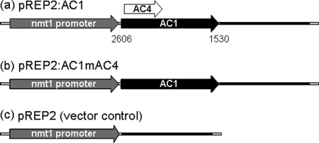
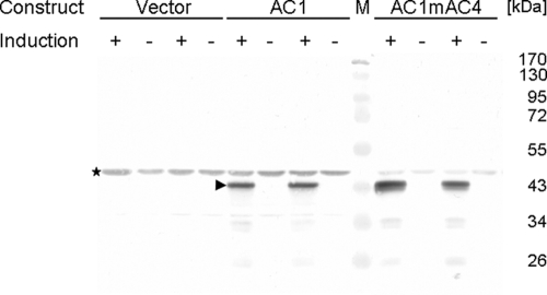
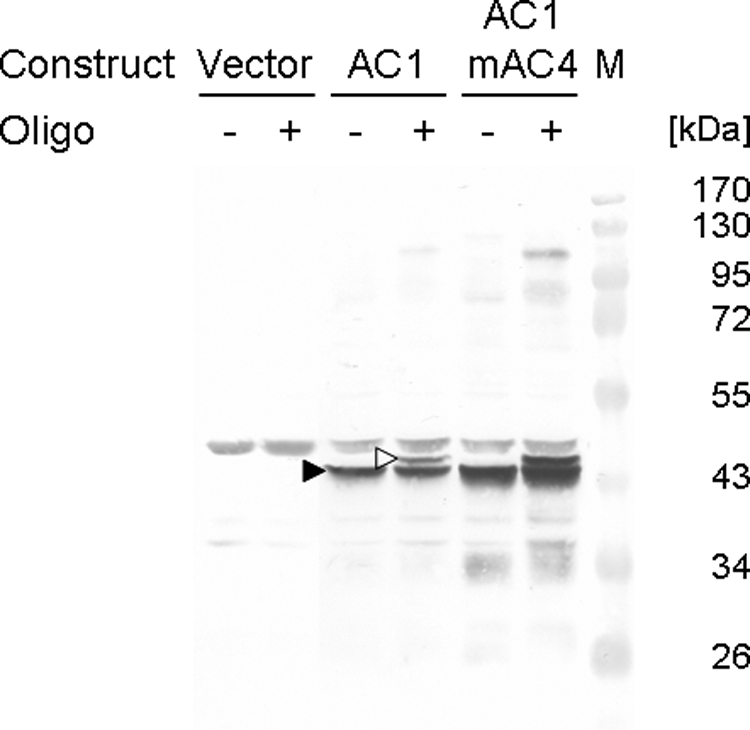
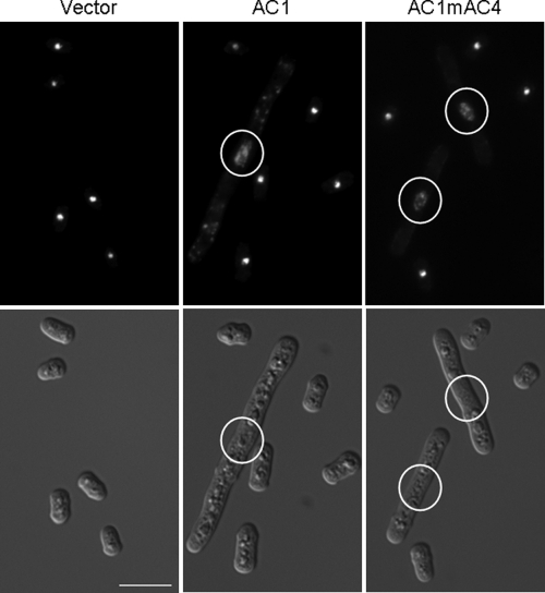
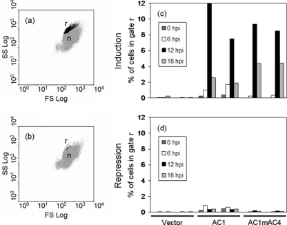
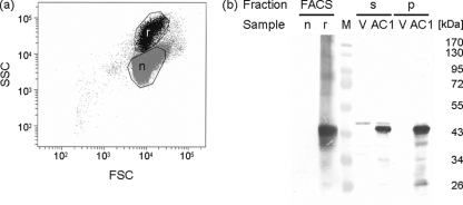
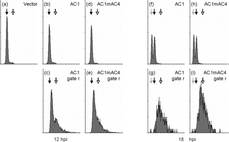
The replication-associated protein (Rep) of geminiviruses, single-stranded DNA viruses of higher plants, is essential for virus replication. Since these viruses do not encode their own polymerases, Rep induces differentiated plant cells to reenter the cell cycle by interacting with the plant homologues of retinoblastoma proteins in order to activate the host DNA synthesis machinery. We have used fission yeast (Schizosaccharomyces pombe) as a model organism to analyze the impact of ectopically expressed African cassava mosaic virus Rep protein on the cell division cycle in closer detail. Upon expression, Rep showed its characteristic DNA cleavage activity, and about 10% of the cells exhibited morphological changes. They were elongated threefold, on average, and possessed a single but enlarged and less compact nucleus in comparison to noninduced or vector-only control cells. Flow cytometry of Rep-expressing cultures revealed a distinct subpopulation of Rep protein-containing cells with aberrant morphology. The other 90% of the cells were indistinguishable from control cells, and no Rep was detectable. Rep-expressing cells exhibited DNA contents beyond 2C, indicating ongoing replication without intervening mitosis. Because a second open reading frame (ORF), AC4, is present within the Rep gene, the role of AC4 was examined by destroying its start codon within the AC1 ORF. The results confirmed that Rep is necessary and sufficient to induce rereplication in fission yeast. The unique potential of this well-investigated model for dissecting the cell cycle control by geminiviral proteins is discussed.


Figures

FIG. 1.
FIG. 1.
Schematic representation of expression constructs used in this study. The AC1 ORF (black arrow) includes the AC4 ORF (white arrow) in a different frame of the plasmid pREP2:AC1 (a). The AC4 start codon was destroyed in the plasmid pREP2:AC1mAC4 (b), preventing a potential expression of AC4 from this construct. As a control, the vector pREP2 (c) was used. First and last nucleotides of the AC1 ORF according to GenBank accession number AJ427910 are given below the arrow in panel a. Gray arrows and black bars denote the nmt1 promoter and nmt1 terminator, respectively. All features are drawn to scale. The backbone of the vector is not shown.
FIG. 2.
FIG. 2.
Western blot analysis of AC1 protein expression. Cell extract supernatants were prepared 12 h after cells were shifted to minimal medium without (+ induction) or with (− induction) thiamine. For each of the constructs pREP2 (Vector), pREP2:AC1 (AC1), and pREP2:AC1mAC4 (AC1mAC4), the proteins of two independent clones are shown. The position of the AC1 protein is marked by an arrowhead; the asterisk indicates the position of a cross-reacting S. pombe protein. Marker proteins (M) with their molecular masses (kDa) are shown.
FIG. 3.
FIG. 3.
Western blot analysis of the reaction products after in vitro cleavage. Extracts of cells with either plasmid pREP2 (Vector), pREP2:AC1 (AC1), or pREP2:AC1mAC4 (AC1mAC4) were prepared 12 h after induction of protein expression and incubated with (+) or without (−) oligonucleotide 5 (Table 1) comprising a cleavage substrate for the Rep protein. Arrowheads mark the positions of AC1 protein (filled) and generated adduct (open). Marker proteins (M) with their molecular masses (kDa) are shown.
FIG. 4.
FIG. 4.
Influence of Rep protein on cell morphology. Induced yeast cells carrying either plasmid pREP2, pREP2:AC1, or pREP2:AC1mAC4 were analyzed by light microscopy. Cells were fixed and stained with DAPI. DAPI images (upper row) show the same area as differential interference contrast images (lower row). Enlarged nuclei of elongated cells are encircled. Bar, 10 μm.
FIG. 5.
FIG. 5.
Flow cytometric analysis of cells expressing Rep protein. Induced (a) and noninduced (b) cells harboring plasmid pREP2:AC1 were fixed at 12 hpi, stained with propidium iodide, and analyzed by flow cytometry. A distinct subpopulation of cells (black dots; r) with increased forward scatter (FS Log) and side scatter (SS Log) properties compared to normal cells (gray dots; n) was gated for further analysis. Induced (c) and noninduced (d) cells harboring either empty plasmid (Vector), pREP2:AC1 (AC1), or pREP2:AC1mAC4 (AC1mAC4) were analyzed at different time points. Aliquots of cells were withdrawn after 0, 6, 12, and 18 h; treated as described before; and analyzed by flow cytometry. Cells of the subpopulation present in cultures expressing the Rep protein (r) were gated to determine the fraction of cells in this gate for the different cultures. For each construct, the results of two independent clones are shown.
FIG. 6.
FIG. 6.
(a) AC1 protein is detected solely in cells forming a distinct subpopulation with altered properties in flow cytometry. Induced cells harboring pREP2:AC1 were fixed at 18 hpi. Two samples were obtained by sorting cells of the Rep-expressing subpopulation (black dots; r) and the normal population (gray dots; n) and analyzed by SDS-PAGE and Western blotting. (b) Cells of the same culture used for sorting were harvested at 18 hpi (AC1) as well as control cells with plasmid pREP2 (V). Cell lysates of these samples were separated in a supernatant (s) and a pellet (p) fraction by low-speed centrifugation and used as controls for presence or absence of Rep protein. Marker proteins (M) with their molecular masses (kDa) are shown. SSC, side scatter; FSC, forward scatter.
FIG. 7.
FIG. 7.
DNA contents of cells expressing Rep protein. Cells transformed with either plasmid pREP2 (Vector; a), pREP2:AC1 (AC1; b and f), or pREP2:AC1mAC4 (AC1mAC4; d and h) were grown under inducing conditions for protein expression. Cells were fixed, stained with propidium iodide, and measured for DNA contents by flow cytometry at 12 hpi (a to e) and 18 hpi (f to i). The subpopulation of cells present in cultures expressing the Rep protein, as shown in Fig. 5 (black dots), was analyzed separately (c and g, AC1 gate r; e and i, AC1mAC4 gate r). Gray, black, and open arrows in the histograms indicate 1C, 2C, and 4C DNA contents, respectively. The x axis shows increasing DNA contents as measured by fluorescence. The y axis is scaled to 4,096 counts in a, b, c, e, g, and i; to 128 counts in d and f; and to 16 counts in h and j. The experiment was repeated three times with similar results (data not shown).

Similar articles

Cited by

References

    1. Aberle, H. J., M. L. Rütz, M. Karayavuz, S. Frischmuth, C. Wege, D. Hülser, and H. Jeske. 2002. Localizing BC1 movement proteins of Abutilon mosaic geminivirus in yeasts by subcellular fractionation and freeze-fracture immunolabelling. Arch. Virol. 147103-107. - PubMed
    1. Ach, R. A., T. Durfee, A. B. Miller, P. Taranto, L. Hanley-Bowdoin, P. C. Zambryski, and W. Gruissem. 1997. RRB1 and RRB2 encode maize retinoblastoma-related proteins that interact with a plant D-type cyclin and geminivirus replication protein. Mol. Cell. Biol. 175077-5086. - PMC - PubMed
    1. Alfa, C., P. Fantes, J. Hyams, M. McLeod, and E. Warbrick. 1993. Experiments with fission yeast: a laboratory course manual. Cold Spring Harbor Press, Cold Spring Harbor, NY.
    1. Arguello-Astorga, G., L. Lopez-Ochoa, L. J. Kong, B. M. Orozco, S. B. Settlage, and L. Hanley-Bowdoin. 2004. A novel motif in geminivirus replication proteins interacts with the plant retinoblastoma-related protein. J. Virol. 784817-4826. - PMC - PubMed
    1. Aronson, M. N., A. D. Meyer, J. Györgyey, L. Katul, H. J. Vetten, B. Gronenborn, and T. Timchenko. 2000. Clink, a nanovirus-encoded protein, binds both pRB and SKP1. J. Virol. 742967-2972. - PMC - PubMed

Publication types

LinkOut - more resources