. 2009 Jul;83(13):6464-76.
doi: 10.1128/JVI.02295-08. Epub 2009 Apr 15.

Disulfide bond formation at the C termini of vaccinia virus A26 and A27 proteins does not require viral redox enzymes and suppresses glycosaminoglycan-mediated cell fusion

Affiliations

Disulfide bond formation at the C termini of vaccinia virus A26 and A27 proteins does not require viral redox enzymes and suppresses glycosaminoglycan-mediated cell fusion

Yao-Cheng Ching et al. J Virol. 2009 Jul.

Abstract

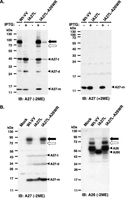
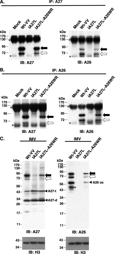
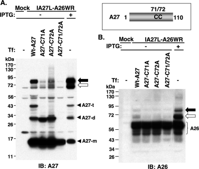
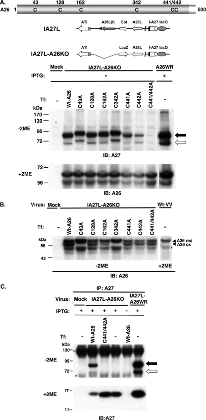
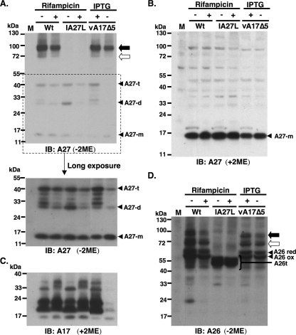
Vaccinia virus A26 protein is an envelope protein of the intracellular mature virus (IMV) of vaccinia virus. A mutant A26 protein with a truncation of the 74 C-terminal amino acids was expressed in infected cells but failed to be incorporated into IMV (W. L. Chiu, C. L. Lin, M. H. Yang, D. L. Tzou, and W. Chang, J. Virol 81:2149-2157, 2007). Here, we demonstrate that A27 protein formed a protein complex with the full-length form but not with the truncated form of A26 protein in infected cells as well as in IMV. The formation of the A26-A27 protein complex occurred prior to virion assembly and did not require another A27-binding protein, A17 protein, in the infected cells. A26 protein contains six cysteine residues, and in vitro mutagenesis showed that Cys441 and Cys442 mediated intermolecular disulfide bonds with Cys71 and Cys72 of viral A27 protein, whereas Cys43 and Cys342 mediated intramolecular disulfide bonds. A26 and A27 proteins formed disulfide-linked complexes in transfected 293T cells, showing that the intermolecular disulfide bond formation did not depend on viral redox pathways. Finally, using cell fusion from within and fusion from without, we demonstrate that cell surface glycosaminoglycan is important for virus-cell fusion and that A26 protein, by forming complexes with A27 protein, partially suppresses fusion.


Figures

FIG. 1.
FIG. 1.
A26 and A27 proteins formed protein complexes through disulfide bond formation in BSC40 cells. (A) Immunoblot (IB) analysis of A27 protein in virus-infected cell lysates following boiling and separation by reducing (with 2ME) and nonreducing (without 2ME) SDS-PAGE. The expression of the A27L ORF in wild-type vaccinia virus (Wt-VV) was constitutive, whereas in IA27L and IA27L-A26WR viruses it was induced by 5 mM IPTG. (B) Immunoblot analysis of A27 and A26 proteins in virus-infected cells cultured in medium containing IPTG. Lysates were boiled, separated on nonreducing (without 2ME) SDS-PAGE, and probed with anti-A26 (1:1,000) and anti-A27 (1:1,000) Abs. The 90- and 70-kDa protein complexes are indicated by black and white arrows, respectively. Three protein bands representing A27 monomers (A27-m), dimers (A27-d), and trimers (A27-t) also were detected. A26t, truncated A26 protein (aa 1 to 426).
FIG. 2.
FIG. 2.
Immunoprecipitation (IP) of A26-A27 protein complexes from cell lysates. BSC40 cells were infected with viruses as shown at the top at an MOI of 10 PFU/cell and were harvested at 24 h p.i. Cell lysates were immunoprecipitated with anti-A27 Ab (1:100) (A) or anti-A26 Ab (1:50) (B). The immunoprecipitates were washed, boiled, and separated on SDS-PAGE and analyzed by immunoblotting using anti-A26 and anti-A27 Abs as described for panel A. The black and white arrows indicate the 90- and 70-kDa protein complexes, while small asterisks indicate Ab heavy and light chains. (C) A26-A27 protein complexes were stably incorporated in IMV particles. Equivalent amounts of each IMV were separated by SDS-PAGE and analyzed by immunoblotting with anti-A26 (1:1,000), anti-A27 (1:1,000), and anti-H3 (1:1,000) Abs. The black and white arrows indicate the 90- and 70-kDa protein complexes, respectively. The asterisk represents the degraded form of A26 protein. IB, immunoblot.
FIG. 3.
FIG. 3.
Cysteines 71 and 72 of A27 protein are required for the formation of A26-A27 protein complexes. Schematic representation of the cysteine residues 71 and 72 on A27 protein. BSC40 cells were mock infected or infected with IA27L-A26WR and subsequently transfected with individual plasmids encoding wild-type A27 or C71A, C72A, or C71/72A double mutant proteins. Cells were harvested at 24 h p.i., boiled, and separated on nonreducing SDS-PAGE for immunoblot (IB) analyses using anti-A27 Ab (A) or anti-A26 Ab (B). The black and white arrows indicate the 90- and 70-kDa protein complexes, respectively. A27-m, A27-d, and A27-t represent monomers, dimers, and trimers of A27 protein, respectively. Tf, transfection.
FIG. 4.
FIG. 4.
(A) Cysteines 441 and 442 of A26 protein are required for the intermolecular disulfide bond formation of A26-A27 protein complexes. All of the six cysteine residues of A26 protein as well as the genome representations of IA27L and IA27L-A26KO viruses are shown at the top. BSC40 cells were mock infected or infected with IA27L-A26KO or IA27L-A26WR, subsequently transfected with individual plasmids encoding wild-type A26 or C43A, C128A, C162A, C342A, C441A, C442A, and C441/442A double mutant proteins, harvested at 24 h p.i., and then boiled to denature proteins for immunoblot analyses (IB) on nonreducing (without 2ME) gel using anti-A27 Ab (1:1,000) or on reducing (with 2ME) SDS-PAGE using anti-A26 (1:1,000) Ab. The black and white arrows indicate the 90- and 70-kDa protein complexes, respectively. (B) C43 and C342 form an intramolecular disulfide bond in A26 protein. The cell lysates shown in panel A were boiled and separated by nonreducing (without 2ME) SDS-PAGE and stained with anti-A26 (1:1,000) Ab. The top migrating band is the reduced form of full-length A26 protein (A26 red). The middle band is the oxidized form of A26 protein (A26 ox), while the asterisk indicates the degraded product of A26 protein. (C) A26 C441/442A mutant protein failed to form disulfide bonds with A27 protein. The immunoprecipitation (IP) of cell lysates described in panel A was performed using anti-A27 Ab (1:100) and was analyzed by SDS-PAGE on nonreducing (without 2ME) or reducing (with 2ME) gels using anti-A27 Ab (1:1,000). The asterisks (left) indicate Ig heavy and light chains. Tf, transfection.
FIG. 5.
FIG. 5.
A26-A27 protein complexes formed prior to virion assembly and did not require A17 protein. (A) Immunoblot analyses (IB) of 70-/90-kDa complex in the infected cells using anti-A27 Ab. BSC40 cells were infected with individual vaccinia viruses as shown at the top at an MOI of 10 PFU per cell. Wt, wild-type vaccinia virus (WR). Cell lysates were boiled and separated by SDS-PAGE on nonreducing (without 2ME) gels and probed with anti-A27 Abs. The bottom portion shows a longer exposure of a portion of the blot (marked by the dotted line). The black and white arrows indicate the 90- and 70-kDa protein complexes, respectively. A27-m, A27-d, and A27-t represent monomers, dimers, and trimers of A27 protein, respectively. (B) Samples were treated with reducing condition (with 2ME) and loaded in the same order as that used for panel A and were probed with anti-A27 Ab. (C) Samples were loaded in the same order as that for panel A and were treated with reducing conditions (with 2ME) and probed with anti-A17 Ab. (D) Samples were loaded in the same order as that used for panel A and were probed with anti-A26 Ab. A26 red, reduced form of A26 protein; A26 ox, oxidized form of A26 protein; and A26t, truncated A26 protein (aa 1 to 426) (5); M, mock-infected cells; Wt, wild type virus.
FIG. 6.
FIG. 6.
A26-A27 complex formation in cells infected with vG4Li. (A) BSC40 cells were infected with vG4Li and harvested at 24 h p.i. with 10% TCA precipitation (TCA ppt) and NEM as described in Materials and Methods. Protein samples were added with or without TCEP, boiled, and separated on gels using anti-A27 Ab. (B) Samples were loaded in the same order as that used for panel A and were probed with anti-A26 Ab. The asterisk represents the degraded A26 protein. (C) The expression of HA-tagged G4 protein (HA-G4) was induced by IPTG. (D) The growth of vG4Li virus was regulated by IPTG. IB, immunoblot.
FIG. 7.
FIG. 7.
A26-A27 complex formation in cells infected with vE10Ri. (A) BSC40 cells were infected with vE10Ri, and cells were harvested at 24 h p.i. with 10% TCA precipitation (TCA ppt) and NEM as described in Materials and Methods. Protein samples were added with or without TCEP, boiled, and separated on gels using anti-A27 Ab. (B) Samples were loaded in the same order as that used for panel A and were probed with anti-A26 Ab. The asterisk represents the degraded A26 protein. (C) The growth of vE10Ri virus was regulated by IPTG. (D) Immunoblot analyses (IB) of total L1 protein (top) and the oxidized form of L1 protein (L1 ox). L1 red, reduced form of L1 protein. 2D5 MAb only recognized the oxidized form of L1R on nonreducing gels (24).
FIG. 8.
FIG. 8.
A26 and A27 protein formed protein complexes in transfected 293T cells. (A) 293T cells were either mock transfected or cotransfected with plasmids expressing A26 and A27 protein, and cell lysates were harvested for immunoprecipitation (IP) using anti-A27 Ab (1:100). The immunoprecipitates were washed, boiled, and separated on SDS-PAGE and analyzed by immunoblotting (IB) using anti-A27 Abs (1:1,000) as described for Fig. 2A. (B) Samples were loaded in the same order as that used for panel A and were analyzed by anti-A26 Ab (1:1,000). The black and white arrows indicate the 90- and 70-kDa protein complexes, respectively.
FIG. 9.
FIG. 9.
A27-dependent cell fusion was partially inhibited by A26 protein. (A) Live imaging recording of cell fusion from within of virus-infected BSC40 cells by time-lapse immunofluorescence microscopy. BSC40 cells were infected with Wt-VV, IA27L, and IA27L-A26WR at an MOI of 5 PFU per cell and incubated at 37°C in the presence of 5 mM IPTG for 34 h, and cell images showing cell fusion development were collected at 30 min, 10 h, 20 h, 27 h, and 34 h p.i. The bottom color figures show confocal images of the infected cells fixed at 34 h p.i. and stained with anti-VV (1:5,000) Ab (green), fluorescein isothiocyanate-conjugated goat anti-rabbit Igs, and DAPI (blue). (B) Parental L or sog9 cells were infected with purified IMV virions for 1 h at 37°C, treated with PBS (pH 4.7) at 37°C for 2 min, incubated in normal medium for 1 h to develop cell fusion from without, and subsequently fixed. Plasma membrane was stained with PKH26 (red) and nuclei were stained with Hoechst 33258 (blue), and cells were visualized by confocal microscopy. White arrows refer to fused cells. (C) Quantification of cell fusion from without of L and sog9 cells in medium containing 5 mM IPTG as shown in panel B. The percentage of cell fusion was quantified based on equations described in Materials and Methods. (D) The fused cell population of L cells in panel C were subdivided into three subpopulation for quantification as described in Materials and Methods: small fused cells (2 to 5 nuclei per cell), medium fused cells (6 to 10 nuclei per cell), and large fused cells (>10 nuclei per cell). A total of >300 cells were counted for each virus, and the experiments were independently repeated twice.
FIG. 10.
FIG. 10.
(A) Schematic drawing of A26 and A27 proteins showing all of the cysteine residues (blue) and the positions of the disulfide bonds. (B) Alignment of amino acids in the C-terminal regions of A26 and A27 proteins (red lines). The conserved amino acids are in red, and brown triangles mark the two cysteines on each protein forming intermolecular bonds. The peptide regions predicted to be α-helixes are underlined. The secondary structure prediction was performed using the HNN secondary structure prediction method at the website http://npsa-pbil.ibcp.fr/cgi-bin/npsa_automat.pl?page=npsa_nn.html (9).

Similar articles

Cited by

References

    1. Armstrong, J. A., D. H. Metz, and M. R. Young. 1973. The mode of entry of vaccinia virus into L cells. J. Gen. Virol. 21533-537. - PubMed
    1. Banfield, B. W., Y. Leduc, L. Esford, K. Schubert, and F. Tufaro. 1995. Sequential isolation of proteoglycan synthesis mutants by using herpes simplex virus as a selective agent: evidence for a proteoglycan-independent virus entry pathway. J. Virol. 693290-3298. - PMC - PubMed
    1. Chang, A., and D. H. Metz. 1976. Further investigations on the mode of entry of vaccinia virus into cells. J. Gen. Virol. 32275-282. - PubMed
    1. Chiu, W. L., and W. Chang. 2002. Vaccinia virus J1R protein: a viral membrane protein that is essential for virion morphogenesis. J. Virol. 769575-9587. - PMC - PubMed
    1. Chiu, W. L., C. L. Lin, M. H. Yang, D. L. Tzou, and W. Chang. 2007. Vaccinia virus 4c (A26L) protein on intracellular mature virus binds to the extracellular cellular matrix laminin. J. Virol. 812149-2157. - PMC - PubMed

Publication types

LinkOut - more resources