

doi: 10.1128/JVI.02299-08.
Epub 2009 Apr 15.
Dissecting the functional domains of a nonenveloped virus membrane penetration peptide
Affiliations
- PMID: 19369344
- PMCID: PMC2698515
- DOI: 10.1128/JVI.02299-08
Item in Clipboard
Dissecting the functional domains of a nonenveloped virus membrane penetration peptide
J Virol.
2009 Jul.
Abstract
Recent studies have established that several nonenveloped viruses utilize virus-encoded lytic peptides for host membrane disruption. We investigated this mechanism with the "gamma" peptide of the insect virus Flock House virus (FHV). We demonstrate that the C terminus of gamma is essential for membrane disruption in vitro and the rescue of immature virus infectivity in vivo, and the amphipathic N terminus of gamma alone is not sufficient. We also show that deletion of the C-terminal domain disrupts icosahedral ordering of the amphipathic helices of gamma in the virus. Our results have broad implications for understanding membrane lysis during nonenveloped virus entry.
Figures
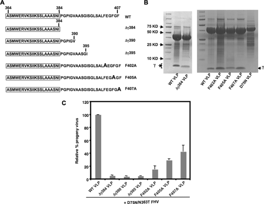
C-terminal region of gamma required for trans-complementation during the entry of maturation-defective FHV. (A) Schematic of truncations or single mutations in the gamma region of FHV capsid protein α in VLPs. The sequence of gamma in each of the mutated VLPs is shown, with the N-terminal amphipathic region boxed. The single F→A mutations in F402A, F405A, and F407A are indicated in boldface. (B) A total of 5 μl of WT, Δγ384, phenylalanine mutant, or D75N VLPs, at a concentration of 5 mg/ml, was subjected to SDS-PAGE on a 4 to 20% Tris-glycine gel (Invitrogen) and stained with Coomassie brilliant blue. The position of gamma is indicated. The cleavage-defective D75N VLPs do not have gamma. (C) Drosophila DL-1 cells (1 × 10⁸) were coinfected with 1.5 × 10³ particles/cell of D75N/N363T FHV and 9 × 10³ particles/cell of WT, Δγ384, Δγ390, Δγ395, F402A, F405A, or F407A VLPs. [35S]methionine-cysteine-labeled progeny virus was quantified, with the amount of progeny produced during coinfection with D75N/N363T FHV and WT VLPs normalized at 100%. The standard deviation was calculated from three replicates.
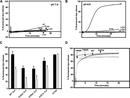
Disruption of liposomes and fluorescent dye release by VLPs in vitro. In each case, total fluorescence is normalized to dye release achieved by the addition of 0.1% Triton X-100 to liposomes under the same conditions. In the case of panels A, B, and D, closely similar results were obtained in three different experiments, whereas the standard deviations in panel C were calculated from three replicates. Fluorescence measurements were carried out at excitation/emission maxima of 492/514 nm for 6-carboxyfluorescein and 535/585 nm for SulfoB. (A) Kinetic study of 6-carboxyfluorescein release from DOPC-treated liposomes upon the addition of 6.37 × 10¹¹ particles (lipid/particle molar ratio, 481:1) of WT, gamma-truncated, or maturation-defective D75N VLPs to liposomes in 50 mM HEPES (pH 7.0). (B) SulfoB release from DOPC-treated liposomes in 50 mM Bis-Tris (pH 6.0) upon the addition of 6.37 × 10¹¹ particles of VLPs. (C) SulfoB release from DOPC-treated liposomes by 6.37 × 10¹¹ particles of WT, F402A, F405A, or F407A VLPs after 1 h at pH 7.0 (black) or by 2 × 10¹¹ particles (lipid/particle molar ratio, 1,387:1) of each of the VLPs after 15 min at pH 6.0 (gray). (D) SulfoB fluorescence upon the addition of heat-released gamma peptide with the WT sequence or with phenylalanine mutations at F402, F405, and F407 to dye-filled liposomes.
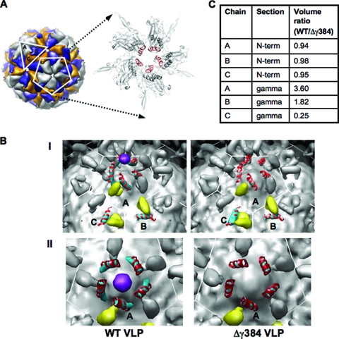
CryoEM image reconstructions of WT and Δγ384 VLPs. (A) Model of WT FHV showing the iASU consisting of the A, B, and C subunits, as well as the A subunits at the fivefold axis of symmetry. An expanded view of the fivefold axis shows the amphipathic region of the gamma peptides (red) forming a pentameric helical bundle. (B) Panel I, inner surface of WT VLP and Δγ384 VLP image reconstructions contoured at 0.4σ. The coloring is as follows: density for the capsid protein N termini (residues 59 to 72) in the iASU is in yellow, density for the gamma amphipathic helices (residues 364 to 381) is in cyan, and a potential pocket factor is in purple. Modeled into the density for the gamma amphipathic helices are the corresponding residues from the FHV crystal structure (PDB entry 2Z2Q) in red. Panel II, inner view, looking down on the fivefold axis of symmetry of WT and Δγ384 VLP image reconstructions. The density for the gamma amphipathic helices from the A subunits is in cyan, with the corresponding residues from the FHV crystal structure modeled in red. A pocket factor is in purple. (C) Volume ratios corresponding to the gamma amphipathic helices and the capsid protein N termini for subunits A, B, and C in the iASU. The reported ratio for each volume pair is the WT VLP density to the corresponding Δγ384 VLP density.
Similar articles
-
Rescue of maturation-defective flock house virus infectivity with noninfectious, mature, viruslike particles.J Virol. 2008 Feb;82(4):2025-7. doi: 10.1128/JVI.02278-07. Epub 2007 Dec 12. J Virol. 2008. PMID: 18077727 Free PMC article.
-
Structure and function of a genetically engineered mimic of a nonenveloped virus entry intermediate.J Virol. 2010 May;84(9):4737-46. doi: 10.1128/JVI.02670-09. Epub 2010 Feb 17. J Virol. 2010. PMID: 20164221 Free PMC article.
-
Non-Enveloped Virus Entry: Structural Determinants and Mechanism of Functioning of a Viral Lytic Peptide.J Mol Biol. 2016 Aug 28;428(17):3540-56. doi: 10.1016/j.jmb.2016.06.006. Epub 2016 Jun 16. J Mol Biol. 2016. PMID: 27320388
-
Flock house virus: a model system for understanding non-enveloped virus entry and membrane penetration.Curr Top Microbiol Immunol. 2010;343:1-22. doi: 10.1007/82_2010_35. Curr Top Microbiol Immunol. 2010. PMID: 20407886 Review.
-
Breach: Host Membrane Penetration and Entry by Nonenveloped Viruses.Trends Microbiol. 2018 Jun;26(6):525-537. doi: 10.1016/j.tim.2017.09.010. Epub 2017 Oct 25. Trends Microbiol. 2018. PMID: 29079499 Review.
Cited by
-
Strategies for antiviral screening targeting early steps of virus infection.Virol Sin. 2010 Aug;25(4):281-93. doi: 10.1007/s12250-010-3135-z. Epub 2010 Jul 28. Virol Sin. 2010. PMID: 20960301 Free PMC article. Review.
-
Atomistic dynamics of a viral infection process: Release of membrane lytic peptides from a non-enveloped virus.Sci Adv. 2021 Apr 14;7(16):eabe1761. doi: 10.1126/sciadv.abe1761. Print 2021 Apr. Sci Adv. 2021. PMID: 33853772 Free PMC article.
-
Folding a viral peptide in different membrane environments: pathway and sampling analyses.J Biol Phys. 2018 Jun;44(2):195-209. doi: 10.1007/s10867-018-9490-y. Epub 2018 Apr 11. J Biol Phys. 2018. PMID: 29644513 Free PMC article.
-
Influence of membrane composition on the binding and folding of a membrane lytic peptide from the non-enveloped flock house virus.Biochim Biophys Acta Biomembr. 2017 Jul;1859(7):1190-1199. doi: 10.1016/j.bbamem.2017.04.002. Epub 2017 Apr 7. Biochim Biophys Acta Biomembr. 2017. PMID: 28395954 Free PMC article.
-
Evolution in action: N and C termini of subunits in related T = 4 viruses exchange roles as molecular switches.Structure. 2010 Jun 9;18(6):700-9. doi: 10.1016/j.str.2010.03.010. Structure. 2010. PMID: 20541507 Free PMC article.
References
-
- Banerjee, M., and J. E. Johnson. 2008. Activation, exposure and penetration of virally encoded, membrane-active polypeptides during non-enveloped virus entry. Curr. Protein Pept Sci. 916-27. - PubMed
-
- Bong, D. T., C. Steinem, A. Janshoff, J. E. Johnson, and M. Reza Ghadiri. 1999. A highly membrane-active peptide in Flock House virus: implications for the mechanism of nodavirus infection. Chem. Biol. 6473-481. - PubMed
Publication types
MeSH terms
Substances
Grants and funding
LinkOut - more resources
Full Text Sources
