. 2009 Jul;83(13):6435-45.
doi: 10.1128/JVI.00018-09. Epub 2009 Apr 15.

Role of the lymphoreticular system in prion neuroinvasion from the oral and nasal mucosa

Affiliations

Role of the lymphoreticular system in prion neuroinvasion from the oral and nasal mucosa

Richard A Bessen et al. J Virol. 2009 Jul.

Abstract

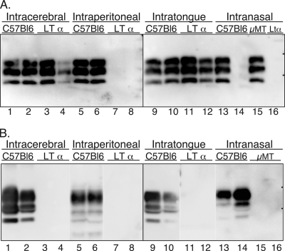
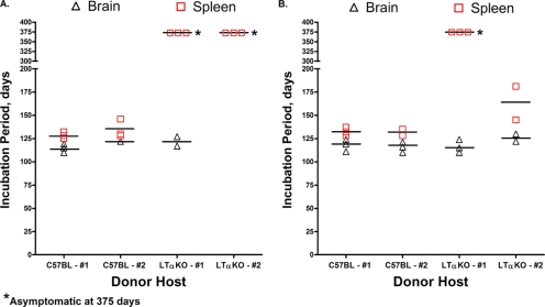
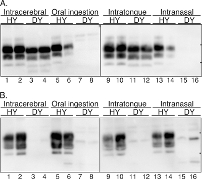
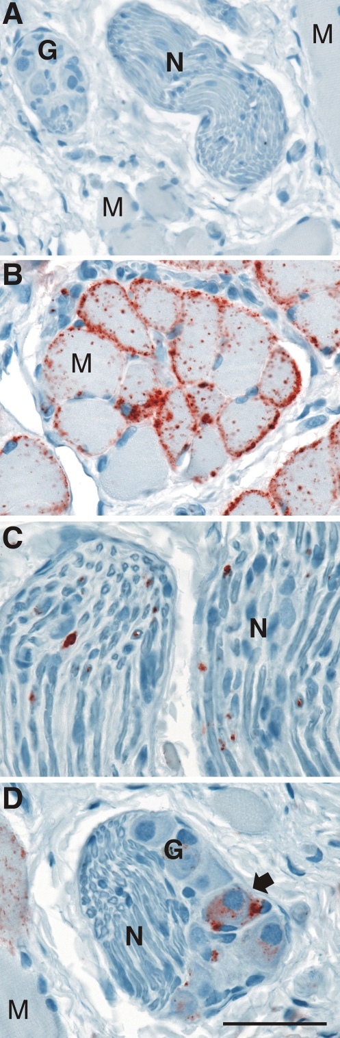
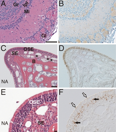
Prion neuroinvasion from peripheral tissues involves agent replication in the lymphoreticular system (LRS) prior to entry into the nervous system. This study investigated the role of the LRS in prion neuroinvasion from the oral and nasal mucosa in wild-type and immunodeficient mice and in hamsters infected with the HY and DY strains of the transmissible mink encephalopathy (TME) agent. Following inoculation at neural sites, all hosts were susceptible to prion disease and had evidence of prion infection in the brain, but infection of the LRS was found only in scrapie-infected wild-type mice and HY TME-infected hamsters. In the LRS replication-deficient models, prion neuroinvasion was not observed following intraperitoneal or oral inoculation. However, immunodeficient mice, which have impaired follicular dendritic cells, were susceptible to scrapie following intratongue and intranasal inoculation despite the absence of PrP(Sc) in the tongue or the nasal cavity. For DY TME, hamsters were susceptible following intratongue but not intranasal inoculation and PrP(Sc) was limited to nerve fibers of the tongue. These findings indicate that neuroinvasion from the tongue and nasal cavity can be independent of LRS infection but neuroinvasion was partially dependent on the strain of the prion agent and/or the host species. The paucity of PrP(Sc) deposition in the oral and nasal mucosa from LRS replication-deficient hosts following neuroinvasion from these tissues suggests an infection of nerve fibers that is below the threshold of PrP(Sc) detection and/or the transport of the prion agent along cranial nerves without agent replication.


Figures

FIG. 1.
FIG. 1.
Distribution of PrPSc in wild-type and immunodeficient mice following neural and extraneural routes of inoculation. C57BL/6J wild-type, LTα null, and muMT (μMT) mice were inoculated by the i.c., i.p., i.t., or i.n. route with the RML scrapie agent, and PrPSc was examined in brain homogenates (0.4 mg) (A) or enriched from spleen tissue (25 mg) (B) of terminally ill or aged asymptomatic mice. Tissues from two representative mice from each inoculation group are illustrated in each panel, except for the i.n. route, for which different mice are illustrated in lanes 14 and 16 in panels A and B. In panel A, lanes 14 and 16 were from mice that did not develop clinical disease, whereas in panel B, these lanes contain preparations from clinically ill mice. Sodium dodecyl sulfate-polyacrylamide gel electrophoresis and Western blot with anti-PrP 6H4 monoclonal antibody were performed as described in Materials and Methods. The short horizontal bars on the right edge of each panel indicate the 20- and 30-kDa markers.
FIG. 2.
FIG. 2.
Incubation period for C57BL/6J wild-type mice following i.c. inoculation of brain or spleen homogenates from C57BL/6J or LTα null mice. C57BL/6J mice or LTα null mice were either i.c. (A), i.t. (B), or i.p. (data not shown) inoculated with the RML scrapie agent as described in Table 1. At clinical disease or in asymptomatic aged mice, the brain and spleen were collected, homogenized, and i.c. inoculated into recipient C57BL/6J mice as a 1% or 10% (wt/vol) homogenate. Symbols (▵ and □) represent the incubation periods of individual animals, and the horizontal bars are the mean incubation period for a particular tissue and route of inoculation. i.c. inoculated C57BL/6J mice surviving to 375 days postinoculation were asymptomatic, and PrPSc was not found in the brains of these aged mice by Western blot (data not shown). The brains and spleens collected at 375 days postinoculation from LTα null mice that were i.p. inoculated with a mock or RML scrapie brain homogenate also did not cause scrapie when inoculated into recipient C57BL/6J mice (data not shown). In most cases, detection of scrapie infection in tissues from at least two mice (#1, #2) was assayed for each route of inoculation.
FIG. 3.
FIG. 3.
PrPSc deposition in tongues of mice and hamsters following i.t. inoculation of the scrapie or TME agent. In panel A, C57BL/6J wild-type, LTα null, and muMT (μMT) mice were inoculated in the tongue with the RML scrapie agent, and PrPSc was enriched from tongue tissue (100 mg) of clinically ill mice. Brain (Br) was used as a positive control. For panel B, tongue tissue (50 mg) was enriched for PrPSc from clinically ill hamsters following i.t. inoculation with the HY TME or DY TME agent. Samples were analyzed by SDS-PAGE and Western blot with anti-PrP 6H4 (A) or 3F4 (B) monoclonal antibody as described in Materials and Methods. Molecular mass markers in kilodaltons are indicated to the right of the panel.
FIG. 4.
FIG. 4.
Distribution of PrPSc in hamsters infected with the HY and DY strains of the TME agent following neural and extraneural routes of inoculation. Syrian golden hamsters were inoculated by the i.c., oral ingestion, i.t., or i.n. route, and in terminally ill or aged hamsters, PrPSc was examined in brain homogenates (0.25 mg) (A) or enriched from spleen (25 mg) (B). Tissues from two representative hamsters from each inoculation group are illustrated in each panel. SDS-PAGE and Western blot with anti-PrP 3F4 antibody were performed as described in Materials and Methods. In the lanes with spleen tissue of hamsters infected with the DY TME agent, weak immunoreactive polypeptides of various molecular masses were sometimes found, but these were considered to represent nonspecific PrP immunoreactivity based on repeated analysis of samples. The short horizontal bars on the right edge of each panel indicate the 20- and 30-kDa markers.
FIG. 5.
FIG. 5.
Distribution of PrPSc following i.t. inoculation of the TME agent. Mock-infected (A) or HY TME-infected (B, C, and D) hamsters were examined at clinical disease for the distribution of PrPSc (red chromogen product) in skeletal muscle (A, B, and D), nerve bundles (A, C, and D), and ganglia (A and D) by immunohistochemistry with anti-PrP 3F4 antibody as described in Materials and Methods. PrPSc deposits were preferentially located at the sarcolemma of muscle cells (M) but were also present in a cytoplasmic distribution in striated muscle cells. PrPSc aggregates were sparsely distributed in nerve fibers (N), while in ganglia (G) it was localized to the cell bodies of neurons (arrow). Tissue was counterstained with hematoxylin. Scale bar is 50 μm.
FIG. 6.
FIG. 6.
Distribution of PrPSc in the olfactory system following i.n. inoculation of the prion agent. Serial tissue sections (A and B, C and D, and E and F) were stained with anti-PrP antibody for PrPSc (B, D, and F) or hematoxylin and eosin stain (A, C, and E). PrPSc immunohistochemistry (B) of the olfactory bulb from a muMT mouse with clinical disease following i.n. inoculation of the RML scrapie agent is shown. PrPSc deposition (brown color) was most prominent in the granular layer (Gr), internal plexiform layer (Ipl), and glomerular layer (Gl) in the olfactory bulb (counterstained with hematoxylin). PrPSc deposition was less prominent in the mitral cell layer (Mi) of the olfactory bulb. In a single hamster following i.n. inoculation of the HY TME agent, PrPSc was found in the olfactory sensory epithelium (OSE) (D and F) and olfactory bulb (data not shown) but not in axon bundles (*) that transverse the subepithelial layer (SE) of the nasal mucosa. PrPSc deposition was prominent in the olfactory receptor neuron layer of the OSE (black arrows) and at the edge of the OSE layer in a location consistent with the dendritic knobs of olfactory neurons (white arrows with black outline). Panels D and F were viewed by differential interference contrast microscopy. NA, nasal airway; B, bone. Scale bar is 100 μm.

References

    1. Banks, T. A., B. T. Rouse, M. K. Kerley, P. J. Blair, V. L. Godfrey, N. A. Kuklin, D. M. Bouley, J. Thomas, S. Kanangat, and M. L. Mucenski. 1995. Lymphotoxin-alpha-deficient mice. Effects on secondary lymphoid organ development and humoral immune responsiveness. J. Immunol. 1551685-1693. - PubMed
    1. Bartz, J. C., J. M. Aiken, and R. A. Bessen. 2004. Delay in onset of prion disease for the HY strain of transmissible mink encephalopathy as a result of prior peripheral inoculation with the replication-deficient DY strain. J. Gen. Virol. 85265-273. - PubMed
    1. Bartz, J. C., C. Dejoia, T. Tucker, A. E. Kincaid, and R. A. Bessen. 2005. Extraneural prion neuroinvasion without lymphoreticular system infection. J. Virol. 7911858-11863. - PMC - PubMed
    1. Bartz, J. C., A. E. Kincaid, and R. A. Bessen. 2003. Rapid prion neuroinvasion following tongue infection. J. Virol. 77583-591. - PMC - PubMed
    1. Benestad, S. L., P. Sarradin, B. Thu, J. Schonheit, M. A. Tranulis, and B. Bratberg. 2003. Cases of scrapie with unusual features in Norway and designation of a new type, Nor98. Vet. Rec. 153202-208. - PubMed

Publication types