. 2009 May;136(10):1727-39.
doi: 10.1242/dev.029140. Epub 2009 Apr 15.

Notch signaling controls liver development by regulating biliary differentiation

Affiliations

Notch signaling controls liver development by regulating biliary differentiation

Yiwei Zong et al. Development. 2009 May.

Abstract

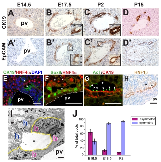
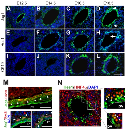
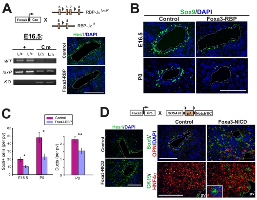
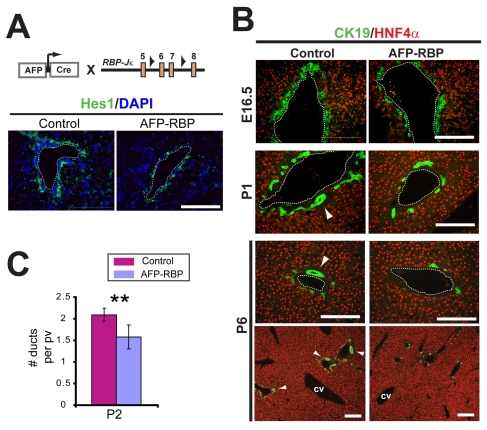
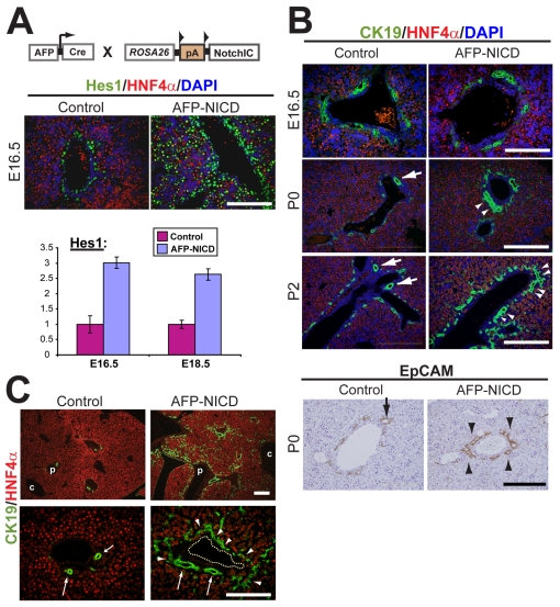
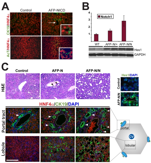
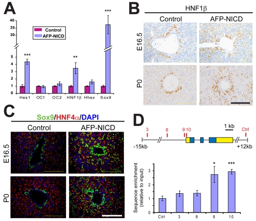
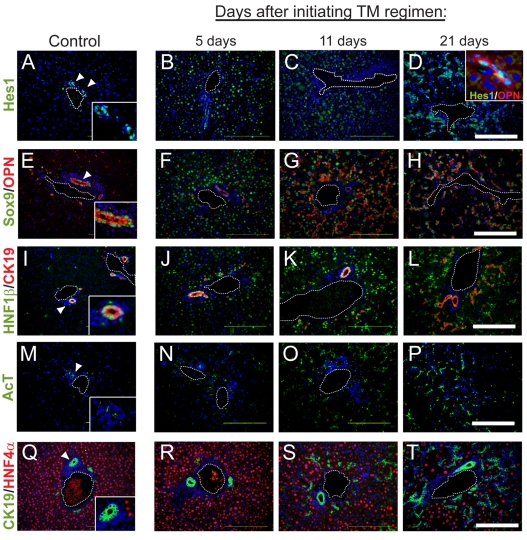
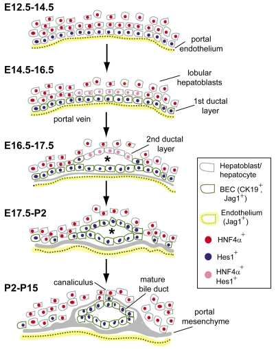
In the mammalian liver, bile is transported to the intestine through an intricate network of bile ducts. Notch signaling is required for normal duct formation, but its mode of action has been unclear. Here, we show in mice that bile ducts arise through a novel mechanism of tubulogenesis involving sequential radial differentiation. Notch signaling is activated in a subset of liver progenitor cells fated to become ductal cells, and pathway activation is necessary for biliary fate. Notch signals are also required for bile duct morphogenesis, and activation of Notch signaling in the hepatic lobule promotes ectopic biliary differentiation and tubule formation in a dose-dependent manner. Remarkably, activation of Notch signaling in postnatal hepatocytes causes them to adopt a biliary fate through a process of reprogramming that recapitulates normal bile duct development. These results reconcile previous conflicting reports about the role of Notch during liver development and suggest that Notch acts by coordinating biliary differentiation and morphogenesis.


Figures

Fig. 1.
Fig. 1.
Biliary tubules arise as asymmetric structures in the ductal plate. (A-D′) Timecourse of mouse intrahepatic bile duct (IHBD) formation. Ck19+ (A-D) and Epcam+ (A′-D′) ductal plate precursor cells arise between E14.5 (A,A′) and E17.5 (B,B′). Tubules initially form as asymmetric structures (E17.5, insets), in which cells on the portal side express Ck19 (B) and Epcam (B′), whereas cells on the parenchymal side do not express these markers (arrowheads). Bile ducts achieve symmetry early in postnatal life (P2, C,C′ insets). During the first 2 weeks of life, most ductal plate cells that are not integrated into a duct regress, leaving behind mature bile ducts (D,D′). (E-H) Nascent tubules (asterisks) at E16.5 are lined by cells that express Ck19, Sox9, acetylated tubulin (AcT, arrowheads) and Hnf1β within the inner portal layer, and Hnf4α within the outer parenchymal layer (arrows). In E, note the presence of numerous Hnf4α-negative nuclei, reflecting the preponderance of hematopoietic and other `non-parenchymal' cells in the embryonic liver. (I) Transmission electron micrograph of an E17.5 asymmetric primitive ductal structure. Outer layer cells (h) and inner layer cells (b) can be distinguished by the presence of glycogen in the former (arrowheads). (J) Quantitation of ductal asymmetry during liver development (±s.e.m.). pv, portal vein; e, endothelial cell. Scale bars: 20 μm in E-H; 4 μm in I.
Fig. 2.
Fig. 2.
Notch signaling is active during biliary development. (A-L) Immunofluorescence (green) for Jag1 (A-D), Hes1 (E-H) and Ck19 (I-L) demonstrates stepwise expression of Notch signaling components. Jag1 is expressed in the portal vein endothelium from E12.5 onward, whereas Hes1 is expressed in peri-portal cells (and endothelial cells) after E14.5. Expression of Ck19, a marker of terminal biliary differentiation, is observed at E16.5. Both Jag1 and Hes1 are expressed in mature ductal structures at E18.5 (arrows). All sections have been counterstained with DAPI (blue). (M) Jag1 staining is detected in portal endothelium (adjacent to dotted lines) and in cells on the portal side of primitive ductal structures at E16.5 (asterisks), where it overlaps with the expression of Sox9 and Ck19 (arrowheads). No Jag1 staining is observed in cells on the parenchymal side of asymmetric tubules. (N) Hes1 is expressed in endothelial cells and in both layers of asymmetric tubules at E16.5; in the outer layer, co-expression of Hes1 and Hnf4α is detected (arrowheads). Scale bars: 50 μm in A-L; 25 μm in M,N.
Fig. 3.
Fig. 3.
Notch signaling controls embryonic biliary fate. (A) Deletion of Rbpj was achieved by creating Foxa3-Cre; RbpjloxP/Δ (Foxa3-RBP) mice, in which one allele of Rbpj has been deleted and the other allele contains loxP sites flanking crucial coding sequences. PCR for the wild-type, mutant and deleted alleles, using liver DNA as template, shows deletion of the Rbpj gene in Foxa3-RBP animals at E16.5. Since as many as half of the cells in the E16.5 liver are hematopoietic in origin (see Fig. 1E), the observed reduction in RbpjloxP PCR product reflects efficient deletion in hepatoblasts at this stage. A reduction in the number of Hes1+ cells in the peri-portal region of Foxa3-RBP mice confirms the loss of ductal plate Notch signaling at this stage. (B,C) Foxa3-RBP mutants exhibit a reduced number of Sox9+ BECs at E16.5 and P0 and a reduced number of bile ducts at P0. Bar charts show the mean (±s.e.m.) of Sox9+ cells or ducts per portal vein (pv). Each bar represents measurements from four independent animals. *P<0.05, **P<0.01. (D) Activation of Notch signaling in Foxa3-Cre; RosaNICD (Foxa3-NICD) mice results in an expansion of the Hes1 expression domain and ectopic biliary differentiation at E16.5; structures resembling mature bile ducts are found in Foxa3-NICD lobules (inset; nuclei are counterstained with DAPI). pv, portal vein. Scale bars: 100 μm.
Fig. 4.
Fig. 4.
Late deletion of Rbpj preserves ductal plate formation but results in abnormal tubulogenesis. (A) Deletion of the transcriptional regulator Rbpj in AFP-Cre; RbpjloxP/loxP results in a more modest reduction in peri-portal Hes1+ cells at E16.5 than is seen in Foxa3-RBP mice (see Fig. 3). (B) AFP-RBP mutants have normal ductal plate development (E16.5) but have fewer mature bile ducts postnatally (arrowheads). cv, central vein. (C) Bar chart showing the mean number (±s.e.m.) of bile ducts per portal vein (pv); each bar represents the scoring of at least 50 portal regions (n=3 animals for each genotype). **P<0.01. Scale bars: 100 μm.
Fig. 5.
Fig. 5.
Notch signaling promotes ectopic biliary differentiation in the periportal region. (A) Real-time PCR or staining for Hes1 (n=3 animals of each genotype per time point) shows increased expression throughout the hepatic lobule in AFP-NICD mouse embryos. (B) Ductal plate precursor cells appear at E16.5 in the correct peri-portal location in AFP-NICD embryos. An increase in peri-portal BECs at P0 and P2 is associated with more elongated and numerous ducts in AFP-NICD livers (arrowheads) compared with controls (arrows). (C) Ectopic biliary cells persist in AFP-NICD livers. (Left) By P15, control portal tracts exhibit near-complete regression of the ductal plate; most remaining Ck19+ cells are incorporated into bile ducts (arrows). (Right) Age-matched AFP-NICD livers exhibit abundant Ck19+ cells arranged in a ductal plate conformation (arrowheads). The peri-portal concentration of these cells, resulting in a portal-to-central gradient, can be appreciated at low magnification (top panels). Note the presence of enlarged bile duct lumens in AFP-NICD livers (arrows). p, portal vein; c, central vein. Scale bars: 200 μm in B (P0 and P2); 100 μm for all others.
Fig. 6.
Fig. 6.
Notch promotes dose-dependent tubulogenesis at postnatal day 2. (A) Ectopic tubules exhibit expression of Ck19 and AcT. (Top) Lobular tubules in AFP-NICD mice (arrow) are lined by cells expressing either Hnf4α+ or Ck19+ (inset shows high magnification). (Bottom) AcT, a cilia marker normally confined to ductal plate progenitor cells, is ectopically expressed in the lobular tubules of AFP-NICD animals (inset shows high magnification). (B) Mice having one (AFP-NICD) or two (AFP-NICD/NICD) copies of RosaNICD exhibit graded levels of Notch1 transcripts by real-time PCR (top; n=3 animals for each genotype) and Hes1 protein by western blot analysis (bottom). (C) NICD gene dosage affects tubulogenesis at P2. Hematoxylin and Eosin staining reveals an increase in the number of lobular tubules with increasing RosaNICD gene dosage (H&E, top). A parallel increase in the size and number of peri-portal bile ducts (arrowheads) and lobular asymmetric tubules (bottom) is evident by Ck19 staining. Most cells surrounding the tubules express Hes1. Since over 90% of hepatic cells exhibit recombination at the Rosa26 locus prior to birth (see Fig. S5 in the supplementary material), these changes are likely to reflect increased `per-cell' activity of NICD rather than an increase in the number of cells expressing NICD. Scale bar: 100 μm.
Fig. 7.
Fig. 7.
Notch induces Hnf1β and Sox9 expression. (A) Real-time PCR measuring transcript levels (mean±s.e.m.) for several transcription factors involved in biliary development, comparing control and AFP-NICD mice at P0 (n=3 for each group). Hnf1b and Sox9 are the only two (other than Hes1) that exhibit a significant increase. Data are representative of two independent experiments. (B,C) Immunostaining for Hnf1β (B) and Sox9 (C) confirms widespread expression of both transcription factors throughout the mutant lobule as early as E16.5. (D) Schematic view of the Sox9 gene (5′ and 3′ untranslated regions in yellow, coding sequence in blue) showing the location of putative Rbpj binding sites and the control site. Chromatin prepared from AFP-NICD livers (P10) was subjected to immunoprecipitation with a Notch1 antibody followed by real-time PCR amplification using primers specific for each candidate binding site (see Materials and methods). The data represent the mean (±s.e.m.) of five independent experiments. *P<0.05, **P<0.01, ***P<0.001; all other differences were not significant at the P=0.05 level. Scale bars: 100 μm.
Fig. 8.
Fig. 8.
Notch activation in differentiated hepatocytes promotes biliary differentiation. (A-T) Albumin-CreER; RosaNICD mice were given five doses of tamoxifen (TM) (6 mg) starting at P6 and examined by immunofluorescence for the indicated markers 5, 11 or 21 days after the first dose. Expression of Hes1, Sox9, Opn, Hnf1β, AcT and Ck19 was initially restricted to the portal tract (arrowheads, left column; insets show high magnification). TM treatment leads to rapid induction of Hes1, Sox9 and Hnf1β, but a more gradual induction of AcT, Opn and Ck19. Note the rearrangement of ectopic biliary cells from a diffuse lobular distribution to a more organized ductal configuration. Clones of ectopic biliary cells generated by administering a low dose of TM are found in the lobule and show a tight correspondence between Hes1 and Opn expression (inset, upper right panel). All sections are counterstained with DAPI (blue). Scale bars: 100 μm.
Fig. 9.
Fig. 9.
Model of bile duct development. Early in liver development (E12.5-14.5), endothelial-derived Jag1 (yellow) activates Notch signaling and Hes1 expression in adjacent hepatoblasts (blue nuclei), resulting in formation of the first ductal plate layer at E14.6-16.5 (cells outlined in green). Between E16.5 and E17.5, tubulogenesis occurs at discrete sites of active Notch signaling in adjacent hepatocytes (pink nuclei), giving rise to a primitive ductal structure (asterisk). Cells comprising the second (outer) layer of this asymmetric tubule undergo biliary differentiation between E17.5 and P2. Subsequent growth of the portal mesenchyme and loss of unincorporated BECs leads to the formation of a mature portal tract by P15. Note that BECs in both the first and second layers of the ductal plate express Jag1. See text for details.

Similar articles

Cited by

References

    1. Ader, T., Norel, R., Levoci, L. and Rogler, L. E. (2006). Transcriptional profiling implicates TGFbeta/BMP and Notch signaling pathways in ductular differentiation of fetal murine hepatoblasts. Mech. Dev. 123, 177-194. - PubMed
    1. Alagille, D., Estrada, A., Hadchouel, M., Gautier, M., Odievre, M. and Dommergues, J. P. (1987). Syndromic paucity of interlobular bile ducts (Alagille syndrome or arteriohepatic dysplasia): review of 80 cases. J. Pediatr. 110, 195-200. - PubMed
    1. Antoniou, A., Raynaud, P., Cordi, S., Zong, Y., Tronche, F., Stanger, B. Z., Jacquemin, P., Pierreux, C. E., Clotman, F. and Lemaigre, F. P. (2009). Intrahepatic bile ducts develop according to a new mode of tubulogenesis regulated by the transcription factor SOX9. Gastroenterology (in press). - PMC - PubMed
    1. Blokzijl, A., Dahlqvist, C., Reissmann, E., Falk, A., Moliner, A., Lendahl, U. and Ibanez, C. F. (2003). Cross-talk between the Notch and TGF-beta signaling pathways mediated by interaction of the Notch intracellular domain with Smad3. J. Cell Biol. 163, 723-728. - PMC - PubMed
    1. Bolos, V., Grego-Bessa, J. and de la Pompa, J. L. (2007). Notch signaling in development and cancer. Endocr. Rev. 28, 339-363. - PubMed

Publication types