. 2009 May 1;23(9):1052-62.
doi: 10.1101/gad.520509. Epub 2009 Apr 15.

Drosophila motor axons recognize and follow a Sidestep-labeled substrate pathway to reach their target fields

Affiliations

Drosophila motor axons recognize and follow a Sidestep-labeled substrate pathway to reach their target fields

Matthias Siebert et al. Genes Dev. .

Abstract

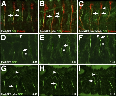
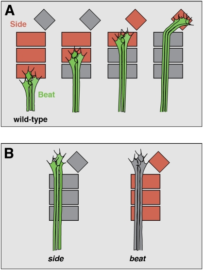
During development of the Drosophila nervous system, migrating motor axons contact and interact with different cell types before reaching their peripheral muscle fields. The axonal attractant Sidestep (Side) is expressed in most of these intermediate targets. Here, we show that motor axons recognize and follow Side-expressing cell surfaces from the ventral nerve cord to their target region. Contact of motor axons with Side-expressing cells induces the down-regulation of Side. In the absence of Side, the interaction with intermediate targets is lost. Misexpression of Side in side mutants strongly attracts motor axons to ectopic sites. We provide evidence that, on motor axons, Beaten path Ia (Beat) functions as a receptor or part of a receptor complex for Side. In beat mutants, motor axons no longer recognize Side-expressing cell surfaces. Furthermore, Beat interacts with Side both genetically and biochemically. These results suggest that the tracing of Side-labeled cell surfaces by Beat-expressing growth cones is a major principle of motor axon guidance in Drosophila.


Figures

Figure 1.
Figure 1.
Motor axons follow Sidestep-labeled cell surfaces. (A–I) FasIIGFPMue397 embryos stained with anti-GFP and anti-Side antibodies to visualize the spatiotemporal relationship of motor axons and Side-expressing cells. (A–C) Ventral view of a stage 12 embryo. (A) FasIIGFPMue397 is expressed in a cluster of four to five cells in each neuromere, including the aCC and pCC neurons. Arrowhead marks the ventral midline. (B) Sidestep is expressed in a belt-like pattern flanking the ventral midline. (C) FasIIGFPMue397-positive cells are located next to Side-expressing cells (cf. arrows in A–C). Arrowheads mark the tracheal precursors. Anterior is left. (D–F) Ventrolateral view of a stage 13 embryo. (D) Pioneering axons of the ISN (arrow) project toward the exit junction and developing ganglionic branch of the trachea (arrowhead). (E) Sidestep is expressed in a triangular area (small arrows) in the CNS that points toward the trachea. (F) Motor axons follow Sidestep-positive cell surfaces toward the trachea (arrows in D–F). Small arrows in F mark the close proximity of the tip of the triangle to the trachea. Asterisk indicates the earliest expression of Side in sensory neurons of the dorsal cluster. (G–I) Lateral view of a stage 14 embryo. (G) ISN motor axons migrate dorsally between the trachea and the developing muscle field. (H) Sensory axons grow ventrally along similar routes. (I) Sensory axons and motor axons fasciculate in the ventrolateral region of the embryo (arrows in G–I). The asterisk in I shows that Side is not detectable any more in the triangular area of the CNS (cf. F).
Figure 2.
Figure 2.
The ISN detaches from sensory axons and shows migratory delays in side mutants. (A–C) Motor axons are stained with anti-GFP and sensory axons with anti-Futsch antibodies. (A) In stage 15 wild-type embryos, the ISN is tightly associated with sensory axons of the anterior fascicle (arrows) until it reaches the dbd neuron (arrowhead). The dbd neuron stains only weakly with anti-Futsch antibodies at this stage, as do motor axons. (B) The ISN is partially detached from sensory axons (arrows) prior to the dbd (arrowhead) in sideC137/sideI1563 mutant embryos. (C) Wild-type embryos overexpressing Side in muscles using Mef2-Gal4 show severe detachments of motor and sensory axons (arrows). For quantification of the detachment phenotypes see Table 1. (D–F) Stills from Supplemental Movie 2-1 showing the migration of the ISN in the lateral body wall of a FasIIGFPMue397 embryo at stage 14. The growth cone of the ISN (arrows) travels between the transverse connective (downward arrowheads) and the myogenic field (asterisks). The growth cone marked with arrows makes a small detour but corrects its path. Time is indicated in hours and minutes (h:min). (G–I) Stills from Supplemental Movie 2-2 showing the advance of the ISN in a sideC137/sideI1563 mutant embryo at stage 16. While one of the ISN nerves has already crossed the dorsal trunk (arrowheads), others are lacking behind (arrows). Delayed growth cones are larger and sprout more filopodia.
Figure 3.
Figure 3.
Sidestep is sufficient to direct the path of motor axons. (A,B) Side attracts motor axons to tracheal branches. Axons are stained with anti-FasII antibodies, the tracheal lumen is stained with a chitin-binding probe coupled to Rhodamine. (A) The ISN growth cones are not in direct contact with tracheal branches in a side mutant embryo at stage 16 (arrows) (genotype: sideC137/sideI1563). (B) Ectopic expression of Side in trachea of side mutants strongly attracts motor axons to tracheal branches, the only source of Side in these embryos (arrows) (genotype: btl-Gal4/+; UAS-side, sideC137/sideI1563). (C,D) Side attracts motor axons to hemocytes. (C) Stage 16 side mutant embryo expressing transmembrane CD8GFP in hemocytes (red). ISN growth cones misproject but are not in direct contact with hemocytes (arrows) (genotype: UAS-mCD8GFP, sideC137/sideI1563, Serpent-Gal4). (D) FasIIGFPMue397 embryo mutant for side and expressing exogenous Side in hemocytes. In the absence of endogenous Side, almost every growth cone of the ISN is in contact with a hemocyte (arrows) (genotype: FasIIGFPMue397; UAS-side, sideC137/sideI1563, Serpent-Gal4). (E–H) Still images of Supplemental Movie 3-1 (time in minutes and seconds) showing strong adhesive interactions between an ISN growth cone (arrow) and a Side-expressing hemocyte (arrowhead) in a side mutant embryo at stage 16 (genotype: FasIIGFPMue397; UAS-side, sideC137/sideI1563, Serpent-Gal4). (I,J) Stills of Supplemental Movie 3-2 (time in hours and minutes) showing ISN growth cones in a stage 15 FasIIGFPMue397 embryo overexpressing Side in muscles (genotype: FasIIGFPMue397; Mef2-Gal4/UAS-side). The growth cone of the ISN splits into several directions, preventing its dorsal migration and deflecting its route along presumptive muscle fibers (arrows).
Figure 4.
Figure 4.
Genetic evidence that Beat and Side function in a common pathway. (A–H) Confocal images of muscle pairs 1/9 and 2/10 in third instar larvae expressing the post-synaptic marker CD8-GFP-Sh. The percentages of noninnervated muscles in the respective genotypes are quantified in Table 2. (A) In wild-type larvae, dorsal-most muscles are innervated by centrally localized NMJs (arrows). (B,C) sideC137/sideI1563 (B) and beat3/beatC163 (C) mutant larvae show similar innervation defects on a variety of muscles and frequently lack NMJs on muscles 1/9. (D) Ectopic expression of Beat in muscles of wild-type larvae using Mef2-Gal4 does not affect the innervation of dorsal muscles. (E) Ectopic expression of Side in muscles results in complete lack of NMJs on dorsal-most muscles. (F) Coexpression of Beat and Side in muscles suppresses the innervation defects caused by overexpression of only Side, resulting in a wild-type innervation pattern on dorsal muscles. (G) Phenotypic strength is not increased in beat3/beatC163; sideC137/sideC137 double mutants when compared with single mutants of beat or side. (H) Overexpression of Side in muscles of beat3/beatC163 mutant animals does not increase phenotypic strength of beat mutants, leading to the absence of NMJs only on muscles 1/9.
Figure 5.
Figure 5.
Expression of Beat in neurons but not in muscles regulates the expression level of Side. (A) At stage 16, Side is no longer detected on peripheral nerves of wild-type embryos but remains weakly expressed in muscles and the neuropil. (B) In beat3 mutant embryos, Side remains highly expressed on peripheral nerves and the ventral neuropil until the end of embryogenesis. (C) Expression of wild-type Beat in a beat3 mutant embryo using Elav-Gal4 rescues the constitutive expression phenotype and induces the down-regulation of Side on peripheral nerves. A few Side-positive particles, however, remain (arrow). (D) Expression of wild-type Beat in muscles of beat3 mutant embryos using Mef2-Gal4 fails to down-regulate Side on peripheral nerves, indicating that Beat does act cell-autonomously.
Figure 6.
Figure 6.
Beat interacts with Side. (A–F) S2 cell aggregation assays. (A–C) Mock transfected (A), Beat-myc transfected (B), or Side-GFP transfected (C) S2 cells do not form homophilic cell aggregates. (D) S2 cells cotransfected with Beat-myc and Side-GFP form large cell aggregates. (E) S2 cells transfected with Beat-myc or Side-GFP in discrete culture dishes and mixed together in a common dish form large cell aggregates (Mix experiment). Bar, 30 μm. (F) S2 cells individually transfected with Beat–Cherry or Side-GFP were mixed together (Mix experiment). Cell aggregates are comprised of both cell types. Bar, 15 μm. (G,H) Immunoprecipitation of protein complexes containing Beat and Side. (G) S2 cells transiently transfected with Beat-myc and/or Side-GFP were immunoprecipitated with anti-GFP antibodies. Immunoblots of the immunoprecipitates (IP) were developed with anti-myc and anti-Side antibodies. Beat-myc is precipitated only in the presence of Side-GFP. (H) S2 cells transiently transfected with Beat-GFP and/or Side-myc were immunoprecipitated with anti-GFP antibodies. Immunoblots of the precipitates were developed with anti-GFP and anti-Side antibodies. Side-myc is precipitated only in the presence of Beat-GFP. Note that molecular weights depend on the tags used.
Figure 7.
Figure 7.
Model: Beat-expressing motor axons follow a Side-labeled substrate pathway. (A) In wild-type embryos, Beat-expressing motor axons (green) recognize and follow Side-labeled cell surfaces (red). Contact with motor axons induces the down-regulation of Side (gray). Growth cones migrate until the end of the Side expression domain. Developmentally controlled up-regulation of Side in another tissue induces growth cone turning. (B, left) In side mutants, growth cones fail to turn, as substrates are not labeled. Other possible phenotypes such as delays or detours are not depicted. (Right) In beat mutants, the Side-labeled pathway is constitutively marked but cannot be recognized, thereby preventing growth cone turning.

Comment in

Similar articles

Cited by

References

    1. Aberle H., Haghighi A.P., Fetter R.D., McCabe B.D., Magalhaes T.R., Goodman C.S. wishful thinking encodes a BMP type II receptor that regulates synaptic growth in Drosophila. Neuron. 2002;33:545–558. - PubMed
    1. Araujo S.J., Tear G. Axon guidance mechanisms and molecules: Lessons from invertebrates. Nat. Rev. Neurosci. 2003;4:910–922. - PubMed
    1. Bate C.M. Pioneer neurones in an insect embryo. Nature. 1976;260:54–56. - PubMed
    1. Bazan J.F., Goodman C.S. Modular structure of the Drosophila Beat protein. Curr. Biol. 1997;7:R338–R339. doi: 10.1016/S0960-9822(06)00168-0. - DOI - PubMed
    1. Buszczak M., Paterno S., Lighthouse D., Bachman J., Planck J., Owen S., Skora A.D., Nystul T.G., Ohlstein B., Allen A., et al. The carnegie protein trap library: A versatile tool for Drosophila developmental studies. Genetics. 2007;175:1505–1531. - PMC - PubMed

Publication types

MeSH terms

LinkOut - more resources