. 2009 Jun;20(12):2932-42.
doi: 10.1091/mbc.e08-12-1193. Epub 2009 Apr 15.

Nuclear fusion and genome encounter during yeast zygote formation

Affiliations

Nuclear fusion and genome encounter during yeast zygote formation

Alan Michael Tartakoff et al. Mol Biol Cell. 2009 Jun.

Abstract

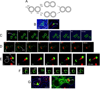
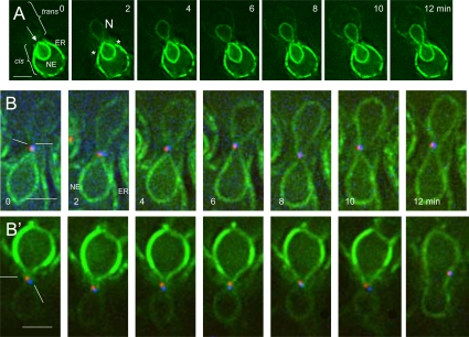
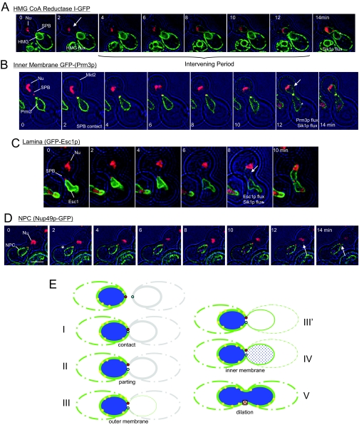
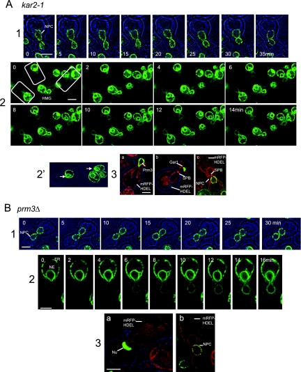
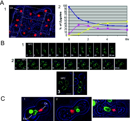
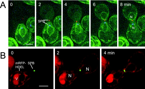
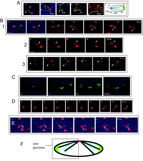
When haploid cells of Saccharomyces cerevisiae are crossed, parental nuclei congress and fuse with each other. To investigate underlying mechanisms, we have developed assays that evaluate the impact of drugs and mutations. Nuclear congression is inhibited by drugs that perturb the actin and tubulin cytoskeletons. Nuclear envelope (NE) fusion consists of at least five steps in which preliminary modifications are followed by controlled flux of first outer and then inner membrane proteins, all before visible dilation of the waist of the nucleus or coalescence of the parental spindle pole bodies. Flux of nuclear pore complexes occurs after dilation. Karyogamy requires both the Sec18p/NSF ATPase and ER/NE luminal homeostasis. After fusion, chromosome tethering keeps tagged parental genomes separate from each other. The process of NE fusion and evidence of genome independence in yeast provide a prototype for understanding related events in higher eukaryotes.


Figures

Figure 1.
Figure 1.
Overview of NE Fusion. (A) Models of fusion of organelles with double membranes. According to both models, the two nuclei diagrammed at the left accomplish fusion of both outer and inner membranes, generating a single nucleus with a continuous envelope. In the trans-first model, the two outer membranes fuse with each other before the two inner membranes. In the cis-first model, the inner and outer membranes of each nucleus first fuse with each other. The cis-first model is considered in the Discussion. (B) Polarization of nuclei of cells after 3-h exposure to α-factor. Left, a cell expressing Spc42p-mRFP and Nup49p-GFP (ATY1816). Right, a cell expressing Htb2p-mRFP and nucleolar Gar1p-GFP (ATY2594). (C) Cross between strains that express Nup49p-GFP (ATY3405 × ATY3358). The nexus is established just after t = 0 min and persists until just before the 40-min time point. (D) Cross between a strain that expresses Mid2p-GFP, Nup49p-GFP, Sik1p-mRFP, and Spc42p-mRFP (ATY1897) with a strain that expresses Mid2p-GFP, Nup49p-GFP, and Spc42p-mRFP (ATY1916). The nexus persists from +5 until +30 min. Note that the flux of Sik1p-mRFP is detected before dilation of the nexus. Disengagement of the SPB from one face of the NE is evident at the last time point. (E) A cross between a strain expressing Gar1p-GFP and Htb2p-mRFP (ATY2416), with a strain expressing Spc42p-CFP (ATY1455). The corresponding nucleus is circled. Note that contact of the SPB (blue) with the trans nucleus (red) is followed by a pause before the initial transfer of Gar1p-GFP (and Htb2p-mRFP), and a further delay before dilation of the nexus and disengagement of the SPB is seen at 22 min. (F) Delay of SPB coalescence. Cross between a strain that expresses Nup49p-GFP and Spc42p-mRFP (ATY1817) and a strain that expresses Spc42p-CFP (ATY1455). The two SPBs, although adjacent, remain separate throughout. The first images precede establishment of contact. (G) Left, cross between kar1-1 expressing mRFP-HDEL (ATY3004) and a strain that expresses Spc42p-mRFP and Gar1p-GFP (ATY3198). Images were collected after 3 h. Note the absence of Gar1p-GFP from the kar1-1 nucleus, implying that this protein does not shuttle to the cytoplasm and that its continued synthesis during the cross does not generate a de novo signal in the trans nucleus. Right, similar observations were made following Sik1p-mRFP in crosses with kar1-1 that expresses GFP-HDEL (ATY3373 × ATY1513).
Figure 2.
Figure 2.
Transit of HMG1-GFP. (A) Time course of transfer of HMG1-GFP from (ATY1529) to a nonfluorescent recipient (ATY2112). Note the parting of the apex of the cis-NE (arrow), followed by the appearance of signal in the trans-NE before it acquires equal intensity in the trans-cortical ER. The increase in intensity of HMG1-GFP in the trans-NE is gradual. See Supplemental Figure S2 for a further example. The asterisks indicate the abrupt interruption of the cortical ER signal. (B and B′) Crosses between a strain expressing HMG1-GFP and Spc42p-mRFP (ATY3525) with a strain expressing Spc42p-CFP (ATY1455). Note the position of the SPB sentinels of distinct parentage during HMG1-GFP transfer. In many images they flank the axis of transfer.
Figure 3.
Figure 3.
Outer membrane fusion precedes fusion of the inner membrane and transfer of NPCs. (A) Transfer of HMG1-GFP compared with Sik1p-mRFP (and Spc42p-mRFP). Note that the HMG1-GFP is detected in the trans-nucleus long before Sik1p-mRFP (asterisk, arrow; ATY1917 × ATY1528). (B) Transfer of the tagged inner membrane protein, GFP-Prm3p, compared with Sik1p-mRFP (and Spc42p-mRFP). GFP-Prm3p and Sik1p-mRFP are detected in the trans-nucleus at essentially the same time (asterisk, arrow). The cell that expresses the mRFP-tagged proteins also expresses Mid2p-GFP (ATY1919 × ATY3149). Note: GFP-Prm3p was not preinduced. (C) Transfer of the tagged protein of the periphery of the nucleoplasm, GFP-Esc1p, compared with Sik1p-mRFP (and Spc42p-mRFP). GFP-Esc1p and Sik1p-mRFP are detected in the trans-nucleus at approximately the same time (asterisk, arrow; ATY1917 × ATY1550). (D) A cell expressing Sik1p-mRFP and Spc42p-mRFP (ATY1917) was crossed with a cell expressing Nup49p-GFP (ATY2226). Sik1p-mRFP arrives in the trans-nucleus well before the NPCs (asterisk), and the tagged NPCs invade the trans-NE (arrow) when dilation of the nexus is seen. Dilation of the nexus is accompanied by separation of the tagged SPBs from one face of the NE. Note: Experiments that follow a mRFP-tagged inner membrane protein along with HMG1-GFP also indicate initial fusion of the outer membrane, but the mRFP signal is not sufficiently strong to allow definitive imaging. (E) Summary diagram of the sequential events of NE fusion. The steps are (I) SPB contact, (II) appearance of the apical discontinuity of the NE, (III) initial transfer of outer membrane proteins, (III′) continued transfer of outer membrane and spreading of the trans-NE signal to the cortical ER, (IV) transfer of inner membrane proteins and the lamina, as well as nucleoplasmic continuity, and (V) visible dilation of the nexus, transfer of NPCs and disengagement of the SPB from one face of the NE. There are obvious delays between steps III and IV and between IV and V, suggesting that, at the molecular level, these intervening periods allow appropriate preparations for the succeeding events. HMG1-GFP, green; NPCs, yellow.
Figure 4.
Figure 4.
NE Fusion in kar2-1 and prm3Δ crosses. (A) kar2-1 × wt. (1) Nuclear encounter of kar2-1 and wt, which express Nup49p-GFP (ATY1713 × ATY3359). (2) Transfer of HMG1-GFP from wt to kar2-1. Note the more uniform timing of arrival of signal in the trans-NE and trans-cortical ER than in wt. (2′) The parting of the apex of the cis-NE in the same preparation (ATY3197 × ATY1528). Unlike wt crosses, when HMG1-GFP arrives in the trans-NE of kar2-1, its intensity in the trans-ER is nearly equal to that of the trans-NE. Thus, arrival of HMG1-GFP in the trans-NE could be via the cortical ER in kar2-1 crosses. (3) To learn whether the inner membranes fuse in kar2-1× wt crosses, we have performed experiments in which one of the parental strains expresses GFP-Prm3p, Gar1p-GFP, or Nup49p-GFP (which would be expected to transfer only if both membranes fused). In each case, the kar2-1 cell expresses mRFP-HDEL. Crosses were examined after 3 h to follow GFP-Prm3p: panel a, ATY3197 × ATY3149; a nucleolar protein, Gar1p-GFP: panel b, ATY3197 × ATY3004; or tagged NPCs, Nup49p-GFP: panel c, ATY3197 × ATY1916. As shown, none are transferred. The partners that express Gar1p-GFP or Nup49p-GFP also express Spc42p-mRFP. ATY1916 additionally expresses Mid2p-GFP. GFP-Prm3p itself concentrates at the nexus (panel a). (B) prm3Δ× prm3Δ. (1) Encounter of two prm3Δ strains that express Nup49p-GFP (ATY3130 x ATY3131). (2) Transfer of HMG1-GFP (ATY3277 × ATY2782). Note that the intensity of the trans-NE signal generally exceeds that of the trans-cortical ER and note the focal gap. As in wt × wt crosses, during arrival of HMG1-GFP in the trans-cell, the intensity in the NE exceeds that of the cortical ER. (3) prm3Δ x prm3Δ crosses were examined after 3 h to learn whether nucleolar Gar1p-GFP (a: ATY3216 × ATY3298) or tagged NPCs (b: ATY3216 × ATY3131) are transferred. The MATa partner expresses mRFP-HDEL in both cases. Note the lack of transfer in each case.
Figure 5.
Figure 5.
NE fusion assay. (A) Two-step assays to identify factors required for karyogamy. (1) Two strains that express Htb2p-mRFP (ATY2835 × ATY2289) were crossed for 3 h on a nocodazole plate and examined. Note the designated prezyogotes in which the nuclei are separate. Ch, chromatin. (2) Congression and karyogamy of the wt strains illustrated in panel 1 after recovery from a nocodazole plate and reincubation at 23°C. Individual zygotes were classified as showing separate nuclei (blue), nuclei in contact with each other (pink), or fused nuclei (yellow). Some wt crosses yield a higher efficiency of fusion and more rapid kinetics than this example. (B) Karyogamy: novel conditions cause arrest. (1) Time lapse of two sec18-1 strains that express Nup49-GFP (ATY2538 × ATY2138) which were crossed on a nocodazole plate at 23°C, recovered, and reincubated at 37°C. (2) Time lapse of two wt strains that express Nup49p (ATY3405 × ATY3359). The strains were crossed on a nocodazole plate and reincubated with 2 mM DTT. (3) Two wt strains that express Nup49p (ATY3405 x ATY3359) were crossed on a nocodazole plate and reincubated with 100 μg/ml cycloheximide for 2 h. (C) Evaluation of inner membrane fusion in novel conditions. To learn whether inner membrane fusion occurs in sec18-1× sec18-1 crosses, in the presence of DTT or cycloheximide, we have followed a copy of GFP-Esc1p that is preinduced from a galactose-inducible promoter and silenced by the glucose in the nocodazole plates. We observe that GFP-Esc1p is restricted to the cis-nucleus in each case. Thus, inner membrane fusion does not occur. (1) sec18-1 strains that express either GFP-Esc1p or Htb2p-mRFP (ATY3522 × ATY3384) were crossed, recovered from a nocodazole plate, and reincubated at 37°C for 2 h. ATY3522 was preinduced. Note that both the green signal and the red histone are confined to one nucleus, indicating lack of inner membrane fusion. V, vacuole; Ch, chromatin. (2) Wild-type strains that express GFP-Esc1p or Htb2p-mRFP (ATY2102 × ATY2289) were crossed, recovered from a nocodazole plate, and reincubated for 2 h with 2 mM DTT. Note that each signal is confined to one nucleus. ATY2102 was pregrown overnight in galactose medium, and the cross was conducted in glucose medium. (3) As in panel 2, with the 2-h chase in the presence of 100 μg/ml cycloheximide. Note 1: To determine whether the outer membrane can fuse in sec18-1 crosses or in the presence of DTT or cycloheximide, one might express HMG1-GFP in one parent and follow its distribution through time using two-step protocols. We observe that HMG1-GFP already surrounds both nuclei upon removal of nocodazole. It therefore is not possible to judge whether flux through the nexus occurs. Note 2: Expression of GFP-Esc1p often distorts the contour of the NE. Cell growth is only slightly slowed. Note 3: In experiments equivalent to those shown, we have followed Htb2p-GFP and observe that it also does not pass between nuclei after arrest in sec18-1 crosses or after treatment with DTT or cycloheximide.
Figure 6.
Figure 6.
Transfer of content before membrane proteins. (A) Cross between a strain that expresses Spc42p-mRFP and a strain that expresses GFP-HDEL and Spc42p-CFP (ATY1774 × ATY3365). mRFP-HDEL transfers before SPB contact. (B) Cross between a strain that expresses Spc42p-GFP (ATY1454) and a strain that expresses mRFP-HDEL (ATY3196). Note redistribution of the HDEL signal before nuclear contact. V, Vacuole.
Figure 7.
Figure 7.
Spatial separation of parental genomes in prezygotes. (A) Polarization of the haploid genome. MATa cells after 3-h exposure to α-factor. Cell outlines (brightfield) are in blue. In panels a–e the approximate position of the SPB is designated by an arrowhead. (a) Htb2p-mRFP-tagged chromatin compared with a pan-kinetochore marker, Cse4p-GFP (Kin). Note that kinetochores are at the apex (ATY3083); (b) the telomere-associated protein, Rap1p-GFP and Htb2p-mRFP-tagged chromatin (Ch). Note the broad distribution of Rap1p (ATY3138); (c) Tub1p-GFP and Spc42p-mRFP (SPB). Note the microtubule cables extending both toward the shmoo tip and into the interior of the nucleus (ATY3034); (d) Tub1p-GFP and Htb2p-mRFP. Note that the microtubule cables in the nucleus extend as far as the red dots that mark rDNA (*) (ATY2524); (e) Tub1p-GFP and mRFP-HDEL. Note that the microtubule cables in the nucleus contact the NE (ATY3342). (f) Model of genome polarization. Because rDNA is organized around the right arm of chromosome XII, chromosome XII may fold back from the nucleolus toward the interior of the nucleus (Loidl, 2003). (B) Crosses between strains that express lacO-tagged loci and lacI-GFP with strains that express Htb2p-mRFP. In each case, the presence of Htb2p-mRFP in the trans volume shows that nuclear fusion has occurred. In (1) the recipient strain also expresses Nup49p-GFP. (1) The lacO-repeat is 11 kb from TELXIVL (ATY1460 × ATY2937). (2) The lacO-repeat is 10 kb from CENIII (ATY1456 × ATY2289). Continuity between the two domains of the nucleus appears interrupted because the focal plane was adjusted to capture the lacO signal. (3) The lacO repeat is adjacent to rDNA (ATY2597 × ATY2289). Note in each case that the tagged locus can move but remains in the parental domain from which it originated. (C) Two bundles of microtubules extend toward the two parental nuclei after karyogamy. Cross between cells that express Tub1p-GFP and Spc42p-mRFP (ATY3004) or Gar1p-GFP (ATY3220). Note the persistence of intranuclear microtubule bundles long after the flux of Gar1p-GFP is detected. (D) An ARS plasmid can pass cis-to-trans after karyogamy. A cell expressing Htb2p-mRFP, a lacO-tagged ARS plasmid, and lacI-GFP (ATY2549) was crossed with an unlabeled wt cell (W303). Note the entry of Htb2p-mRFP, which is followed by the plasmid. (E) Loss of kinetochore function allows chromosome motion. Two ndc10-1 mutants were crossed, one of which carries a tetO insertion adjacent to CENV (Romao et al., 2008; ATY3768) and one of which expresses Htb2p-mRFP (ATY2784). After 2 h at room temperature to initiate zygote formation, they were shifted to 37°C and examined over 1 h, focusing on the ∼50% cells in which the labeled locus was no longer at the apex of the nucleus, indicating that Ndc1p function had been lost. Transit from one to the other domain was seen in ∼20% of these zygotes examined over 30 min. (F) Model indicating genome territoriality after karyogamy.

Comment in

  • Mol Biol Cell. 20:2809.

Similar articles

Cited by

References

    1. Baur T., Ramadan K., Schlundt A., Kartenbeck J., Meyer H. H. NSF- and SNARE-mediated membrane fusion is required for nuclear envelope formation and completion of nuclear pore complex assembly in Xenopus laevis egg extracts. J. Cell Sci. 2007;120:2895–2903. - PubMed
    1. Beilharz T., Egan B., Silver P. A., Hofmann K., Lithgow T. Bipartite signals mediate subcellular targeting of tail-anchored membrane proteins in Saccharomyces cerevisiae. J. Biol. Chem. 2003;278:8219–8223. - PubMed
    1. Belgareh N., Doye V. Dynamics of nuclear pore distribution in nucleoporin mutant yeast cells. J. Cell Biol. 1997;136:747–759. - PMC - PubMed
    1. Bernales S., Papa F. R., Walter P. Intracellular signaling by the unfolded protein response. Annu. Rev. Cell Dev. Biol. 2006;22:487–508. - PubMed
    1. Brandriff B. F., Gordon L. A., Segraves R., Pinkel D. The male-derived genome after sperm-egg fusion: spatial distribution of chromosomal DNA and paternal-maternal genomic association. Chromosoma. 1991;100:262–266. - PubMed

Publication types

MeSH terms

Substances