. 2009 Jun;20(11):2785-95.
doi: 10.1091/mbc.e08-11-1138. Epub 2009 Apr 15.

LKB1 catalytic activity contributes to estrogen receptor alpha signaling

Affiliations

LKB1 catalytic activity contributes to estrogen receptor alpha signaling

Suchita Nath-Sain et al. Mol Biol Cell. 2009 Jun.

Abstract

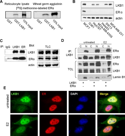
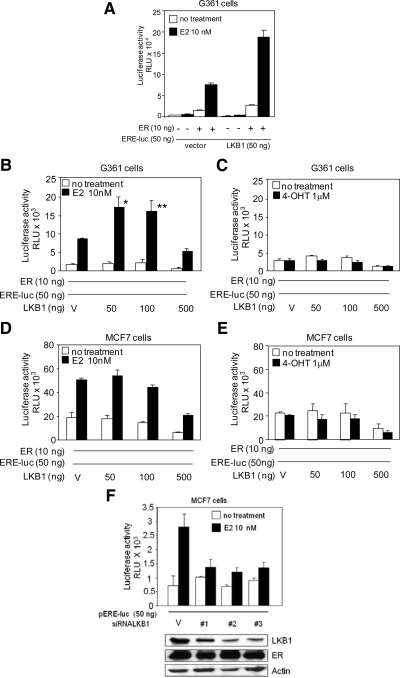
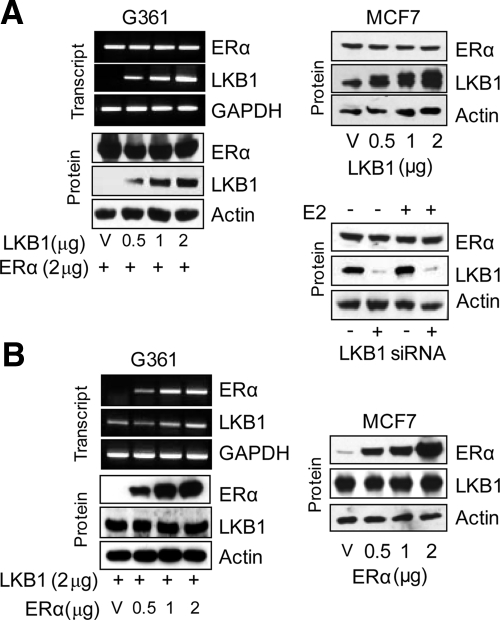
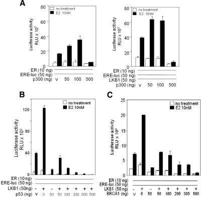
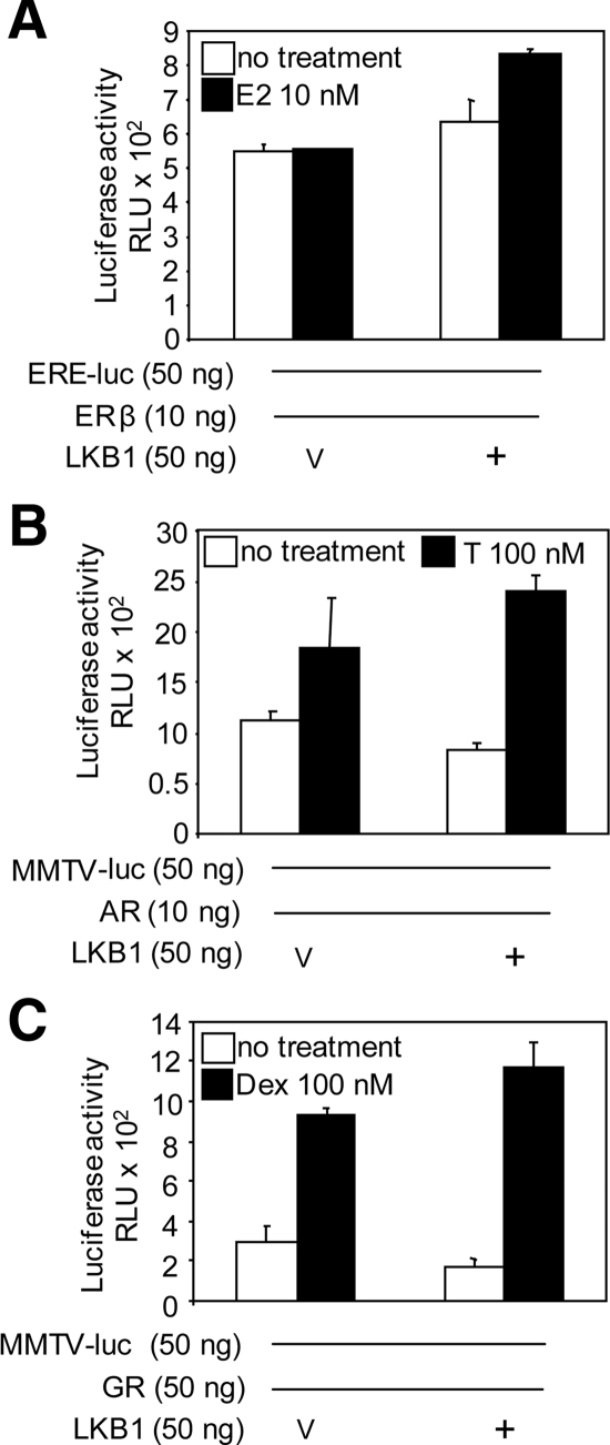
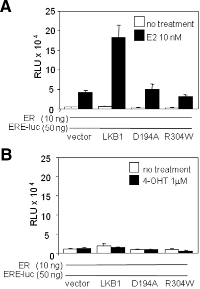
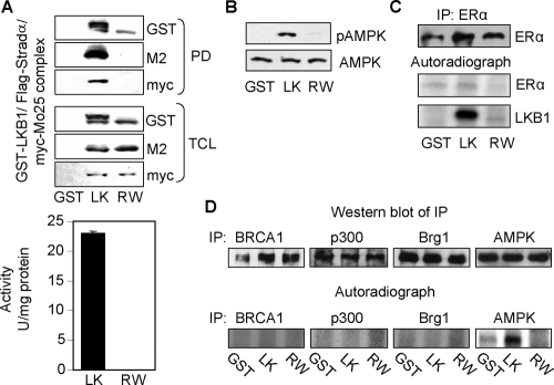
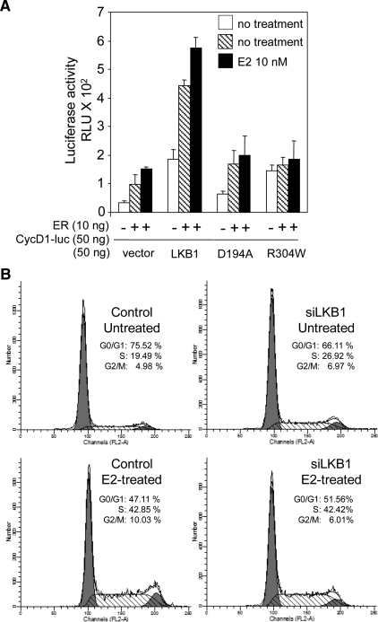
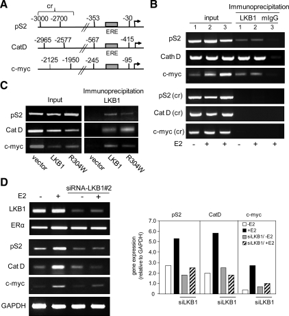
The tumor suppressor serine-threonine kinase LKB1 is mutated in Peutz-Jeghers syndrome (PJS) and in epithelial cancers, including hormone-sensitive organs such as breast, ovaries, testes, and prostate. Clinical studies in breast cancer patients show low LKB1 expression is related to poor prognosis, whereas in PJS, the risk of breast cancer is similar to the risk from germline mutations in breast cancer (BRCA) 1/BRCA2. In this study, we investigate the role of LKB1 in estrogen receptor alpha (ERalpha) signaling. We demonstrate for the first time that LKB1 binds to ERalpha in the cell nucleus in which it is recruited to the promoter of ERalpha-responsive genes. Furthermore, LKB1 catalytic activity enhances ERalpha transactivation compared with LKB1 catalytically deficient mutants. The significance of our discovery is that we demonstrate for the first time a novel functional link between LKB1 and ERalpha. Our discovery places LKB1 in a coactivator role for ERalpha signaling, broadening the scientific scope of this tumor suppressor kinase and laying the groundwork for the use of LKB1 as a target for the development of new therapies against breast cancer.


Figures

Figure 1.
Figure 1.
LKB1 binds to ERα. (A) Left, GST pull-down assays were conducted using 35S-ERα incubated with GST and GST-LKB1 (50 pmol) fusion proteins and visualized by autoradiography. (B) Western blot analysis of LKB1 and ERα expression in different cancer cell lines was confirmed by western blot analysis using antibodies specific for LKB1, ERα, and actin. (C) Anti-LKB1, -ERα, and IgG (control) antibodies were used to IP endogenous proteins from MCF7 cells, followed by Western blot analysis for IP LKB1, ERα, and TCL in which actin was used as loading control. (D) MCF cells were transfected with pcDNA3.1, ERα and LKB1 expression plasmids. Cells were treated with E2 (100 nM) followed by cellular fractionation: nuclear (N) and cytoplasmic (C). LKB1 was IP using anti-LKB1 antibody, followed by Western blot analysis in which membranes were probed for LKB1, ERα, and lamin B1 (nuclear protein marker). (E) Representative immunofluorescent images of MCF7 cells left untreated or treated with E2 (100 nM), fixed, and incubated with anti-LKB1 (green) and anti-ERα (red) antibodies and appropriate secondary antibodies. Nuclei were stained with DAPI. Results are representative of three experiments.
Figure 2.
Figure 2.
LKB1 enhances ERα activity in the presence of 17-β estradiol. (A) G361 cells were transfected with LKB1, ERα, ERE-luc, and pRL-tk expression plasmids or vector (V), left untreated or treated with E2 as described in Materials and Methods, followed by reporter assays. Data representative of two experiments in triplicate mean ± SD. (B) G361 cells were transfected with ERα, ERE-luc, and pRL-tk expression plasmids, and increasing concentrations of LKB1 expression plasmid, left untreated, or treated with E2 or (C) 4-OHT as described in Materials and Methods followed by reporter assays. Results are three experiments in triplicate. Data are mean ± SEM. *p < 0.02 compared with 0 ng; **p < 0.05 compared with 0 ng. (D and E) MCF7 cells were transfected and treated as described in B and C. Results are three experiments in triplicate. Data are mean ± SEM. (F) MCF7 cells were transfected with three different siRNA-LKB1 duplexes, ERE-luc, pRL-tk, and treated with E2 as described in Materials and Methods followed, by reporter assays. Western blot analysis confirms abrogation of LKB1 expression by using anti-LKB1, -ERα, and -actin antibodies. Results are representative of two experiments in triplicate mean ± SD.
Figure 3.
Figure 3.
ERα expression is not altered by LKB1. (A) Left, G361 cells were transfected with expression plasmids or vector (V) as indicated. LKB1 and ERα expression were determined by PCR and Western blot analysis. Top, right, Western blot analysis of MCF7 cells transfected with increasing concentrations of LKB1. Bottom, right, Western blot analysis of LKB1 and ERα expression in MCF7 cell transfected with LKB1 siRNA-#2 duplex (100 nM). (B) Cells were transfected with expression plasmids as indicated. LKB1 and ERα expression were determined by PCR and Western blot analysis. Results are representative of three experiments.
Figure 4.
Figure 4.
LKB1 and p300 synergize to enhance ERα activity. (A) Left, G361 cells were transfected with increasing concentrations of p300 expression plasmids as indicated; ERα, ERE-luc, and pRL-tk, or vector (V); left untreated or treated with E2 for 24 h followed by reporter gene assays. (A) Right, G361 cells were transfected with increasing concentrations of p300 expression plasmids, a constant concentration of LKB1 as indicated; ERα, ERE-luc, and pRL-tk or V; left untreated or treated with E2 for 24 h followed by reporter gene assays. Results are two experiments in triplicate. Data are mean ± SD (B) G361 cells were transfected with pRL-tk, ERα, ERE-luc, and increasing concentrations of p53, or LKB1 expression plasmids, left untreated or treated with E2 for 24 h followed by reporter gene assays. Data representative of three experiments in triplicate. Data are mean ± SD. (C) Transfection conditions similar to B; however, in the presence of increasing concentrations of BRCA1. Data are representative of three experiments in triplicate mean ± SD.
Figure 5.
Figure 5.
LKB1 does not alter ERβ, GR, or AR activity. (A) G361 cells were transfected with LKB1, ERβ, ERE-luc, and pRL-tk expression plasmids or vector (V). Cells were left untreated or treated with E2 (10 nM) for 24 h before harvesting for reporter assay. (B) Cells were transfected with LKB1, AR, MMTV-luc, and pRL-tk expression plasmids or V, followed by testosterone (T; 100 nM) treatment. (C) Cells were transfected with LKB1, GR, MMTV-luc, and pRL-tk expression plasmids or V followed by treatment with dexamethasone (Dex; 100 nM). Results are three experiments in triplicate mean ± SEM.
Figure 6.
Figure 6.
LKB1 catalytic activity is necessary for ERα transactivation. (A and B) G361 cells were transfected with LKB1, D194A, R304W, ERα, ERE-luc, and pRL-tk expression plasmids or vector (V), left untreated or treated with E2 or 4-OHT for 24 h before harvesting for reporter assay. Results are representative of two experiments in triplicate. Data are mean ± SD.
Figure 7.
Figure 7.
LKB1 does not phosphorylate ERα. (A) Expression of purified GST-LKB1/FLAG-STRADα/Myc-MO25α complex from HEK293 cells was confirmed by Western blot analysis (top). Confirmation of catalytic activity of complex toward LKBtide peptide (bottom) and (B) purified recombinant human AMPK incubated with complex followed by Western blot analysis using anti-pAMPK(T172) and anti-AMPK for total expression. (C) ERα was IP using anti-ERα antibody from MCF7 cells followed by incubation with purified complex in the presence of [γ-32P]ATP. Top panel confirms IP of ERα by Western blot analysis; bottom panels are corresponding autoradiographs. (D) BRCA1, p300, Brg1 (negative control), and AMPK (positive control) were immunoprecipitated from MCF7 cells by using the appropriate antibodies followed by incubation with complex described in A, in the presence of [γ-32P]ATP. Top panel confirms IP of BRCA1, p300, Brg1, and AMPK by Western blot analysis. Bottom panel represent autoradiographs. Results are representative of three separate experiments. Purified complexes: LK, GST-LKB1/STRADα/Myc-MO25α; RW, GST-R304W/STRADα/Myc-MO25α.
Figure 8.
Figure 8.
LKB1 enhances nonclassical ERα signaling. (A) G361 cells were transfected with LKB1, D194A and R304W, ERα, cyclin D1-luc and pRL-tk, expression plasmids, or vector. Cells were left untreated or treated with E2 (10 nM) 24 h before harvesting for reporter assay. Results are representative of three experiments in triplicate. Data are mean ± SD. (B) Cells were transfected with siLKB1#2 duplex and GFP-empty vector, left untreated or treated with E2 (100 nM) for 24 h, followed by FACS analysis. DNA profiles for PI/GFP-positive cells were recorded and analyzed. Data as shown are representative of three experiments.
Figure 9.
Figure 9.
Recruitment of LKB1 to promoters. (A) Schematic representation of pS2, cathepsin D (Cat D), and c-myc promoters and control regions (cr). (B) ChIP analysis using anti-LKB1 antibody showing recruitment of LKB1 to the promoters of pS2, Cat D, and c-myc in the presence and absence of E2 treatment (100 nM) in MCF-7 cells. Control region (cr) of pS2(−3000 to −2700), Cat D (−2965 to −2577), and c-Myc (−2125 to −1950) (C) G361 cells lacking endogenous LKB1 and ERα expression were transfected with ERα, plus LKB1 or R304W expression plasmids followed by ChIP using anti-LKB1 antibody. (D) MCF7 cells were transfected with siLKB1#2 duplex, left untreated or treated with E2 (100 nM). Expression of ERα-responsive genes pS2, Cat D, and c-myc was determined by RT-PCR (left) and expressed relative to GAPDH as determined by densitometry (right). Results are representative of three separate experiments.

Similar articles

Cited by

References

    1. Augereau P., Miralles F., Cavailles V., Gaudelet C., Parker M., Rochefort H. Characterization of the proximal estrogen-responsive element of human cathepsin D gene. Mol. Endocrinol. 1994;8:693–703. - PubMed
    1. Avizienyte E., et al. LKB1 somatic mutations in sporadic tumors. Am. J. Pathol. 1999;154:677–681. - PMC - PubMed
    1. Barnes D., Gillett C. Cyclin D1 in breast cancer. Breast Cancer Res. Treat. 1998;52:1–15. - PubMed
    1. Batsche E., Desroches J., Bilodeau S., Gauthier Y., Drouin J. Rb enhances p160/SRC coactivator-dependent activity of nuclear receptors and hormone responsiveness. J. Biol. Chem. 2005;280:19746–19756. - PubMed
    1. Bignell G. R., Barfoot R., Seal S., Collins N., Warren W., Stratton M. R. Low frequency of somatic mutations in the LKB1/Peutz-Jeghers syndrome gene in sporadic breast cancer. Cancer Res. 1998;58:1384–1386. - PubMed

Publication types

MeSH terms