. 2009 Jun;20(11):2639-49.
doi: 10.1091/mbc.e08-11-1104. Epub 2009 Apr 15.

Identification of the neuroblastoma-amplified gene product as a component of the syntaxin 18 complex implicated in Golgi-to-endoplasmic reticulum retrograde transport

Affiliations

Identification of the neuroblastoma-amplified gene product as a component of the syntaxin 18 complex implicated in Golgi-to-endoplasmic reticulum retrograde transport

Takehiro Aoki et al. Mol Biol Cell. 2009 Jun.

Abstract

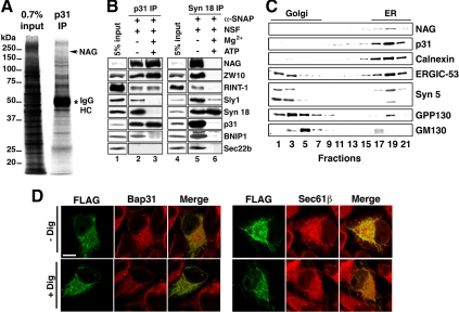
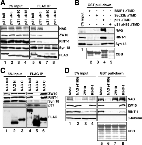
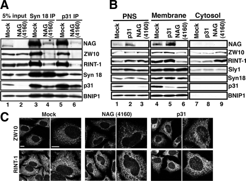
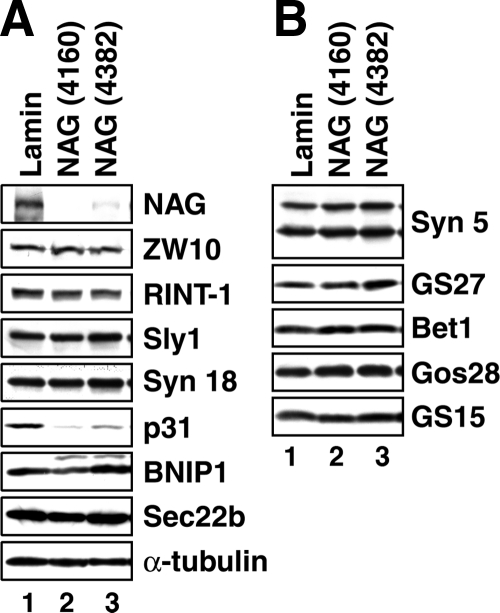
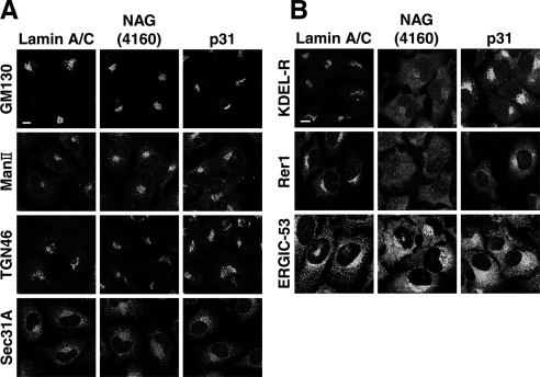
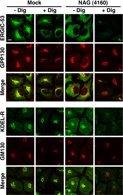
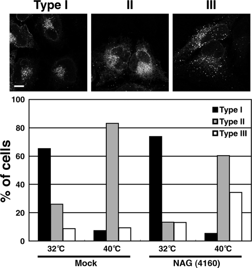
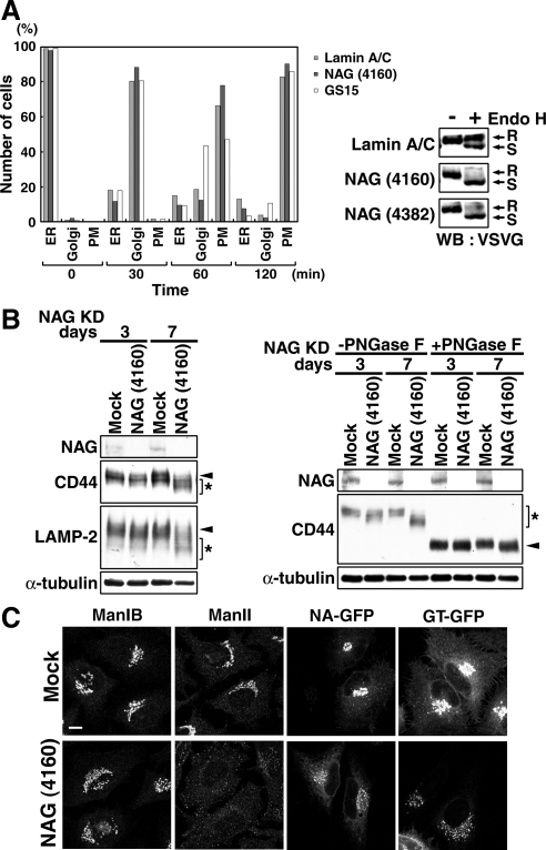
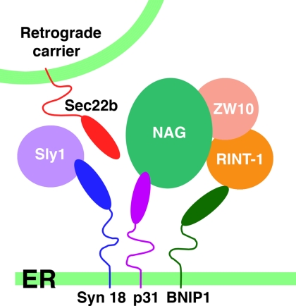
Syntaxin 18, a soluble N-ethylmaleimide-sensitive factor (NSF) attachment protein receptor (SNARE) protein implicated in endoplasmic reticulum (ER) membrane fusion, forms a complex with other SNAREs (BNIP1, p31, and Sec22b) and several peripheral membrane components (Sly1, ZW10, and RINT-1). In the present study, we showed that a peripheral membrane protein encoded by the neuroblastoma-amplified gene (NAG) is a subunit of the syntaxin 18 complex. NAG encodes a protein of 2371 amino acids, which exhibits weak similarity to yeast Dsl3p/Sec39p, an 82-kDa component of the complex containing the yeast syntaxin 18 orthologue Ufe1p. Under conditions favoring SNARE complex disassembly, NAG was released from syntaxin 18 but remained in a p31-ZW10-RINT-1 subcomplex. Binding studies showed that the extreme N-terminal region of p31 is responsible for the interaction with NAG and that the N- and the C-terminal regions of NAG interact with p31 and ZW10-RINT-1, respectively. Knockdown of NAG resulted in a reduction in the expression of p31, confirming their intimate relationship. NAG depletion did not substantially affect Golgi morphology and protein export from the ER, but it caused redistribution of Golgi recycling proteins accompanied by a defect in protein glycosylation. These results together suggest that NAG links between p31 and ZW10-RINT-1 and is involved in Golgi-to-ER transport.


Figures

Figure 1.
Figure 1.
Identification of the NAG protein as a component of the syntaxin 18 complex. (A) Triton X-100 extracts of 293T cells were immunoprecipitated with an anti-p31 antibody (mAb 5C3) attached to protein G-Sepharose 4B. The coprecipitated proteins were resolved by SDS-PAGE, stained with silver, and analyzed by LC-MS/MS. An asterisk denotes immunoglobulin heavy chain. (B) Triton X-100 extracts of 293T cells were incubated at 16°C for 60 min with 10 μg/ml NSF and 5 μg/ml α-SNAP in the presence or absence of 8 mM Mg2+ and 0.5 mM ATP. After incubation, the samples were immunoprecipitated with a mAb against p31 (lanes 2 and 3) or syntaxin 18 (lanes 5 and 6). The precipitated proteins were separated by SDS-PAGE and analyzed by immunoblotting with the indicated antibodies. (C) Cell homogenates were subjected to Nycoprep density gradient centrifugation and analyzed by immunoblotting with the indicated antibodies. (D) HeLa cells were transfected with the plasmid encoding FLAG-NAG. At 24 h after transfection, the cells were left untreated (top row; − Dig) or treated with digitonin (bottom row; + Dig), and double-stained with antibodies against FLAG and Bap31 (left) or Sec61β (right). Bar, 20 μm.
Figure 2.
Figure 2.
NAG links between p31 and ZW10-RINT-1 through an extreme N-terminal region of p31. (A) 293T cells were transfected with the plasmids encoding full-length or N-terminally truncated versions of FLAG-p31. At 24 h after transfection, the cells were lysed, and immunoprecipitation experiments were carried out using an anti-FLAG antibody. The precipitated proteins were separated by SDS-PAGE and analyzed by immunoblotting with the indicated antibodies. (B) Recombinant GST-BNIP1ΔTMD, GST-Sec22bΔTMD, GST-p31ΔTMD, and GST-p31ΔΝ15ΔTMD immobilized onto glutathione-Sepharose 4B were incubated with lysates of 293T cells at 4°C overnight. Pull-down experiments were conducted, and the proteins bound to the resin were separated by SDS-PAGE and visualized by immunoblotting or Coomassie Brilliant Blue (CBB) staining. (C) Lysates of 293T cells transfected with the plasmids encoding full-length or truncated versions of FLAG-NAG were immunoprecipitated using an anti-FLAG antibody. The precipitated proteins were separated by SDS-PAGE and analyzed by immunoblotting with the indicated antibodies. (D) Recombinant GST-p31ΔTMD immobilized onto glutathione-Sepharose 4B was incubated at 4°C overnight with lysates of HeLa cells depleted of NAG, ZW10, or RINT-1. Pull-down experiments were conducted, and the proteins bound to the resin were separated by SDS-PAGE and visualized by immunoblotting or CBB staining.
Figure 3.
Figure 3.
Knockdown of NAG causes the release of ZW10-RINT-1 from membranes as well as from p31. HeLa cells were mock-transfected or transfected with NAG (4160) or p31 siRNA. (A) At 72 h after transfection, cell lysates were prepared and immunoprecipitated with an antibody against syntaxin 18 (lanes 3 and 4) or p31 (lanes 5 and 6). The precipitated proteins were separated by SDS-PAGE and analyzed by immunoblotting with the indicated antibodies. (B) At 72 h after transfection, the cells were homogenized by passage 40 times through a 27-gauge needle. The homogenate was centrifuged at 1100 × g for 5 min to obtain the postnuclear supernatant (PNS; lanes 1–3) and then at 135,000 × g for 30 min to separate the membrane (lanes 4–6) and cytosol (lanes 7–9). (C) At 72 h after transfection, the cells were stained with the indicated antibodies. Note that many and some RINT-1-negative large puncta were observed in NAG- and p31-depleted cells, respectively (bottom row). Bars, 10 μm.
Figure 4.
Figure 4.
Depletion of NAG induces a reduction in the expression level of p31. HeLa cells were transfected with lamin A/C siRNA, NAG (4160), or NAG (4382). At 72 h after transfection, the cells were solubilized in phosphate-buffered saline with 0.5% SDS, and the lysates (15 μg each) were separated by SDS-PAGE and analyzed by immunoblotting with the indicated antibodies.
Figure 5.
Figure 5.
Depletion of NAG changes the distribution of recycling proteins without affecting the Golgi apparatus. HeLa cells were transfected with lamin A/C siRNA, NAG (4160), or p31 siRNA. At 72 h after transfection, the cells were fixed and immunostained with antibodies against proteins in the Golgi or ER exit sites (A) or recycling proteins (B). Bars, 10 μm.
Figure 6.
Figure 6.
Digitonin sensitivity of recycling proteins in NAG-depleted cells. HeLa cells were mock transfected (left two columns) or transfected with NAG (4160) (right two columns). At 72 h after transfection, the cells were permeabilized with digitonin and double-stained with antibodies against ERGIC-53 (top row) and GPP130 (second row) or KDEL-R (fourth row) and GM130 (fifth row). Bar, 10 μm.
Figure 7.
Figure 7.
Retrograde transport of VSVG-KDEL-R is impaired in NAG-depleted cells. The experiments were conducted as described in Materials and Methods. Expressed VSVG-KDEL-R-YFP was predominantly in the perinuclear region (type I), in the ER (type II), or in diffuse distribution (type III). Bottom, quantitative data. The average of three independent experiments is shown. For each sample, >200 cells were evaluated. Bar, 10 μm.
Figure 8.
Figure 8.
Depletion of NAG causes a defect in glycosylation of secretory and lysosomal proteins. (A) HeLa cells were successively transfected with lamin A/C siRNA, NAG (4160), or GS15 siRNA and then with the plasmid for VSVG-GFP. Transport of VSVG-GFP was monitored as described in Materials and Methods. The number of cells in which VSVG-GFP was localized in the ER, the Golgi apparatus, and plasma membrane (PM) were counted. Alternatively, lysates of cells double transfected were prepared after 120-min incubation at the permissive temperature and treated with endoglycosidase H (Endo H). The samples were subjected to SDS-PAGE and analyzed by immunoblotting with an anti-VSVG antibody. R, Endo H-resistant bands; S, Endo H-sensitive bands. (B, left) HeLa cells were mock treated or treated with NAG (4160) for 3 or 7 d, solubilized in phosphate-buffered saline with 0.5% SDS, separated by SDS-PAGE, and analyzed by immunoblotting with the indicated antibodies. Arrowheads and asterisks indicate fully glycosylated and underglycosylated proteins, respectively. (B, right) Lysates of HeLa cells depleted of NAG for 3 or 7 d were treated with PNGase F. Samples were loaded on SDS-PAGE and analyzed by immunoblotting with the indicated antibodies. Asterisk and arrowheads indicate the positions of glycosylated and deglycosylated forms of CD44, respectively. (C) Distribution of glycosylation enzymes in HeLa cells treated with NAG (4160) for 7 d. In the expression of N-acetylglucosaminyltransferase I-GFP (NA-GFP) and β-1,4-galactosyltransferase 1-GFP (GT-GFP), the plasmids encoding GFP fusion proteins were transfected into HeLa cells at 6 d after transfection with NAG (4160). Bar, 10 μm.
Figure 9.
Figure 9.
Schematic representation of the syntaxin 18 complex. NAG associates with p31 and ZW10-RINT-1 through its N-terminal and C-terminal regions, respectively. RINT-1 may weakly interact with BNIP1.

References

    1. Andag U., Neumann T., Schmitt H. D. The coatomer-interacting protein Dsl1p is required for Golgi-to-endoplasmic reticulum retrieval in yeast. J. Biol. Chem. 2001;276:39150–39160. - PubMed
    1. Andag U., Schmitt H. D. Dsl1p, an essential component of the Golgi-endoplasmic reticulum retrieval system in yeast, uses the same sequence motif to interact with different subunits of the COPI vesicle coat. J. Biol. Chem. 2003;278:51722–51734. - PubMed
    1. Aoki T., Kojima M., Tani K., Tagaya M. Sec22b-dependent assembly of endoplasmic reticulum Q-SNARE proteins. Biochem. J. 2008;410:93–100. - PubMed
    1. Appenzeller-Herzog C., Hauri H. P. The ER-Golgi intermediate compartment (ERGIC): in search of its identity and function. J. Cell Sci. 2006;119:2173–2183. - PubMed
    1. Arasaki K., Taniguchi M., Tani K., Tagaya M. RINT-1 regulates the localization and entry of ZW10 to the syntaxin 18 complex. Mol. Biol. Cell. 2006;17:2780–2788. - PMC - PubMed

Publication types

MeSH terms