Comparative Study
. 2009 Apr 15;29(15):4697-707.
doi: 10.1523/JNEUROSCI.6130-08.2009.

Axonal mRNA in uninjured and regenerating cortical mammalian axons

Affiliations
Comparative Study

Axonal mRNA in uninjured and regenerating cortical mammalian axons

Anne M Taylor et al. J Neurosci. .

Abstract

Using a novel microfluidic chamber that allows the isolation of axons without contamination by nonaxonal material, we have for the first time purified mRNA from naive, matured CNS axons, and identified the presence of >300 mRNA transcripts. We demonstrate that the transcripts are axonal in nature, and that many of the transcripts present in uninjured CNS axons overlap with those previously identified in PNS injury-conditioned DRG axons. The axonal transcripts detected in matured cortical axons are enriched for protein translational machinery, transport, cytoskeletal components, and mitochondrial maintenance. We next investigated how the axonal mRNA pool changes after axotomy, revealing that numerous gene transcripts related to intracellular transport, mitochondria and the cytoskeleton show decreased localization 2 d after injury. In contrast, gene transcripts related to axonal targeting and synaptic function show increased localization in regenerating cortical axons, suggesting that there is an increased capacity for axonal outgrowth and targeting, and increased support for synapse formation and presynaptic function in regenerating CNS axons after injury. Our data demonstrate that CNS axons contain many mRNA species of diverse functions, and suggest that, like invertebrate and PNS axons, CNS axons synthesize proteins locally, maintaining a degree of autonomy from the cell body.


Figures

Figure 1.
Figure 1.
The compartmentalized microfluidic chamber is used to isolate pure axonal mRNA from mammalian cortical neurons. A, A schematic of the microfluidic chamber (∼22 mm square). B, Fluorescent micrograph of neurons cultured within the microfluidic chamber showing the ability to isolate axons free of somata and dendrites (MAP2 = red; tau = green). Due to the length of the microgrooves, only axons extend into the axonal compartment. Axonal mRNA was harvested from within this axonal compartment. Scale bar, 100 μm. C, rt-PCR analysis of mRNA collected from neurons and axons within the microfluidic chamber showing that H1 histone mRNA is present in cells, but not the harvested axons. β-Actin mRNA is present in both cells and axons.
Figure 2.
Figure 2.
Microarray analysis shows that neuronal and axonal mRNAs isolated from within the microfluidic chamber have distinct profiles. A heat map of RMA-calculated expression values for 2484 transcripts which were present in 70% of axonal microarrays and had recognizable functions. Each column corresponds to a microarray sample (N, neuronal sample; A, axonal sample). Expression values are normalized to sample A1 (batch 3) as this microarray sample was run simultaneously with regenerating axons in a subsequent experiment and was used to evaluate changes in mRNA composition between naive and injured conditions (shown later in Fig. 4). Neuronal and axonal mRNA was harvested from the microfluidic chamber after 13 d in culture.
Figure 3.
Figure 3.
GO analysis shows that within the mRNA population reliably localized to naive cortical axons multiple functions are enriched and that many of these transcripts overlap with adult DRG axons. A, Enriched GO terms include translation (p = 1.7 × 10⁻³⁰), mitochondrion (p = 6.7 × 10⁻¹⁰), intracellular transport (p = 7.5 × 10⁻⁵), and cytoskeleton (p = 6.0 × 10⁻⁴). Notably, there are many transcripts not included in these enriched functional categories. B, Venn diagram showing overlapping transcripts in the naive matured cortical axons and adult injury-conditioned DRG axons identified in (Willis et al., 2007).
Figure 4.
Figure 4.
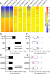
The mRNA pool in regenerating matured cortical axons is distinct from naive matured cortical axons and supports an altered functional role for axonal protein synthesis 2 d following axotomy. A, A heat map showing the RMA-calculated expression level of 866 transcripts identified as >20% changed between naive and regenerating axons. Expression levels are normalized to axonal microarray sample, A1 (batch 3), as this sample was run simultaneously with the regenerating axonal samples (R1–R3). All samples used RNA isolated within the microfluidic chamber at 13 d in vitro. For the regenerating axons, axons were removed at 11 d in vitro, then harvested at 13 d. B, Enriched functional categories showing overall increased localization following injury include cell–cell signaling (p = 3.8 × 10⁻¹⁷), cell differentiation (p = 5.0 × 10⁻¹²), and secretion (p = 2.6 × 10⁻⁵). Criteria for selecting changed transcripts: (>20% change, FDR <0.05). C, Boxplot showing expression values following injury (normalized to naive axons) for all probe sets identified in categories listed in (B). Median expression levels (red lines) are all >1. D, Enriched functional categories showing overall decreased localization following injury include mitochondrion (p = 2.1 × 10⁻⁵), intracellular transport (p = 1.5 × 10⁻⁶), and cytoskeleton (p = 2.5 × 10⁻⁴). E, Boxplot showing expression values following injury for all probe sets identified in categories listed in D. Median expression levels are all <1. For boxplots, red lines indicate medians; tops and bottoms of each “box” are 25th and 75th percentile of the samples; “whiskers” show extent of data; outliers are denoted in red and consist of data >1.5 times interquartile ranges.
Figure 5.
Figure 5.
Fluorescence in situ hybridizations for eukaryotic elongation factor 1α1, β-catenin, and neurexin III shows the localization of their transcripts in matured axons, their change following injury, and validates the findings of the microarray studies. A, Fluorescent micrographs of FISH results showing mRNA puncta (green) present in cortical axons. The axons are also immunolabeled with tubulin (TUBB) (red). ICC, Immunocytochemistry. Scale bar, 5 μm. B, Fluorescent micrographs of cell bodies following FISH for both sense and antisense probes for Ctnnb1. White dashed lines delineate the boundary of the cell body. Scale bar, 5 μm. C, Boxed region is an example of the axonal region used for quantification of transcript levels in (D) and (E). Scale bar, 50 μm. D, Number of puncta per area of naive axons, showing a similar mRNA expression level trend as the microarray analysis. E, Puncta levels changed significantly (*) following injury (normalized to average puncta level in naive axons) (Eef1a1 p = 0.00018; Ctnnb1 p = 0.010; Nrxn3 p = 0.019; two-tailed Student's t test). The percentage of area of β-tubulin protein was not significantly changed between naive and injured axons (data not shown). F, Localization of mRNA and corresponding protein. The protein signal is pseudocolored using the intensity lookup table, “Fire.” Scale bar, 5 μm. Error bars indicate SEM.

Similar articles

Cited by

References

    1. Aschrafi A, Schwechter AD, Mameza MG, Natera-Naranjo O, Gioio AE, Kaplan BB. MicroRNA-338 regulates local cytochrome c oxidase IV mRNA levels and oxidative phosphorylation in the axons of sympathetic neurons. J Neurosci. 2008;28:12581–12590. - PMC - PubMed
    1. Asher RA, Morgenstern DA, Shearer MC, Adcock KH, Pesheva P, Fawcett JW. Versican is upregulated in CNS injury and is a product of oligodendrocyte lineage cells. J Neurosci. 2002;22:2225–2236. - PMC - PubMed
    1. Bamji SX, Shimazu K, Kimes N, Huelsken J, Birchmeier W, Lu B, Reichardt LF. Role of beta-catenin in synaptic vesicle localization and presynaptic assembly. Neuron. 2003;40:719–731. - PMC - PubMed
    1. Bassell GJ, Singer RH, Kosik KS. Association of poly(A) mRNA with microtubules in cultured neurons. Neuron. 1994;12:571–582. - PubMed
    1. Beaudoin GM., 3rd Con-nectin axons and dendrites. J Cell Biol. 2006;174:7–9. - PMC - PubMed

Publication types