Comparative Study
. 2009 Apr 15;29(15):4736-49.
doi: 10.1523/JNEUROSCI.0325-09.2009.

Connexin32 mutations cause loss of function in Schwann cells and oligodendrocytes leading to PNS and CNS myelination defects

Affiliations
Comparative Study

Connexin32 mutations cause loss of function in Schwann cells and oligodendrocytes leading to PNS and CNS myelination defects

Irene Sargiannidou et al. J Neurosci. .

Abstract

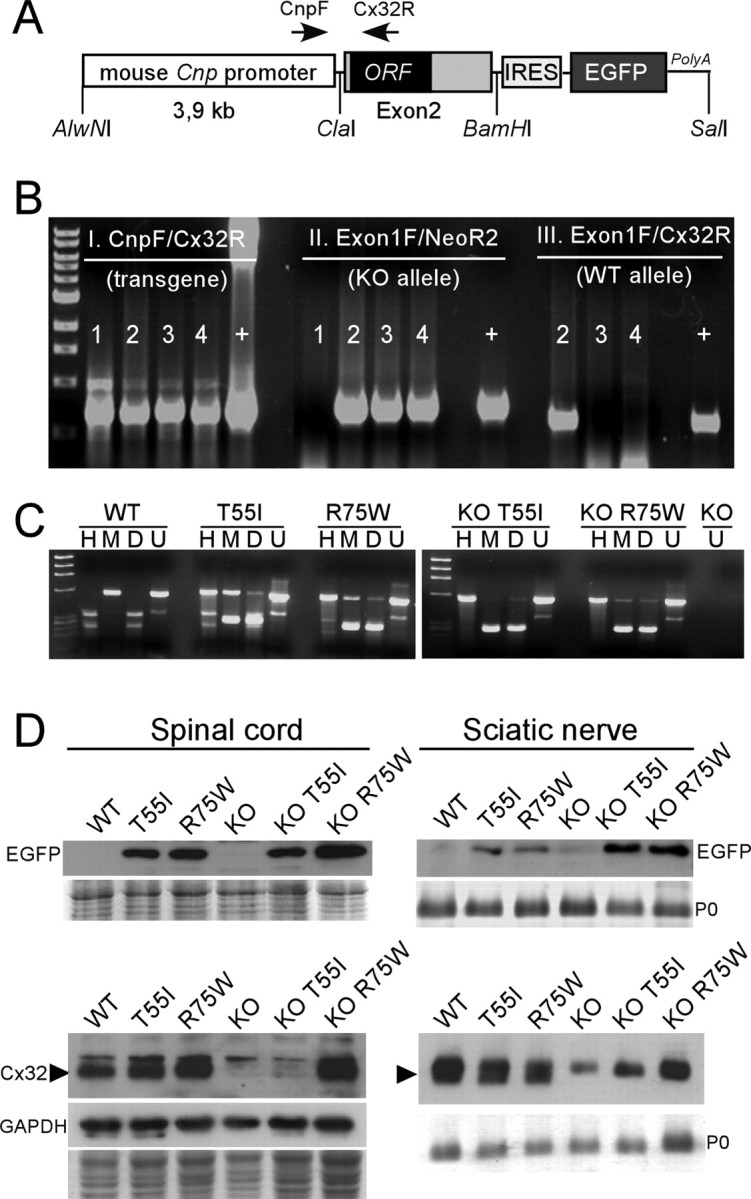
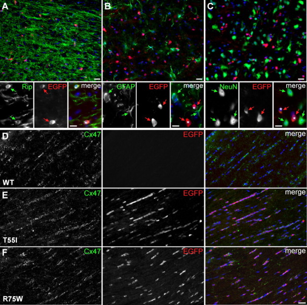
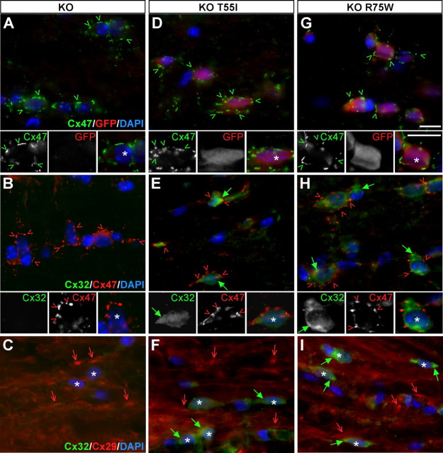
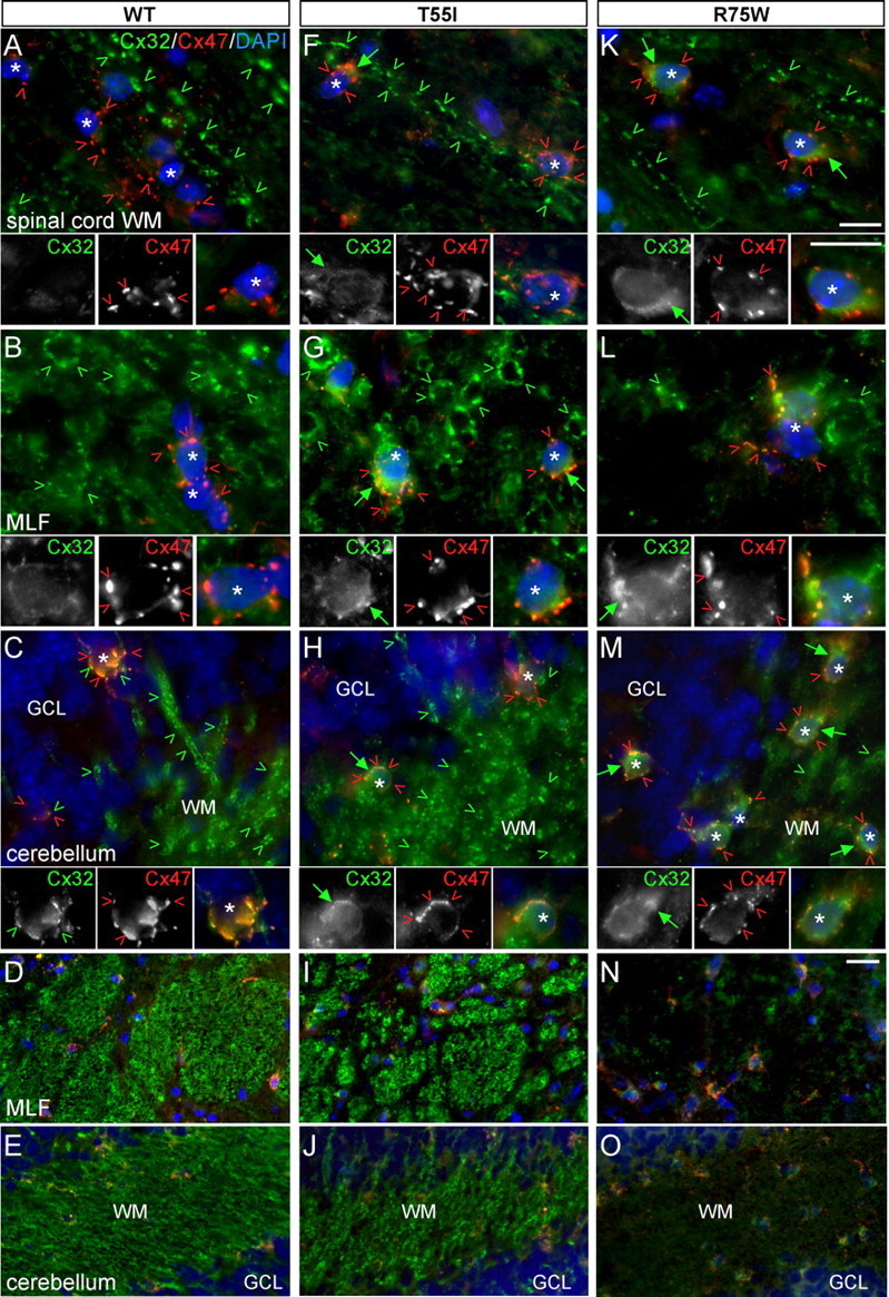
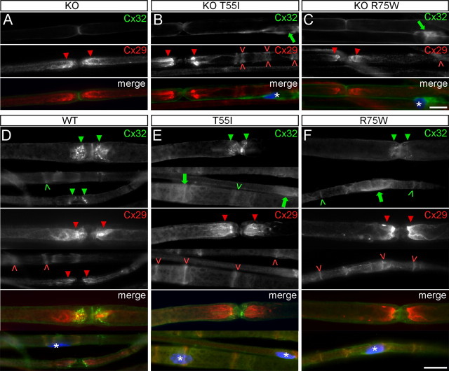
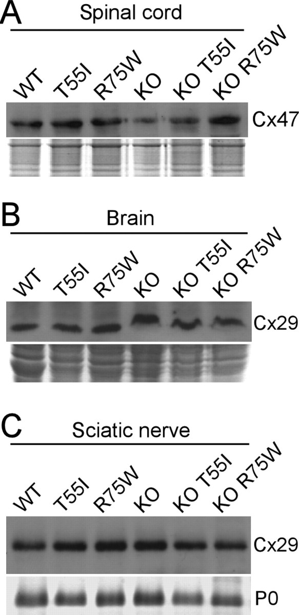
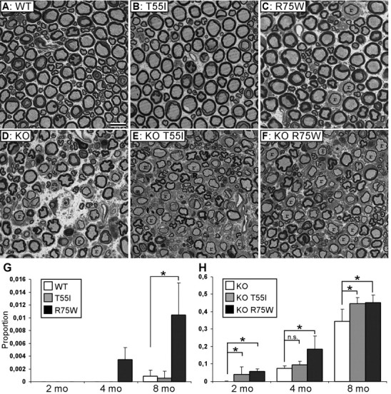
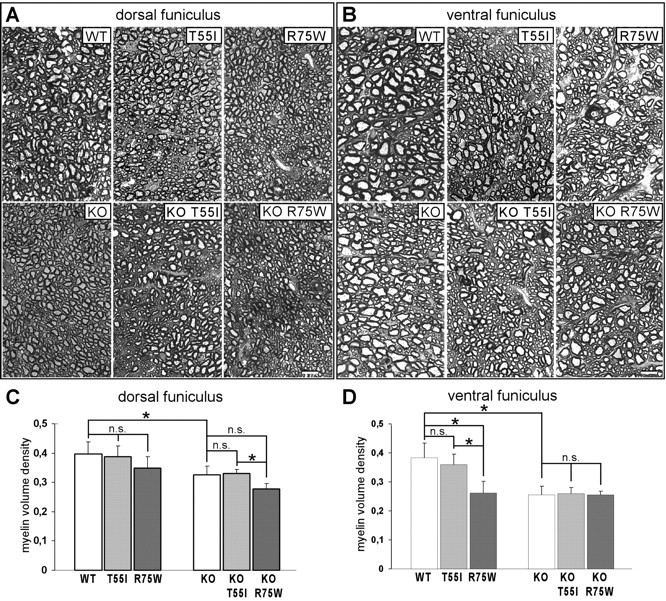
The gap junction (GJ) protein connexin32 (Cx32) is expressed by myelinating Schwann cells and oligodendrocytes and is mutated in X-linked Charcot-Marie-Tooth disease. In addition to a demyelinating peripheral neuropathy, some Cx32 mutants are associated with transient or chronic CNS phenotypes. To investigate the molecular basis of these phenotypes, we generated transgenic mice expressing the T55I or the R75W mutation and an IRES-EGFP, driven by the mouse Cnp promoter. The transgene was expressed in oligodendrocytes throughout the CNS and in Schwann cells. Both the T55I and the R75W mutants were localized in the perinuclear cytoplasm, did not form GJ plaques, and did not alter the expression or localization of two other oligodendrocytic GJ proteins, Cx47 and Cx29, or the expression of Cx29 in Schwann cells. On wild type background, the expression of endogenous mCx32 was unaffected by the T55I mutant, but was partly impaired by R75W. Transgenic mice with the R75W mutation and all mutant animals with Gjb1-null background developed a progressive demyelinating peripheral neuropathy along with CNS myelination defects. These findings suggest that Cx32 mutations result in loss of function in myelinated cells without trans-dominant effects on other GJ proteins. Loss of Cx32 function alone in the CNS causes myelination defects.


Figures

Figure 1.
Figure 1.
A, The structure of the transgene used to express Cx32 in myelinating cells. The 3.9 kb mouse Cnp promoter is joined upstream of the exon 2 (which contains the ORF) of the human GJB1 gene. The IRES-EGFP sequence was cloned downstream of the GJB1 exon 2 to allow coexpression of the EGFP. The positions of the primers used for PCR screening and restriction sites used for cloning are indicated. B, Triple PCR screening of transgenic lines on WT and Gjb1-null (KO) background. Primer pairs amplifying the transgene (CnpF/Cx32R; shown in A), the neomycin gene that replaced the ORF of Gjb1 in Cx32 KO mice (Exon1F/NeoR2), and the WT Gjb1 allele (Exon1F/Cx32R) were used in combination. Mouse 1 is a male transgenic on WT background (TG+X+Y); mouse 2 is a female transgenic heterozygous KO (TG+X+X); mouse 3 is TG+XY, and mouse 4 is TG+XX. “+” is positive control DNA. C, RT-PCR analysis of transgene expression in mouse brain. Mouse and/or human Cx32 cDNA was amplified by RT-PCR (using primers that amplify both) from single brains. The PCR product was digested with MscI (M) (cuts the human Cx32 cDNA) or HhaI (H) (cuts the mouse Cx32 cDNA). D, “Double-cut” with MscI and HhaI; U, uncut. The M-digested human Cx32 cDNA is detectable only in transgenic lines but not in the WT or Cx32 KO mouse, whereas the H-digested mouse Cx32 cDNA is present in WT mouse and in transgenic lines on WT background, but not in the lines on KO background. No human or mouse Cx32 cDNA is present in the Cx32 KO. In the transgenic lines on a WT background, the M-digested product (resulting in two bands of almost same size and therefore merged) gives ∼2-fold higher band intensity than the H-digested product (2 separate bands), indicating that the level of the transgene mRNA is approximately double that of the endogenous/mouse mRNA. D, Immunoblots of spinal cord or sciatic nerve lysates from transgenic lines as well as WT or KO mice, as indicated. Coomassie-stained gels are shown under the blots; P0 is the dense band in the sciatic nerve blots; one blot was reprobed for GAPDH to demonstrate the loading. Note that EGFP (27 kDa band) is detected in the spinal cord and sciatic nerves of all mutants on both WT and KO backgrounds, but not in WT or in KO mice. The specific band for the Cx32 monomer (arrowhead, ∼27 kDa) is detected in the spinal cord and sciatic nerve of WT, T55I, and R75W mice, it is absent from KO mice, reappears in KO R75W mutant mice but is faint (sciatic nerve) or absent (spinal cord) in KO T55I tissues. A faint nonspecific band is present above the Cx32 band in all samples including the KO tissues.
Figure 2.
Figure 2.
Expression of the transgene in oligodendrocytes of CMT1X mice. A–F, These are images of sections of spinal cords (A–C) or optic nerves (D–F) from 4-month-old WT mice or mutant mice in a WT background, double labeled with antibodies against EGFP (red) and cell markers or Cx47 (green) as indicated. Nuclei are visualized with DAPI (blue). In spinal cords, EGFP-positive cells (red arrows and asterisks in merged images) are Rip-positive oligodendrocytes (green arrows in A), and not GFAP-positive astrocytes (green arrows in B) or NeuN-positive neurons (green arrows in C). In optic nerves, EGFP is expressed in Cx47-positive oligodendrocytes in the T55I and R75W lines, but not in WT mice. Scale bars, 20 μm; in insets, 10 μm.
Figure 3.
Figure 3.
Mutant Cx32 does not alter the localization of other gap junction proteins in oligodendrocytes. A–I, These are images of longitudinal sections through the white matter of spinal cords from Cx32 KO mice (A–C) as well as T55I (D–F) and R75W (G–I) mutant mice in a KO background, as indicated. Sections are double labeled with mouse monoclonal antibodies (green) against Cx47 (A, D, G) or Cx32 (B, C; E, F; H, I) and rabbit antisera (red) against GFP (A, D, G), Cx47 (B, E, H), or Cx29 (C, F, I). Nuclei are labeled with DAPI (blue). In mutant mice (D, G), EGFP-positive oligodendrocytes (asterisks) express Cx47, which forms numerous GJ plaques at the perikaryon and proximal processes (green arrowheads), as in Cx32 KO mice (A). Cx32 is absent from Cx32 KO mice (B, C), and mutant Cx32 is localized in the cytoplasm of oligodendrocytes in both KO T55I and KO R75W lines (green arrows in E, F and H, I); in the same cells Cx47 again appears to be normally localized, forming GJ plaques (red arrowheads in B, E, H). Cx29 is also normally localized along thin myelinated fibers (red arrows) in both mutant lines despite the presence of the Cx32 mutants (F, I), as it does in Cx32 KO mice (C). Scale bars (including insets), 10 μm.
Figure 4.
Figure 4.
The R75W mutant but not the T55I mutant alters the expression of endogenous mCx32 on WT background. A–O, These are images of the indicated CNS regions, longitudinal sections of spinal cord white matter (WM) (A, F, K), transverse sections of the medial longitudinal fasciculus (MLF) (B, G, L, and low magnification in D, I, N), and the cerebellar white mater (C, H, M, and low magnification in E, J, O), from WT mice (A–E) as well as T55I (F–J) and R75W (K–O) mutant mice in a WT background. Sections were double labeled with a mouse monoclonal antibody against Cx32 (green) and a rabbit antiserum against Cx47 (red). Cell nuclei are visualized with DAPI (blue). In WT mice (A–E), endogenous/mouse Cx32-immunoreactivity is seen along large myelinated fibers and occasionally in perikarya of cerebellar oligodendrocytes (C, green arrowheads); T55I mice (F–J) have the same pattern of Cx32-immunoreactivity except that oligodendrocyte somata have more pronounced Cx32-immunoreactivity (likely the T55I mutant protein). In contrast, in R75W mice (K–O), Cx32-immunoreactivity is strongly reduced in white matter tracts; this is particularly evident in low-magnification images of MLF (N) and cerebellar white matter (O). In addition, oligodendrocytes (asterisks) have stronger Cx32-immunoreactivity in their perinuclear cytoplasm (likely the R75W mutant protein) than WT or T55I mutant mice. Despite the presence of mutant Cx32, Cx47 is normally expressed in the perikarya and proximal processes of oligodendrocytes (red arrowheads) in all of these CNS areas, as in the WT CNS. Scale bars: in A–C, F–H, K–M (and insets), 10 μm; in D, E, I, J, N, O, 20 μm.
Figure 5.
Figure 5.
Cx32 mutants do not affect the expression of Cx29 in Schwann cells but the R75W mutant alters the expression of endogenous Cx32. A–F, These are images of teased fibers from adult sciatic nerves of Cx32 KO (A), WT (D), or T55I and R75W mutant mice on KO background (B, C) or Cx32 WT background (E, F), double stained with a monoclonal antibody against Cx32 (green) and a rabbit antiserum against Cx29 (red), as indicated. Schwann cell nuclei (asterisks) are stained with DAPI. Cx32 is absent in KO mice (A), and is localized in the perinuclear cytoplasm of Schwann cells of the T55I and R75W mutants on KO background (green arrows in B, C and E, F). Cx32-immunoreactivity appears stronger in the R75W mutant compared with T55I (B, C). In a WT background, endogenous Cx32 is normally localized at paranodes (green arrowheads) and incisures (green carets); this staining is maintained in T55I mutant mice, but appears reduced in the R75W mouse, with a corresponding increase in perinuclear staining (F). In all cases, Cx29 is properly localized at juxtaparanodes (red arrowheads) and incisures (red carets). Scale bars, 10 μm.
Figure 6.
Figure 6.
Cx32 mutants do not affect the expression of other gap junction proteins in myelinating cells. A–C, Immunoblot analysis of Cx47 in the spinal cords (A) and Cx29 in the brains (B) and sciatic nerves (C) of Cx32 WT, Cx32 KO, and T55I or R75W mutant mice either on WT or on KO background, as indicated. In all cases, the levels are similar. Coomassie-stained gels are shown under the blots; P0 is the dense band in the sciatic nerve blot.
Figure 7.
Figure 7.
Cx32 mutant mice develop a progressive demyelinating peripheral neuropathy. A–F, Photomicrographs of semithin sections of femoral motor branches from 8-month-old WT (A), KO (D), as well as T55I and R75W Cx32 mutant mice on WT (B, C) or KO (E, F) background, as indicated. Myelinated axons appear normal in WT and T55I mice (A, B), whereas there are some remyelinated axons (r) in R75W mice (C). In all KO lines (D–F), there many remyelinated (r) and some demyelinated (*) axons. Scale bar, 10 μm. G, H, Quantitative analysis of abnormally myelinated axons in femoral nerves from 2-, 4-, and 8-month-old transgenic mice on WT background (G) and on KO background (H). The average proportion of abnormal fibers is shown; those data are shown in Table 1. On a WT background, the R75W but not the T55I mutant causes significant myelination defects compared with WT (G). All KO lines (H) have progressively increasing number of abnormal fibers with age; the R75W (significant at all ages) and the T55I mutants (significant at 2 and 8 months of age) have more abnormal fibers than do simple KO.
Figure 8.
Figure 8.
Spinal cord myelination defects in Cx32 mutant mice. A–D, Myelin volume density was measured in semithin sections from the dorsal columns (C) and ventral funiculus (D) of 8 month groups (n = 4 in each group); representative images of dorsal columns (A) and ventral funiculi (B) are shown. Note that myelin density is significantly reduced in all lines on KO background compared with WT in both white matter areas. Furthermore, the R75W mutants on WT background show reduced myelin volume density in both areas compared with WT or T55I mutants, more severe in the ventral funiculus (similar to the KO). There are no significant differences between KO and mutants on KO background except for reduced myelin volume density in the KO R75W mice compared with KO T55I mice in dorsal funiculus (see also Table 2). Scale bars, 10 μm.

Similar articles

Cited by

References

    1. Ahn M, Lee J, Gustafsson A, Enriquez A, Lancaster E, Sul JY, Haydon PG, Paul DL, Huang Y, Abrams CK, Scherer SS. Cx29 and Cx32, two connexins expressed by myelinating glia, do not interact and are functionally distinct. J Neurosci Res. 2008;86:992–1006. - PMC - PubMed
    1. Altevogt BM, Paul DL. Four classes of intercellular channels between glial cells in the CNS. J Neurosci. 2004;24:4313–4323. - PMC - PubMed
    1. Altevogt BM, Kleopa KA, Postma FR, Scherer SS, Paul DL. Connexin29 is uniquely distributed within myelinating glial cells of the central and peripheral nervous systems. J Neurosci. 2002;22:6458–6470. - PMC - PubMed
    1. Anzini P, Neuberg DH, Schachner M, Nelles E, Willecke K, Zielasek J, Toyka KV, Suter U, Martini R. Structural abnormalities and deficient maintenance of peripheral nerve myelin in mice lacking the gap junction protein connexin32. J Neurosci. 1997;17:4545–4551. - PMC - PubMed
    1. Bähr M, Andres F, Timmerman V, Nelis ME, Van Broeckhoven C, Dichgans J. Central visual, acoustic, and motor pathway involvement in a Charcot-Marie-Tooth family with an Asn205Ser mutation in the connexin32 gene. J Neurol Neurosurg Psychiatry. 1999;66:202–206. - PMC - PubMed

Publication types

MeSH terms