Comparative Study
. 2009 Apr 15;29(15):4768-81.
doi: 10.1523/JNEUROSCI.5951-08.2009.

Ensheathing glia function as phagocytes in the adult Drosophila brain

Affiliations
Comparative Study

Ensheathing glia function as phagocytes in the adult Drosophila brain

Johnna Doherty et al. J Neurosci. .

Abstract

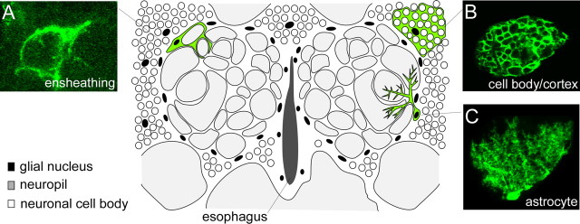
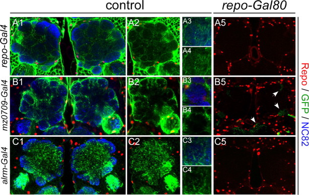
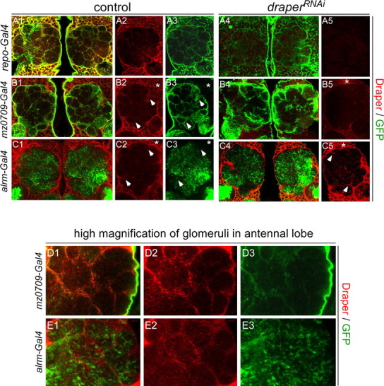
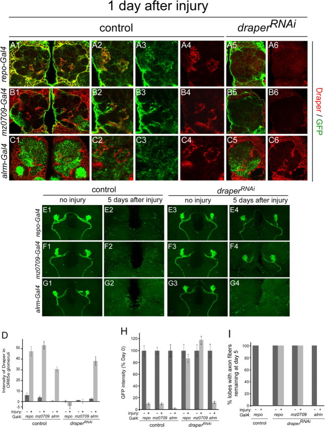
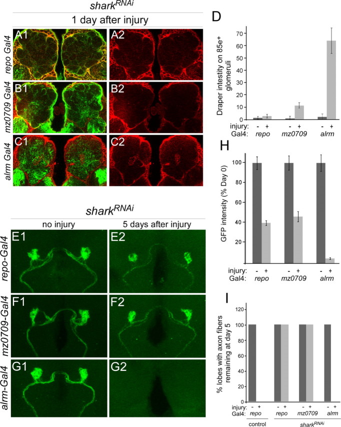
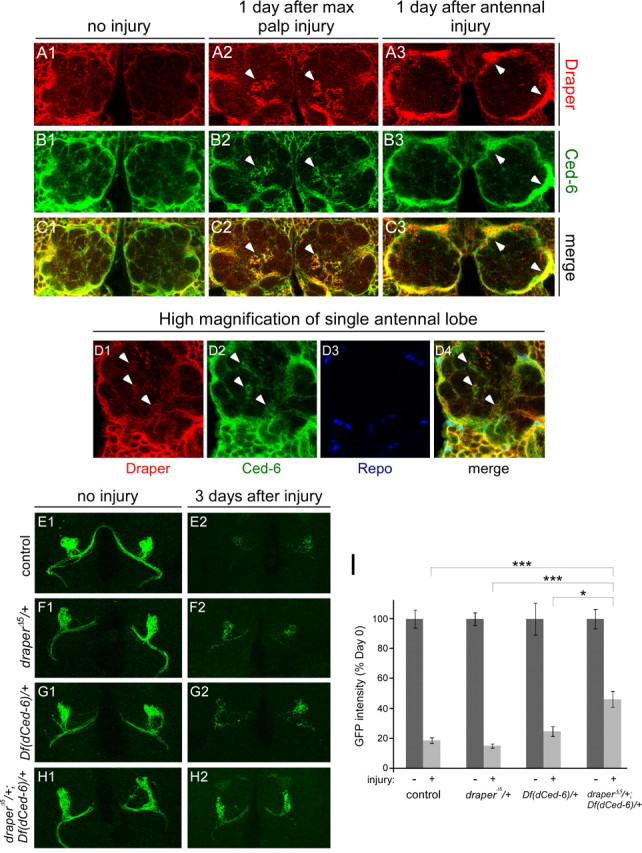
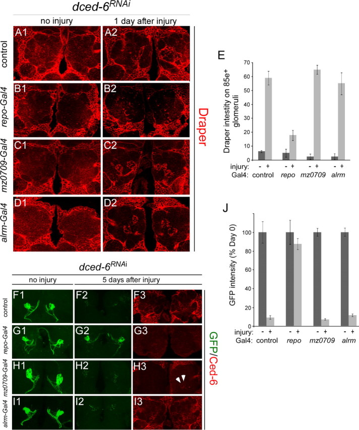
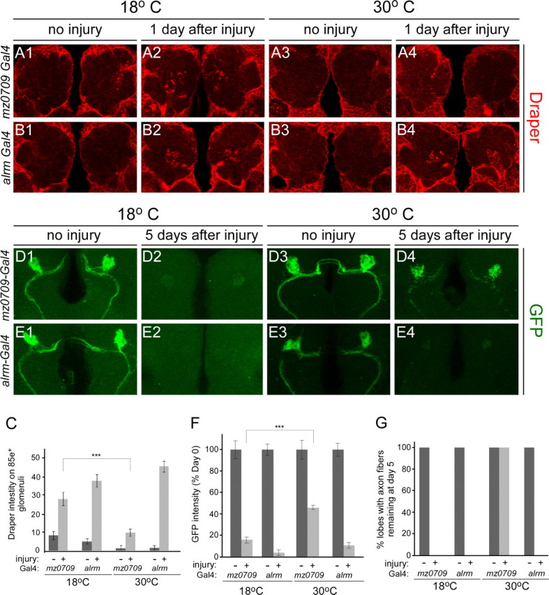
The mammalian brain contains many subtypes of glia that vary in their morphologies, gene expression profiles, and functional roles; however, the functional diversity of glia in the adult Drosophila brain remains poorly defined. Here we define the diversity of glial subtypes that exist in the adult Drosophila brain, show they bear striking similarity to mammalian brain glia, and identify the major phagocytic cell type responsible for engulfing degenerating axons after acute axotomy. We find that neuropil regions contain two different populations of glia: ensheathing glia and astrocytes. Ensheathing glia enwrap major structures in the adult brain, but are not closely associated with synapses. Interestingly, we find these glia uniquely express key components of the glial phagocytic machinery (e.g., the engulfment receptor Draper, and dCed-6), respond morphologically to axon injury, and autonomously require components of the Draper signaling pathway for successful clearance of degenerating axons from the injured brain. Astrocytic glia, in contrast, do not express Draper or dCed-6, fail to respond morphologically to axon injury, and appear to play no role in clearance of degenerating axons from the brain. However, astrocytic glia are closely associated with synaptic regions in neuropil, and express excitatory amino acid transporters, which are presumably required for the clearance of excess neurotransmitters at the synaptic cleft. Together these results argue that ensheathing glia and astrocytes are preprogrammed cell types in the adult Drosophila brain, with ensheathing glia acting as phagocytes after axotomy, and astrocytes potentially modulating synapse formation and signaling.


Figures

Figure 1.
Figure 1.
Identification of morphologically distinct subtypes of glial cells in the adult Drosophila brain. repo-Gal4 was used to label MARCM glial clones with GFP, and the morphology of individual glial cells was analyzed in the adult antennal lobe brain region. We identified three major subtypes of glial cells: ensheathing glia, cortex (or cell body) glia, and astrocytes. Cartoon schematic of the adult antennal lobe brain region depicts the standard position of cell bodies and approximate sizes of each glial cell type within the brain. Confocal Z-stack projections of representative MARCM clones of each glial subtype are shown in A–C. A, Ensheathing glia had a flattened appearance with relatively few branch points, and their membranes appeared to surround and demarcate distinct compartments of the neuropil. B, Cortex glia resided outside the neuropil in the cortex where neuronal cell bodies are found, and appeared to fully ensheath the soma of every brain neuron within its spatial domain. C, Astrocytes projected into the neuropil a major stalk that branched and ramified profusely, ultimately positioning astrocyte membrane processes in close proximity to the synapse-rich regions of the glomeruli.
Figure 2.
Figure 2.
Characterization of Gal4 drivers that uniquely label astrocytes and ensheathing glial subtypes. Individual Gal4 drivers were crossed to UAS-mCD8::GFP, and stained for GFP (α-GFP, green), neuropil (nc82, blue), and glial nuclei (α-Repo, red). Panels show single confocal slices of both antennal lobes (A1, A5, B1, B5, C1, C5), a single antennal lobe without nc82 stain (A2, B2, C2), or a high-magnification view of a single glomerulus (top, with nc82; bottom, without) (A3, A4, B3, B4, C3, C4). A minimum of 10 animals were imaged for each experiment with similar patterns of expression observed. A, repo-Gal4-driven mCD8::GFP labeled all glial membranes (A1, A2), which were seen surrounding and invading glomeruli (A2–A4). All mCD8::GFP expression was suppressed in this genetic background by repo-Gal80 (A5). B, mz0709-Gal4 labeled ensheathing glia that surround glomeruli (B1, B2), but their membranes did not invade glomeruli (B2–B4). Nearly all GFP expression was suppressed by repo-Gal80 (B5), indicating that this driver is largely specific to glia. However, in some brains, a small number of neurons remained GFP labeled (arrowheads). These cells were identified as neurons by tracing axons to Repo neuronal cell bodies. C, alrm-Gal4 labeled only astrocytes (C1, C2), which projected ramified processes that deeply invaded glomeruli (C2–C4), and all GFP expression driven by this Gal4 line was suppressed by repo-Gal80 (C5).
Figure 3.
Figure 3.
The engulfment receptor Draper is expressed in ensheathing and cortex glia but not in astrocytes. Flies carrying UAS-mCD8::GFP were crossed to each glial subtype driver line. Glial membranes were visualized with α-GFP (green) and assayed for colocalization of α-Draper (red). Panels show single confocal sections of merged Draper and GFP images (A1, A4, B1, B4, C1, C4) as well as single antennal lobes showing either GFP or Draper staining alone (A2, A3, A5, B2, B3, B5, C2, C3, C5). A minimum of 10 animals were imaged for each genotype with similar results. A, Pan-glial expression of mCD8::GFP with repo-Gal4 resulted in extensive colocalization of Draper and GFP-labeled membranes in the neuropil and cortex of the brain (A1–A3). No Draper immunoreactivity was detectable when UAS-draperRNAi was expressed in the same genetic background (A4, A5). B, Driving mCD8::GFP with mz0709-Gal4 resulted in extensive colocalization of Draper and GFP-labeled membranes (arrowheads) at the edges of the neuropil and surrounding individual glomeruli, but not in the cortex (asterisk; B1–B3). When mz0709-Gal4 was used to drive UAS-draperRNAi, Draper immunoreactivity was absent immediately surrounding and within the neuropil but was still detectable in the cell cortex (B4, B5; asterisk). C, Labeling of astrocyte membranes with alrm-Gal4-driven mCD8::GFP resulted in no detectable colocalization of Draper and GFP (C1–C3; arrowheads, asterisk), and expression of UAS-draperRNAi in astrocytes had no effect on Draper levels in the cortex or neuropil (C4, C5; arrowheads, asterisk). D, High-magnification view of glomeruli within the antennal lobe shows Draper and mz0709-Gal4-driven GFP colocalizing on membranes that ensheath (D1–D3). E, High-magnification view of glomeruli within the antennal lobe shows distinct staining patterns for Alrm+ membranes and Draper. Alrm+ membranes innervate but do not wrap around glomeruli.
Figure 4.
Figure 4.
Ensheathing glia express Draper, are recruited to severed ORN axons, and phagocytose degenerating axonal debris. A–D, To assay recruitment of each glial subtype to severed axons, we ablated maxillary palps, allowed maxillary palp axons to degenerate for 1 d, and then assayed glial membrane morphology with mCD8::GFP (green) and colocalization of glial membranes with Draper (α-Draper in red). Images are single confocal slices of the antennal lobe region. A, In animals with Repo+ glial membranes labeled with GFP we found that GFP was enriched in glomeruli housing degenerating maxillary palp ORN axons, that these same glomeruli were decorated with Draper, and that Repo+ membranes colocalized perfectly with Draper (A1–A3). Expression of UAS-draperRNAi in Repo+ glia completely suppressed recruitment of glial membranes and Draper to severed axons (A4, A5). B, When glial membranes were labeled with mCD8-GFP using the mz0709-Gal4 driver we found that mz0709+ GFP-labeled glial membranes also colocalized with Draper after maxillary palp ORNs were severed (B1–B3). Moreover, expression of UAS-draperRNAi using the mz0709-Gal4 driver suppressed all recruitment of Draper and GFP-labeled membranes to severed axons 1 d after injury (B4, B5). C, Labeling astrocyte membranes with mCD8::GFP using the alrm-Gal4 driver did not result in the colocalization of GFP-labeled glial membranes with severed axon-associated Draper staining (C1–C3), and expression of UAS-draperRNAi using this driver failed to suppress Draper recruitment to severed axons 1 d after injury (C4, C5). D, Quantification of data from A–C. Error bars represent SEM; n ≥ 10 antennal lobes for each experiment. E–I, To explore the functional requirements for Draper in glial subtypes, we labeled a subset of ORN axons with OR85e-mCD8::GFP, drove UAS-draperRNAi in glial subsets with the indicated drivers, severed axons by ablating maxillary palps, and assayed axon clearance 5 d after injury. All images are confocal Z-stack projections of GFP+ OR85e axonal material. E–G, In control animals (no UAS-draperRNAi), axons developed normally (E1, F1, G1), degenerated, and were cleared from the CNS by 5 d after injury (E2, F2, G2). Driving UAS-draperRNAi with repo-Gal4 (E3, E4) or the ensheathing glial driver mz0709-Gal4 (F3, F4) blocked glial clearance of severed axons. However, expression of UAS-draperRNAi in astrocytes with alrm-Gal4 had no effect on glial clearance of axons from the CNS 5 d after injury (G3, G4). H, Quantification of data from E–G. Error bars represent SEM; n ≥ 10 antennal lobes for each experiment. I, The number of antennal lobes containing GFP-labeled axon debris 5 d after maxillary palp ablation was counted. In control animals (no UAS-DraperRNAi) no GFP-labeled axon debris remained 5 d after ablation. Knockdown of Draper with repo-Gal4 or mz0709-Gal4 resulted in GFP-labeled axon debris present in 100% of the antennal lobes counted 5 d after maxillary palp ablation. Consistent with control animals, driving UAS-draperRNAi with alrm-Gal4 resulted in a complete absence of any GFP-labeled axons 5 d after injury. n ≥ 10 antennal lobes for all.
Figure 5.
Figure 5.
The non-receptor tyrosine kinase Shark functions in ensheathing glia to drive engulfment of ORN axonal debris. We knocked down Shark function in subsets of glia with UAS-SharkRNAi and assayed recruitment of Draper to severed maxillary palp axons 1 d after injury (A–D), and glial engulfment of degenerating axonal debris 5 d after injury (E–I). A1, B1, and C1 show α-Draper (red) and Gal4-driven mCD8::GFP (green); A2, B2, C2 show Draper alone. Representative single confocal slices (A–C) and confocal Z-stacks (E–G) are shown. A, Knockdown of Shark in Repo+ glia suppressed recruitment of Draper to severed maxillary palp ORN axons 1 d after injury. B, Knockdown of Shark in ensheathing glia with mz0709-Gal4 also suppressed recruitment of Draper to severed axons. C, Draper was strongly recruited to severed axons when UAS-SharkRNAi was driven in astrocytes with alrm-Gal4. D, Quantification of data from A–C. Error bars represent SEM; n ≥10 antennal lobes for each experiment. E, SharkRNAi in Repo+ glia suppressed glial clearance of degenerating axonal debris. F, Knockdown of Shark in ensheathing glia also suppressed glial clearance of degenerating axonal debris. G, SharkRNAi treatment of astrocytes failed to suppress glial clearance of degenerating axonal debris. H, Quantification of data from E–G. Error bars represent SEM; n ≥ 10 antennal lobes for each experiment. I, The percentage of antennal lobes containing GFP+ axonal membranes 5 d after maxillary palp ablation. In control animals (no UAS-sharkRNAi), no GFP+ axonal material was present in any samples. SharkRNAi treatment of all glial cells, using repo-Gal4, or ensheathing glial cells, using mz0709-Gal4, resulted in perdurance of GFP+ axonal debris 5 d after maxillary palp ablation in 100% of the samples. Similar to control animals, driving UAS-sharkRNAi with alrm-Gal4 led to a complete loss of GFP+ axonal material 5 d after injury in all antennal lobes. n ≥ 10 antennal lobes for all.
Figure 6.
Figure 6.
dCed-6 is recruited to severed ORN axons after injury and genetically interacts with Draper. A–C, We compared α-dCed-6 (green) and α-Draper (red) localization in three sets of animals: control (no injury), 1 d postmaxillary palp ablation, and 1 d postantennal ablation. Representative images of single confocal slices through the antennal lobe regions are shown. Draper (A1) and dCed-6 (B1) had overlapping patterns of expression (C1) in control animals, including cortex and neuropil regions of the brain. One day after maxillary palp ablation, high levels of Draper (A2) and dCed-6 (B2) immunoreactivity were colocalized (C2) on severed axons (arrowheads). Ablation of antennae resulted in a characteristic dramatic increase in Draper immunoreactivity in glia surrounding the antennal lobes 1 d later (A3). We found that dCed-6 also showed a dramatic increase in immunoreactivity in glia outlining the antennal lobes (white arrowheads) at this time point (B3, C3). D, Single confocal slice of antennal lobe at high magnification stained with Draper (D1), dCed-6 (D2), and Repo (D3), and merged image (D4). Note colocalization of Draper and dCed-6 (arrowheads). E–I, To assay axon clearance of 85e+ maxillary palp ORN axons were labeled with mCD8::GFP, maxillary palps were ablated, and the amount of GFP+ axonal debris was quantified in control, draperΔ5/+, Df(dCed-6)/+, and Df(dCed-6)/+;draperΔ5/+ animals 3 d after axotomy. Representative confocal Z-stack images are shown. Severed axons were largely cleared 3 d after injury in control (E), draperΔ5/+ (F), and Df(dCed-6)/+ (G) animals. Less axonal debris was cleared from the CNS 3 d after injury in Df(dCed-6)/+;draperΔ5/+ (H) animals compared with all other genotypes. I, Quantification of data from E–H. Error bars represent SEM; n ≥ 10 antennal lobes for each experiment; *p < 0.05; ***p < 0.0001.
Figure 7.
Figure 7.
Glial-specific knockdown of dCed-6 suppresses clearance of degenerating ORN axonal debris. dCed-6 was knocked down in glia using a UAS-dced-6RNAi construct and recruitment of Draper to severed axons (A–E) and clearance of degenerating GFP+ axonal debris (F–J) were assayed. Representative single confocal images (A–D) and Z-stack projections (F–I) are shown. A, In control animals (repo-Gal4 driver alone), Draper was recruited at high levels to severed axons 1 d after injury. B, Pan-glial knockdown of dCed-6 using repo-Gal4 suppressed Draper recruitment to severed axons. C, Knockdown of dCed-6 in ensheathing glia using the mz07090-Gal4 driver did not suppress glial recruitment to severed axons. D, Astrocyte-specific knockdown of Draper using alrm-Gal4 did not affect recruitment of Draper to severed axons after axotomy. E, Quantification of data from A–D. Error bars represent SEM; n ≥ 10 antennal lobes for each experiment. F, In control animals (repo-Gal4 driver alone), severed axons were cleared from the CNS 5 d after injury (F1, F2), and dCed-6 was expressed strongly throughout the adult brain (F3). G, Knockdown of dCed-6 in all glia with repo-Gal4 completely suppressed clearance of degenerating axons 5 d after injury (G1, G2), and dCed-6 immunoreactivity was no longer detectable in the adult brain (G3). H, Knockdown of dCed-6 in ensheathing glia with mz0709-Gal4 did not suppress glial clearance of axonal debris (H1, H2), and, notably, dCed-6 staining in the adult brain was only partially reduced in dCed-6RNAi animals (white arrowheads in H3). I, dCed-6 knockdown in astrocytes with alrm-Gal4 had no effect on clearance of axonal debris (I1, I2) or dCed-6 staining in the adult brain (I3). J, Quantification of axon clearance data from (F–I). n ≥ 10 antennal lobes for each experiment, error bars are SEM.
Figure 8.
Figure 8.
Endocytic function is required in ensheathing glia, but not astrocytes, for glial clearance of degenerating ORN axons. The requirements for endocytic activity during phagocytosis of axons in ensheathing glia and astrocytes were determined by expressing UAS-shibirets with mz0709-Gal4 and alrm-Gal4, respectively, and assaying recruitment of Draper to severed axons and clearance of degenerating axonal debris from the brain. A, Expression of Shibirets in ensheathing glia did not block recruitment of Draper to severed maxillary palp ORN axons 1 d after injury when animals were maintained at the permissive temperature of 18°C (A1, A2), but Draper recruitment to severed axons was strongly suppressed when these animals were maintained at the restrictive temperature of 30°C (A3, A4). B, Shibirets expression in astrocytes had no effect on Draper recruitment to severed axons at either 18°C or 30°C (B1–B4). C, Quantification of data from A and B. Error bars represent SEM; n ≥ 10 antennal lobes for each experiment. ***p < 0.0001. D, Expression of Shibirets in ensheathing glia had no effect on glial clearance of severed axons at 18°C (D1, D2). Shifting to the restrictive temperature of 30°C strongly suppressed clearance of degenerating axons 5 d after axotomy (D3, D4). E, Expression of Shibirets in astrocytes with alrm-Gal4 had no effect on glial clearance of degenerating axons 18°C or 30°C (E1–E4). F, Quantification of data from D and E. Error bars represent SEM; n ≥ 10 antennal lobes for each experiment; ***p < 0.0001. G, The number of antennal lobes containing GFP-labeled axon debris 5 d after maxillary palp ablation were counted and expressed as a percentage of the total number. In control animals (entire duration of the experiment performed at the permissive temperature of 18°C) no GFP-labeled axons were present in either the mz0709-Gal4 or the alrm-Gal4 flies. When the flies are shifted to the restrictive temperature of 30°C, 100% of the axons in the mz0709-Gal4 flies are still GFP+, whereas none of the axons in the alrm-Gal4 flies contain any GFP-labeled axons.

Similar articles

Cited by

References

    1. Auld VJ, Fetter RD, Broadie K, Goodman CS. Gliotactin, a novel transmembrane protein on peripheral glia, is required to form the blood-nerve barrier in Drosophila. Cell. 1995;81:757–767. - PubMed
    1. Awasaki T, Tatsumi R, Takahashi K, Arai K, Nakanishi Y, Ueda R, Ito K. Essential role of the apoptotic cell engulfment genes draper and ced-6 in programmed axon pruning during Drosophila metamorphosis. Neuron. 2006;50:855–867. - PubMed
    1. Awasaki T, Lai SL, Ito K, Lee T. Organization and postembryonic development of glial cells in the adult central brain of Drosophila. J Neurosci. 2008;28:13742–13753. - PMC - PubMed
    1. Bainton RJ, Tsai LT, Schwabe T, DeSalvo M, Gaul U, Heberlein U. moody encodes two GPCRs that regulate cocaine behaviors and blood-brain barrier permeability in Drosophila. Cell. 2005;123:145–156. - PubMed
    1. Barres BA. The mystery and magic of glia: a perspective on their roles in health and disease. Neuron. 2008;60:430–440. - PubMed

Publication types

LinkOut - more resources