Comparative Study
. 2009 Apr 15;29(15):4829-35.
doi: 10.1523/JNEUROSCI.5884-08.2009.

Pharmacological analysis demonstrates dramatic alteration of D1 dopamine receptor neuronal distribution in the rat analog of L-DOPA-induced dyskinesia

Affiliations
Comparative Study

Pharmacological analysis demonstrates dramatic alteration of D1 dopamine receptor neuronal distribution in the rat analog of L-DOPA-induced dyskinesia

Amandine Berthet et al. J Neurosci. .

Abstract

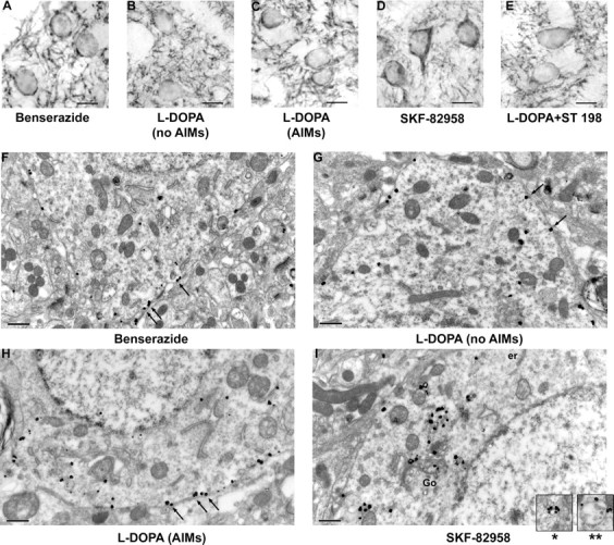
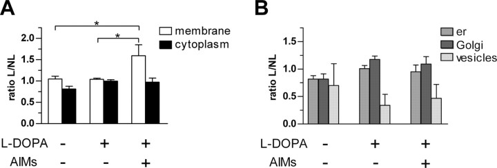
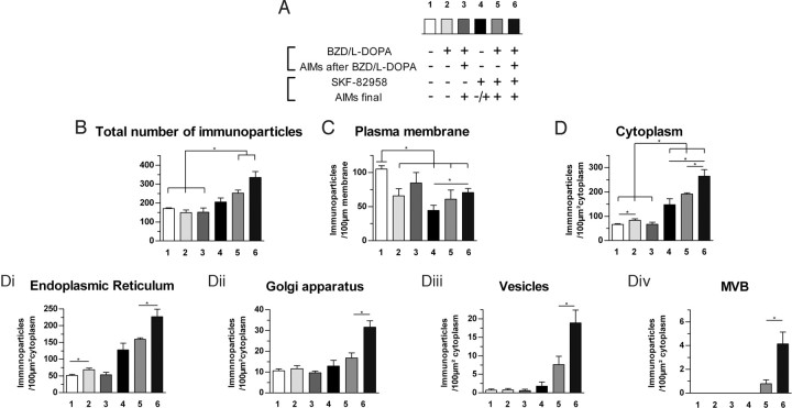
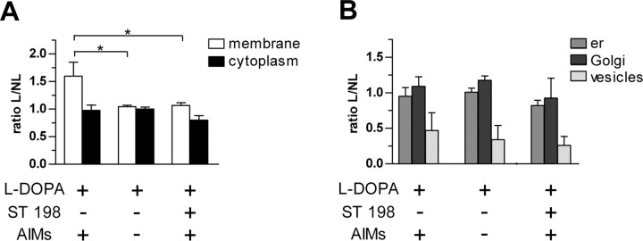
We have associated behavioral, pharmacological, and quantitative immunohistochemical study in a rat analog of l-DOPA-induced dyskinesia to understand whether alterations in dopamine receptor fate in striatal neurons may be involved in mechanisms leading to movement abnormalities. Detailed analysis at the ultrastructural level demonstrates specific alterations of dopamine D(1) receptor (D(1)R) subcellular localization in striatal medium spiny neurons in l-DOPA-treated 6-hydroxydopamine-lesioned rats with abnormal involuntary movements (AIMs). This includes exaggerated D(1)R expression at the plasma membrane. However, D(1)R retains ability of internalization, as a challenge with the potent D(1)R agonist SKF-82958 induces a strong decrease of labeling at membrane in animals with AIMs. Since a functional cross talk between D(1)R and D(3)R has been suggested, we hypothesized that their coactivation by dopamine derived from l-DOPA might anchor D(1)R at the membrane. Accordingly, cotreatment with l-DOPA and the D(3)R antagonist ST 198 restores normal level of membrane-bound D(1)R. Together, these results demonstrate that AIMs are related to abnormal D(1)R localization at the membrane and intraneuronal trafficking dysregulation, and suggest that strategies aiming at disrupting the D(1)R-D(3)R cross talk might reduce l-DOPA-induced dyskinesia by reducing D(1)R availability at the membrane.


Figures

Figure 1.
Figure 1.
Detection of D1R immunoreactivity at light and electron microscopic levels in striatal medium spiny neurons of 6-OHDA unilaterally lesioned rats. All animals bear a 6-OHDA lesion. A–E, Light microscopy, Avidin–biotin DAB Nickel technique (scale bar, 10 μm). A, Control (benserazide only); B, l-DOPA treated without AIMs; C, l-DOPA-treated with AIMs; D, SKF-82958 treated; E, combination of l-DOPA and ST 198. D1R immunoreactivity is mostly located at plasma membrane of cell bodies and in the neuropil in all situations, except after SKF-82958 where immunoreactivity has prominent cytoplasmic localization. F–I, Electron microscopy, Immunogold technique (scale bar, 0.5 μm). F, Control (benserazide only); G, l-DOPA treated without AIMs; H, l-DOPA treated with AIMs; I, combination of l-DOPA and SKF-82958. D1R immunoreactivity shows prominent localization at plasma membrane in all situations (F–H, arrows) but after combination of l-DOPA and SKF-82958 (I) where D1R immunoreactivity is prominently localized in cytoplasmic organelles (er, endoplasmic reticulum; Go, Golgi apparatus). Insets in I show labeling at the periphery of vesicles (*) and multivesicular bodies (**).
Figure 2.
Figure 2.
Effect of l-DOPA during D1R subcellular distribution in striatal neurons. Ratio L/NL (mean ± SEM) represents the number of immunoparticles in lesioned side (L) versus nonlesioned (NL) side at the plasma membrane [Kruskal–Wallis (KW) = 7.423; p < 0.05], in the cytoplasm [KW = 4.269; not significant (ns; A)], and in organelles (B) (er, endoplasmic reticulum; KW = 0.199, ns; Golgi, KW = 0.105, ns; vesicles, KW = 0.675, ns). * indicates a significant difference between related groups; p < 0.05; Mann–Whitney post hoc test.
Figure 3.
Figure 3.
Relationship between D1R subcellular distribution and AIMs: effect of D1R agonist. Data (mean ± SEM) were collected on the 6-OHDA-lesioned side. A, Experimental details of the six groups showing the pharmacological treatments and their behavioral consequences. Number of D1R immunoparticles (B) per neuronal cell body (F(1,23) = 11.6, p < 0.01), per 100 μm of plasma membrane (F(1,23) = 5.5, p < 0.05) (C), and per 100 μm2 of cytoplasm (F(1,23) = 34.4, p < 0.001) (D). The subcellular distribution in cytoplasmic organelles is further broken down into (Di) endoplasmic reticulum (F(1,22) = 39.4, p < 0.001), (Dii) Golgi apparatus (F(1,23) = 8.3, p < 0.01), (Diii) vesicles (F(1,23) = 8.4, p < 0.01), and (Div) multivesicular bodies (F(1,23) = 4.9, p < 0.05). *indicates a significant difference between related groups; p < 0.05, Mann–Whitney post hoc test.
Figure 4.
Figure 4.
Effect of D3R antagonist ST-198 during D1R subcellular distribution in striatal neurons. Ratio L/NL (mean ± SEM) represents the number of immunoparticles in lesioned side (L) versus nonlesioned (NL) side at the plasma membrane [Kruskal–Wallis (KW) = 7.423, p < 0.05], in the cytoplasm [KW = 3.5, not significant (ns)] (A), and in organelles (B) (er, endoplasmic reticulum; KW = 0.167, ns; Golgi, KW = 0.437, ns; vesicles, KW = 0.716, ns). *indicates a significant decrease compared with l-DOPA-treated dyskinetic rats; p < 0.05, Mann–Whitney post hoc test.

References

    1. Aubert I, Guigoni C, Håkansson K, Li Q, Dovero S, Barthe N, Bioulac BH, Gross CE, Fisone G, Bloch B, Bezard E. Increased D1 dopamine receptor signalling in levodopa-induced dyskinesia. Ann Neurol. 2005;57:17–26. - PubMed
    1. Bernard V, Décossas M, Liste I, Bloch B. Intraneuronal trafficking of G-protein-coupled receptors in vivo. Trends Neurosci. 2006;29:140–147. - PubMed
    1. Bezard E, Brotchie JM, Gross CE. Pathophysiology of levodopa-induced dyskinesia: potential for new therapies. Nat Rev Neurosci. 2001a;2:577–588. - PubMed
    1. Bezard E, Dovero S, Prunier C, Ravenscroft P, Chalon S, Guilloteau D, Crossman AR, Bioulac B, Brotchie JM, Gross CE. Relationship between the appearance of symptoms and the level of nigrostriatal degeneration in a progressive 1-methyl-4-phenyl-1,2,3,6-tetrahydropyridine-lesioned macaque model of Parkinson's disease. J Neurosci. 2001b;21:6853–6861. - PMC - PubMed
    1. Bézard E, Ferry S, Mach U, Stark H, Leriche L, Boraud T, Gross C, Sokoloff P. Attenuation of levodopa-induced dyskinesia by normalizing dopamine D3 receptor function. Nat Med. 2003;9:762–767. - PubMed

Publication types

MeSH terms

LinkOut - more resources