Comparative Study
. 2009 Apr 15;29(15):4911-21.
doi: 10.1523/JNEUROSCI.0332-09.2009.

Linking the input to the output: new sets of neurons complement the polarization vision network in the locust central complex

Affiliations
Comparative Study

Linking the input to the output: new sets of neurons complement the polarization vision network in the locust central complex

Stanley Heinze et al. J Neurosci. .

Abstract

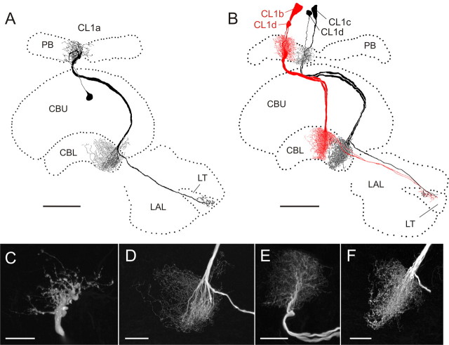
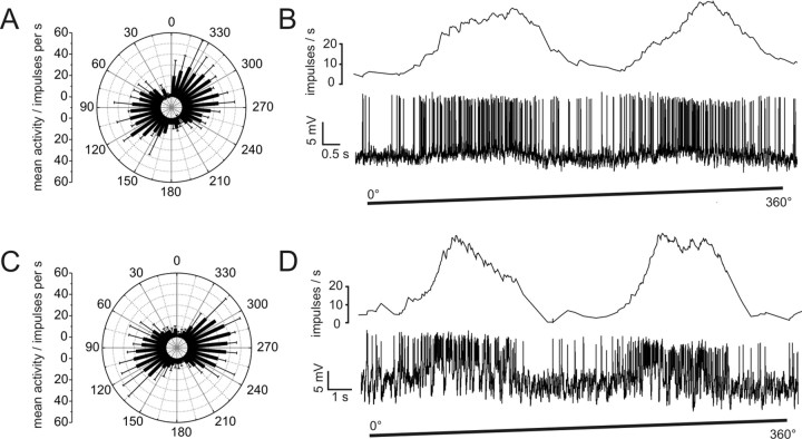
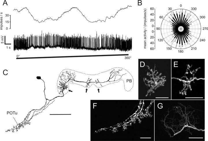
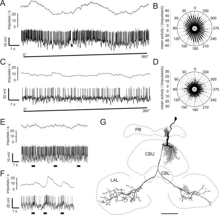
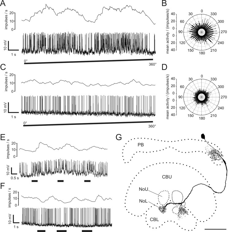
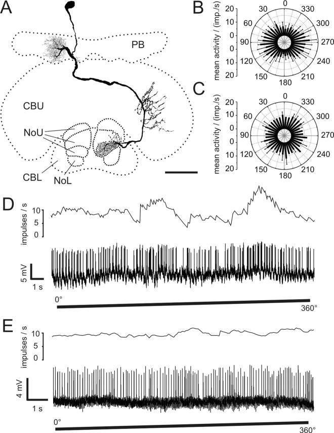
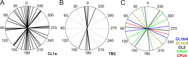
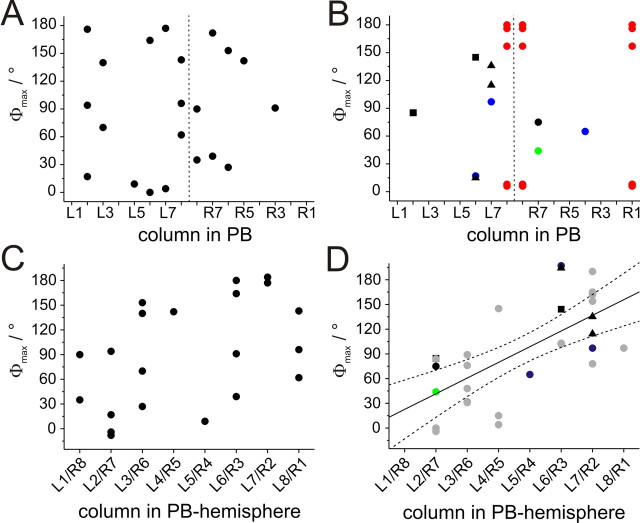
Polarized light is a key feature of the blue sky, used by many animals as a sensory cue for compass navigation. Like other insects, locusts perceive the E-vector orientation of polarized light with a specialized region of their compound eye, the dorsal rim area. Neurons in the brain relay this information through several processing stages to the central complex. The central complex has a modular neuroarchitecture, composed of vertical columns and horizontal layers. Several types of central-complex neurons respond to dorsally presented, rotating E-vectors with tonic modulation of their firing frequency. These neurons were found at the input stage of the central complex, as well as near the proposed output stage, where neurons are tuned to form a compass-like representation of E-vector orientations underlying the columnar organization of the central complex. To identify neurons suited to link input and output elements, we recorded intracellularly from 45 neurons of the central complex. We report several novel types of polarization-sensitive neurons. One of these is suited to fill the gap between input and output stages of the central-complex polarization vision network. Three types of neurons were sensitive to polarized light in only 50% of experiments suggesting that they are recruited to the network depending on behavioral context. Finally, we identified two types of neurons suited to transfer information toward thoracic motor circuits. The data underscore the key role of two subunits of the central complex, the lower division of the central body and the protocerebral bridge, in sky compass orientation.


Figures

Figure 1.
Figure 1.
Morphology of CL1 neurons. A, Frontal reconstruction of a CL1a neuron. It connects the CBL with the PB and the LT in the LAL. B, Frontal reconstruction of a CL1b (red) and a CL1c (black) neuron. Each neuron is joined by a fourth type of CL1 neuron, termed CL1d, with arborizations in the same column, but much smaller soma size. CL1c cells lack the additional axonal process to the LT. C, D, Details of arborization trees from the CL1a neuron shown in A. Maximal-intensity projections of confocal image stacks reveal varicose endings in the PB (C) and a center-surround organization of the arborization tree in the CBL (D). CBL arborizations are of varicose appearance in the center, surrounded by smooth, fine processes in the periphery. E, F, Details of arborizations of the CL1c/d neurons shown in B. Confocal images show that the polarity of these cells is reversed compared with that of CL1a neurons. Arborizations in the PB are of smooth appearance (E), whereas uniformly varicose endings are present in the CBL (F). Scale bars: A, B, 80 μm; C–F, 20 μm.
Figure 2.
Figure 2.
Responses of CL1a and CL1b/d neurons to polarized light. A, Circular diagram of mean frequencies of action potentials of a CL1a neuron plotted against E-vector orientation during dorsal stimulation with a rotating polarizer (n = 2, error bars = SD, bin size 10°, p < 10−12). B, Spike train (lower trace) and mean spiking frequency (upper trace, gliding average with bin size of 1 s) of the same neuron during a clockwise 360° rotation of the polarizer. C, Mean activity of a double-labeled CL1b/d neuron from four 360° rotations of the polarizer (error bars, SD; bin size 10°; p < 10−12). D, Spike train and mean spiking frequency from the same neuron as in C during a clockwise 360° rotation of the polarizer. Postsynaptic potentials are visible throughout the recording.
Figure 3.
Figure 3.
Physiology and morphology of a TB2 neuron. A, Mean activity and intracellular recording trace during clockwise rotation of the polarization filter. B, Mean activity of the neuron during 360° rotations of the polarizer plotted against E-vector orientation (n = 4; error bars, SD; bin size 10°; p < 10−12). C, Frontal reconstruction of the neuron. Three domains of varicose arborizations (arrows) and two wider regions of smooth arborizations are present in the PB. Ramifications in the posterior optic tubercle (POTu) are also varicose. D, E, Detailed views of varicose ramifications in the PB (D, lateral arborizations; E, medial arborizations in left hemisphere; maximal-intensity projections of confocal image stacks). F, Varicose endings in the POTu (maximal-intensity projection). G, Smooth endings in left hemisphere of the PB (maximal-intensity projection). Scale bars: C, 80 μm; D–G, 20 μm.
Figure 4.
Figure 4.
Response characteristics and morphology of conditionally polarization-sensitive CPU2 neurons. A, Spike train (lower trace) and mean spiking frequency (upper trace; gliding average, bin size 1 s) of a polarization-sensitive CPU2 neuron during 360° rotation of the polarizer (clockwise). B, Circular diagram of mean activity during four rotations of the polarizer (same neuron as A, bin size 10°; error bars, SD; p = 2.7 × 10⁻¹¹). C, Activity of a polarization-insensitive CPU2 neuron during rotation of the polarizer (spike train in lower trace, mean spiking frequency in upper trace). D, Circular diagram of mean activity during four rotations of the polarizer (same neuron as C; error bars, SD; bin size 10°). The mean activity of the neuron does not change significantly during rotation of the polarizer (p = 0.68). E, F, Neuronal activity during stimulation with large-field frontal light flashes. E, No response occurred in the polarization-sensitive neuron (same as in A). F, Excitatory responses were present in the polarization-insensitive neuron (same as in C). G, Frontal reconstruction of the CPU2 neuron recorded in A. Arborizations are present in the PB, in all layers of the CBU, and in both ventral shells of the LAL. Scale bar, 80 μm.
Figure 5.
Figure 5.
Conditionally polarization-sensitive CL2 neurons. A, Spike train and mean spiking frequency of a polarization-sensitive CL2 neuron during stimulation with a rotating polarizer. B, Mean response during dorsal stimulation with a rotating polarizer (n = 4; bin size 10°; error bars, SD; p = 0.009). C, Activity of a polarization-insensitive CL2 neuron during rotation of the polarizer. D, Mean spiking frequency from the neuron in C during four rotations of the polarizer (bin size 10°; error bars, SD; p = 0.88). E, F, Responses to unpolarized light stimuli (spike trains and mean activity). E, The polarization-sensitive neuron (shown in A) shows weak excitation during large-field frontal light flashes. F, The polarization-insensitive neuron (shown in C) responds to small, dorsal flashes of unpolarized light with inhibition followed by weak rebound excitation. G, Frontal reconstruction of a CL2 neuron (same as in A, B, and E). The neuron has arborizations in the PB, in the CBL, and in the lower unit of the contralateral nodulus (NoL). NoU, Upper unit of the nodulus. Scale bar, 80 μm.
Figure 6.
Figure 6.
Conditionally polarization-sensitive CPU4 neurons. A, Reconstruction of a CPU4c neuron viewed from posterior. Smooth arborizations are present in the PB, whereas endings in the CBU (layer III) and in layer III of the upper unit of the contralateral nodulus (NoU) are varicose. B, Mean response of the polarization-sensitive neuron in A during four rotations of the polarizer (bin size 10°; error bars, SD; p = 4.2 × 10⁻¹⁰). imp., Impulses. C, Mean spiking activity (two rotations of the polarizer) of a polarization-blind CPU4b neuron (bin size 10°; error bars, SD; p = 0.99). D, Spiking activity and mean frequency of the CPU4c neuron shown in A during clockwise rotation of the polarizer. E, Activity of the CPU4b neuron (same as in C) during stimulation with a rotating polarizer (spike train and mean frequency). NoL, Lower unit of the nodulus. Scale bar, 50 μm.
Figure 7.
Figure 7.
Distribution of Φmax values. A, Φmax values of CL1a neurons cover the complete range of 180° with a distribution not significantly different from randomness (Rao's spacing test, p > 0.1). B, Φmax values of TB2 neurons are clustered around 178° (Rao's spacing test, p < 0.01). C, Φmax values of all remaining neurons are randomly distributed over the range of 180° (Rao's spacing test, p > 0.95).
Figure 8.
Figure 8.
Relation of Φmax values to innervated column in the PB. A, B, Φmax values plotted against the innervated PB columns. Dashed vertical line indicates the midline of the brain. A, CL1a neurons. B, CL1b/d neurons (blue), CL1c/d neurons (green), CL2 neurons (black squares), CPU2 neurons (black triangles), CPU4 neurons (black circles), and TB2 neurons (red) C, For linear regression analysis, Φmax values of all CL1a neurons were combined in one hemisphere and plotted against the innervated PB column. Values near the 0°/180° boundary of E-vectors in medial and lateral columns were shifted by adding or subtracting 180°. Within the resulting data set no significant correlation was observed (significance level of 0.01; t test against slope of 0, p = 0.03; correlation coefficient r = 0.47). D, Data from CL1b/d, CL1c/d, CL2, CPU2, and CPU4 neurons (symbols as in B) combined with previously published data from CPU1, CP1, and CP2 neurons (gray circles) (Heinze and Homberg, 2007) result in a highly significant correlation (significance level: 0.01; t test against slope of 0, p < 0.0001, r = 0.67; y = 19x + 3.6).
Figure 9.
Figure 9.
POL neurons with input arborizations in the LAL. A, Frontal reconstruction of a LAL-pPC neuron. The cell has smooth endings in the left LAL. Bilateral projections to the posterior protocerebrum have varicose terminals. B, Frontal reconstruction of a LAL-LT neuron. The neuron connects the LAL of the left hemisphere with the LT in the right hemisphere. Side branches are also present in the right median olive. The location of the soma could only be inferred by the course of the faintly stained primary neurite. C, Circular plot of mean spiking frequency from the LAL-pPC neuron shown in A (means + SD, n = 6, bin size 10°, p = 4.2 × 10⁻⁸). D, Neuronal activity of the LAL-LT neuron presented in B during 360° rotation of the polarizer. E, Mean spiking activity of the LAL-LT neuron presented in B plotted against E-vector orientation during four rotations of the polarizer (error bars = SD, bin size 10°, p < 10−12). Scale bars, 100 μm.
Figure 10.
Figure 10.
Proposed scheme of the polarization coding network in the central complex. A, Input pathways. Polarization vision information is transferred from the optic lobe to the lower unit of the anterior optic tubercle (AOTu-LU; open blue arrows) and reaches the LT and median olive (MO) via TuLAL1a/b neurons (Pfeiffer et al., 2005). TL2 and TL3 tangential neurons connect the LT and MO to different layers of the CBL (Träger et al., 2008). CL1a neurons are candidates to transmit polarization vision signals from the CBL to the PB. For clarity, this connectivity is only shown for one double column in each hemisphere (CL1a axons to the LT have been omitted). TB1 neurons integrate the signals within the PB and are the first neurons that contribute to the topographic representation of E-vectors in the PB columns (shown for two TB1 cells only) (Heinze and Homberg, 2007). Open, brown arrows indicate the columnar output of the two TB1 neurons. B, Output pathways. The output from TB1 neurons in the PB is likely transferred onto CPU1, CP1, and CP2 columnar neurons projecting to the lateral accessory lobes (LALs, red), the MO (blue), or the LT (violet; Heinze and Homberg, 2007). Of these, CPU1 neurons receive additional input in the CBU. A second pathway connects the CBL directly to the LT via small-diameter axons of CL1a/b neurons, not involving the PB. All terminals of columnar neurons within the LT and MO could potentially provide input to LAL-LT neurons (black), which connect these areas with the contralateral LAL. This neuron type, as well as CPU1 neurons, might synapse onto the LAL-pPC neuron, which provides a connection to the posterior protocerebrum. TB1 neurons provide another possible output from the PB to the posterior optic tubercle (POTu). Descending neurons might receive input from either of these regions (brown, open arrows), and/or from the LAL, to provide information flow to motor control circuits in the thorax. AOTu-UU, Upper unit of the anterior optic tubercle.

Similar articles

Cited by

References

    1. Batschelet E. Circular statistics in biology. London: Academic; 1981.
    1. Clements AN, May TE. Studies on locust neuromuscular physiology in relation to glutamic acid. J Exp Biol. 1974;60:673–705. - PubMed
    1. Eggers A, Gewecke M. The dorsal rim area of the compound eye and polarization vision in the desert locust (Schistocerca gregaria) In: Wiese K, Gribakin FG, Popov AV, Renninger G, editors. Sensory systems of arthropods. Basel: Birkhäuser; 1993. pp. 101–109.
    1. Hanesch U, Fischbach KF, Heisenberg M. Neuronal architecture of the central complex in Drosophila melanogaster. Cell Tissue Res. 1989;257:343–366.
    1. Heinze S, Homberg U. Maplike representation of celestial E-vector orientations in the brain of an insect. Science. 2007;315:995–997. - PubMed

Publication types

LinkOut - more resources