Comparative Study
. 2009 Apr 15;29(15):4953-63.
doi: 10.1523/JNEUROSCI.6020-08.2009.

Substance P mediates excitatory interactions between striatal projection neurons

Affiliations
Comparative Study

Substance P mediates excitatory interactions between striatal projection neurons

Craig P Blomeley et al. J Neurosci. .

Abstract

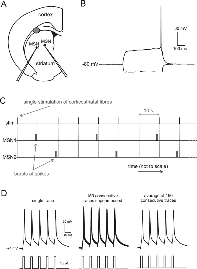
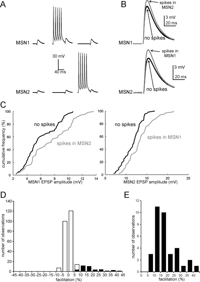
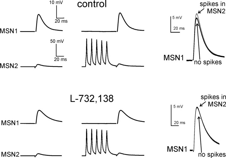
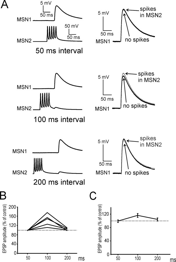
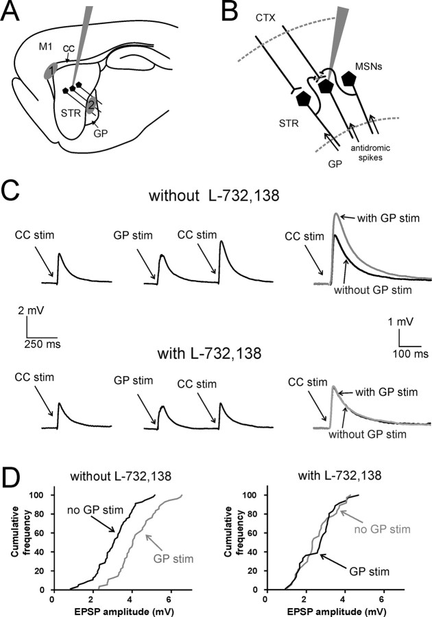
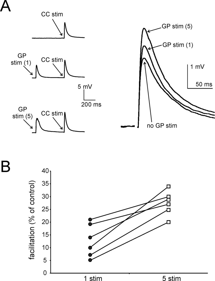
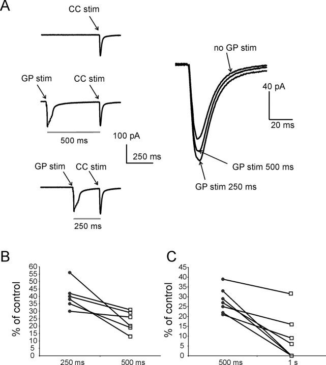
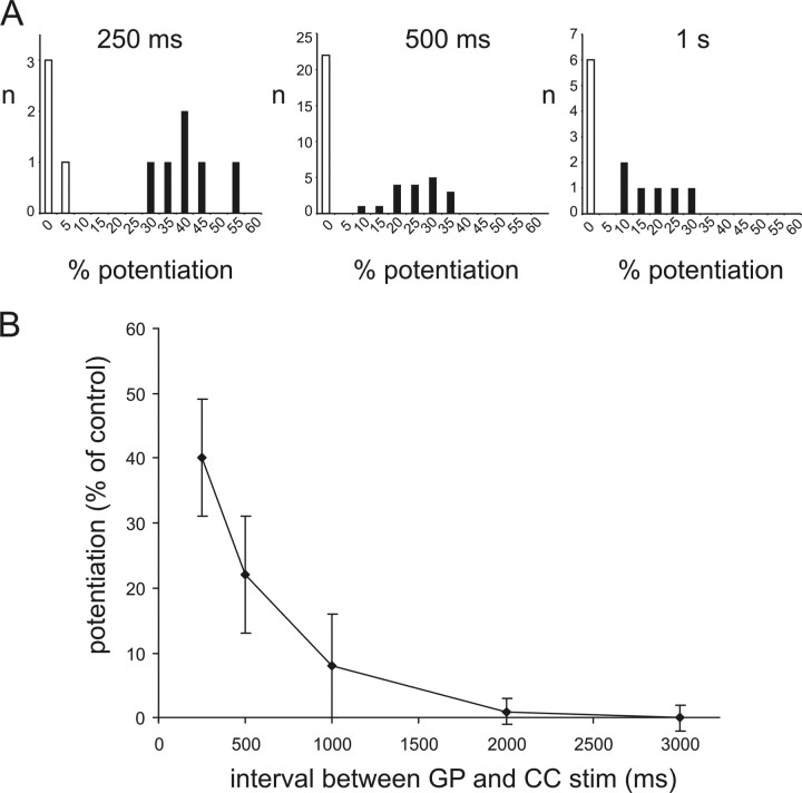
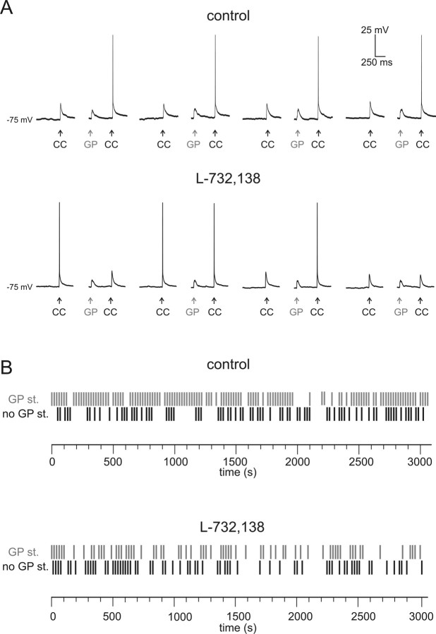
The striatum is the largest nucleus of the basal ganglia, and is crucially involved in motor control. Striatal projection cells are medium-size spiny neurons (MSNs) and form functional GABAergic synapses with other MSNs through their axon collaterals. A subpopulation of MSNs also release substance P (SP), but its role in MSN-MSN communication is unknown. We studied this issue in rat brain slices, in the presence of antagonists for GABA, acetylcholine, dopamine, and opioid receptors; under these conditions, whole-cell paired recordings from MSNs (located <100 microm apart) revealed that, in 31/137 (23%) pairs, a burst of five spikes in a MSN caused significant facilitation (14.2 +/- 8.9%) of evoked glutamatergic responses in the other MSN. Reciprocal facilitation of glutamatergic responses was present in 4 of these pairs. These facilitatory effects were maximal when spikes preceded glutamatergic responses by 100 ms, and were completely blocked by the NK1 receptor antagonist L-732,138. Furthermore, in 31/57 (54%) MSNs, a burst of 5 antidromic stimuli delivered to MSN axons in the globus pallidus significantly potentiated glutamatergic responses evoked 250 or 500 ms later by stimulation of the corpus callosum. These effects were larger at 250 than 500 ms intervals, were completely blocked by L-732,138, and facilitated spike generation. These data demonstrate that MSNs facilitate glutamatergic inputs to neighboring MSNs through spike-released SP acting on NK1 receptors. The current view that MSNs form inhibitory networks characterized by competitive dynamics will have to be updated to incorporate the fact that groups of MSNs interact in an excitatory manner.


Figures

Figure 1.
Figure 1.
Experimental configuration for paired recording experiments. A, Typical positioning of the electrodes used for whole-cell paired recordings from MSNs (located <100 μm apart), and of the stimulating electrode (gray area) used to activate corticostriatal fibers end evoke glutamatergic responses in MSNs in coronal brain slices. B, Typical responses of MSNs to positive and negative current injections (±200 pA). The positive injection was just large enough to elicit one spike. Note the depolarizing ramp causing delayed firing, and the smaller extent of responses to negative steps, indicative of inward rectification. C, Graphical illustration of the protocol used to investigate the effects of spikes in each MSN on the glutamatergic responses of the other MSN. For sake of clarity, time is not represented to scale, as the intervals between single stimuli to corticostriatal fibers (10 s) have been compressed with respect to the duration of the bursts of spikes and the interval between such bursts and the stimulation of the corticostriatal fibers. D, Bursts of precisely timed action potentials were evoked in MSNs using 3 ms current injections separated by 10 ms intervals; this protocol was repeated every 10 s. In this experiment, 150 bursts (each comprising 5 spikes) were evoked. Superimposition of 150 consecutive traces (middle) shows that there was little scattering (<1 ms) in the action potential peak time; the average of the 150 traces is shown on the right.
Figure 2.
Figure 2.
Action potentials in MSNs facilitate glutamatergic responses of neighboring MSNs. A, Average responses of two simultaneously recorded MSNs to stimulation of corticostriatal fibers in the absence of preceding spikes (left); in the presence of a preceding burst of five spikes in MSN1 (middle); and in the presence of a preceding burst of five spikes in MSN2 (right). The first spike of a burst in MSN1 or MSN2 preceded the stimulation of corticostriatal fibers by 100 ms. These three protocols were applied sequentially at 10 s intervals (each protocol was applied 100 times in this experiment). B, Enlargement of average glutamatergic responses in MSN1 and MSN2 (same experiments as in A) in the absence (black trace) or the presence (gray trace) of preceding spikes in the other MSN. In this experiment, spikes in each MSN significantly (p < 0.001) facilitated the responses of the neighboring MSN. C, Cumulative distribution of EPSP amplitude in the same MSNs of A and B, in the absence (black line) or presence (gray line) of preceding action potentials in the other MSN. In both cases, a rightward shift of the distribution in the presence of spikes in the other cell is clearly visible. D, Overall distribution of the effects of a burst of five spikes in a neighboring MSN on the amplitude of glutamatergic responses evoked by stimulation of corticostriatal fibers. Data are from 137 paired recording experiments (for each of which effects in both directions were considered). The white part of each column represents cases in which no significant effects of the spikes were observed; the black part represents cases in which significant (p < 0.05) facilitation was present; note that significant inhibition of evoked responses was never observed under the conditions of this study. E, An enlargement of the distribution of the effects for the cases in which significant facilitation was present.
Figure 3.
Figure 3.
The facilitatory effects of MSN spikes on glutamatergic responses are mediated by NK1 receptors. In this representative example, 5 spikes in MSN2 significantly (p < 0.05) increased EPSP amplitude in MSN1. Traces are averages of 100 consecutive presentations for each protocol. Superimposed traces on the right show the average evoked EPSP in MSN1 with (gray) and without (black) preceding spikes in MSN2 for each pharmacological condition. Subsequent bath application of the NK1 receptor antagonist L-732,138 (5 μm) fully blocked the ability of MSN2 spikes to facilitate MSN1 evoked glutamatergic responses. L-732,138 also significantly decreased the amplitude of basal evoked glutamatergic responses.
Figure 4.
Figure 4.
The facilitatory effects of MSN spikes on glutamatergic responses are maximal at 100 ms intervals. A, In this experiment, a burst of 5 spikes in MSN2 significantly (p < 0.05) increased EPSP amplitude in MSN1 when the interval between the first spike and the glutamatergic response was 100 ms; however, no significant facilitation was present when this interval was 50 or 200 ms. Traces are averages of 100 consecutive presentations for each protocol. Superimposed traces on the right show the average evoked EPSP in MSN1 with (gray) and without (black) preceding spikes in MSN2. B, Effects of a burst of five spikes in a MSN on the evoked glutamatergic responses of a neighboring MSN, for intervals of 50, 100, and 200 ms. Data are from five paired recording experiments in which significant facilitatory effects were observed (in one direction) for intervals of 100 ms. Each point represents the average effect of the spikes for a given interval; data from the same experiment are connected by lines. In all cases, no significant facilitation was present at 50 ms intervals. In two of five cases there was still significant (p < 0.05) facilitation at 200 ms, although this was always smaller than at 100 ms. C, Average effects for the five experiments of B. Error bars represent SDs.
Figure 5.
Figure 5.
Antidromic stimulation of a population of MSNs facilitates evoked corticostriatal glutamatergic responses of individual MSNs through activation of NK1 receptors. A, Typical positioning of the patch electrode used for single whole-cell recordings, and of the stimulating electrodes used to elicit orthodromic activation of corticostriatal fibers and antidromic activation of MSN axons in sagittal slices. Corticostriatal fibers were preferentially stimulated with an electrode placed in the CC, between the cortex and the striatum (area 1). MSN axons were stimulated with an electrode placed in the GP (area 2). B, Simplified diagram illustrating the neurons and axons activated in these experiments. Antidromic spikes travel from the GP to the striatum, invading MSN axon collaterals and releasing neurotransmitters, including substance P, in the striatum. Corticostriatal fibers impinge on the recorded MSN, eliciting glutamatergic responses. C, In a representative experiment, preceding GP stimulation strongly increased (p < 0.001) the amplitude of CC-evoked responses in a MSN. Five stimuli were delivered at 100 Hz in the GP, and were followed by a single stimulation in the CC; the interval between the first GP stimulus and the CC stimulus was 500 ms. This protocol was alternated (every 10 s) with one in which a single stimulus was delivered in the CC without preceding GP stimuli. Each trace is the average of 150 consecutive presentations of a certain protocol. Superimposed traces on the right show the average evoked EPSP with (gray) and without (black) preceding GP stimulation. Subsequent bath application of L-732,138 fully abolished the effects of GP stimulation on corticostriatal responses. L-732,138 also caused a significant decrease in the amplitude of control corticostriatal responses. Traces are the average of 100 consecutive presentations. D, Cumulative distribution of CC-evoked EPSP amplitude for the MSN of C, in the absence (black line) or presence (gray line) of preceding GP stimulation, before (left) and after (right) application of L-732,138. In the absence, but not in the presence, of L-732,138, preceding GP stimulation causes a substantial rightward shift of the cumulative distribution of EPSP amplitude.
Figure 6.
Figure 6.
Five GP stimuli are more effective than one at facilitating CC-evoked responses. A, In this representative experiment, a single CC stimulus (delivered every 10 s) was sequentially preceded by (1) no GP stimulation; (2) one GP stimulus (preceding CC stimulation by 500 ms); and (3) five GP stimuli (also preceding CC stimulation by 500 ms). Traces are averages of 90 consecutive presentations of each protocol. Average CC-evoked responses for the three conditions are enlarged and superimposed on the right. While both one and five preceding GP stimuli significantly facilitate CC-evoked responses, facilitation is significantly larger with five preceding stimuli than with a single one. B, Similar results were found in each of six different experiments. For each experiment, data points representing average facilitation of CC-evoked responses in the presence of one or five preceding GP stimuli are connected by a line.
Figure 7.
Figure 7.
Facilitation of corticostriatal responses induced by GP stimulation is larger at 250 ms than at 500 ms intervals. A, In this representative voltage-clamp experiments (Vholding = −80 mV), a single CC stimulus was delivered every 10 s. These stimuli were sequentially preceded by (1) no GP stimulation; (2) five GP stimuli preceding CC stimulation by 500 ms; and (3) five GP stimuli preceding CC stimulation by 250 ms. Traces are averages of 120 consecutive repetitions for each protocol. Average synaptic currents evoked by CC stimulation for each of these three conditions are enlarged and superimposed on the right. While at both intervals GP stimuli significantly (p < 0.001) facilitated CC-evoked responses, facilitation was significantly (p < 0.05) larger when the GP stimuli preceded CC stimulation by 250 ms than by 500 ms. B, Similar results were found in six different experiments. For each experiment, data points representing average facilitation of CC-evoked responses in the presence of GP stimuli preceding CC stimulation by 250 and 500 ms are connected by a line. C, In a different series of experiments, a similar protocol was used to compare the effects of GP stimuli preceding CC stimulation by 500 and 1000 ms. In each of the seven cases represented in the plot, significant (p < 0.01) facilitation was observed at 500 ms intervals. In three of these cases no facilitation was present at 1000 ms intervals; in the remaining four cases, there was significant (p < 0.05) facilitation of CC-evoked responses at 1000 ms intervals, but this was always significantly (p < 0.05) smaller than at 500 ms intervals.
Figure 8.
Figure 8.
Time course of facilitatory effects of GP stimulation on CC-evoked responses. A, These histograms illustrate the overall distribution of the effects of five preceding GP stimuli on CC-evoked responses for intervals of 250, 500, and 1000 ms. While a similar percentage of cases in which no significant facilitation was present (white columns) is found for all intervals, in cases where significant (p < 0.05) facilitation is present (black columns) its extent is larger for 250 ms than for 500 ms, and for 500 ms than for 1000 ms intervals. B, Average effects of GP stimulation on CC-evoked responses as a function of the interval preceding CC stimulation. Data from all experiments in which significant effects were found for at least one interval are included. Effects are maximal for the shortest interval (250 ms), and decay in an approximately exponential manner. Error bars represent SDs.
Figure 9.
Figure 9.
Antidromic MSN stimulation facilitates spike generation through NK1 receptor activation. A, Glutamatergic responses were evoked in a MSN by a single CC stimulation every 10 seconds; every other CC stimulation was preceded by five GP stimuli (at 100 Hz). Eight consecutive CC responses (four of which preceded by GP stimuli) are shown in the absence (upper traces) and in the presence (lower traces) of L-732,138. In the absence, but not in the presence, of L-732,138, CC stimulation evoked a spike more frequently when it was preceded by GP stimuli. Stimulation intensity was increased in the presence of L-732,138, which had caused a slight decrease in CC-evoked response amplitude. B, Raster plots for the experiment illustrated in A. A black vertical dash indicate that a spike was elicited by CC stimulation not preceded by GP stimuli, while a gray vertical dash indicate that a spike was elicited by CC stimulation preceded by GP stimuli. Three hundred six CC stimuli were delivered in the absence of L-732,138, and 300 CC stimuli were delivered in the presence of L-732,138.

Similar articles

Cited by

References

    1. Albin RL, Young AB, Penney JB. The functional anatomy of disorders of the basal ganglia. Trends Neurosci. 1995;18:63–64. - PubMed
    1. Alexander GE, Crutcher MD. Functional architecture of basal ganglia circuits: neural substrates of parallel processing. Trends Neurosci. 1990;13:266–271. - PubMed
    1. Almeida TA, Rojo J, Nieto PM, Pinto FM, Hernandez M, Martín JD, Candenas ML. Tachykinins and tachykinin receptors: structure and activity relationships. Curr Med Chem. 2004;11:2045–2081. - PubMed
    1. Baranauskas G, Traversa U, Rosati AM, Nistri A. An NK1 receptor-dependent component of the slow excitation recorded intracellularly from rat motoneurons following dorsal root stimulation. Eur J Neurosci. 1995;7:2409–2417. - PubMed
    1. Bar-Gad I, Morris G, Bergman H. Information processing, dimensionality reduction and reinforcement learning in the basal ganglia. Prog Neurobiol. 2003;71:439–473. - PubMed

Publication types