

Direct and indirect connections with upper limb motoneurons from the primate reticulospinal tract
- PMID: 19369568
- PMCID: PMC2690979
- DOI: 10.1523/JNEUROSCI.3720-08.2009
Direct and indirect connections with upper limb motoneurons from the primate reticulospinal tract
Abstract
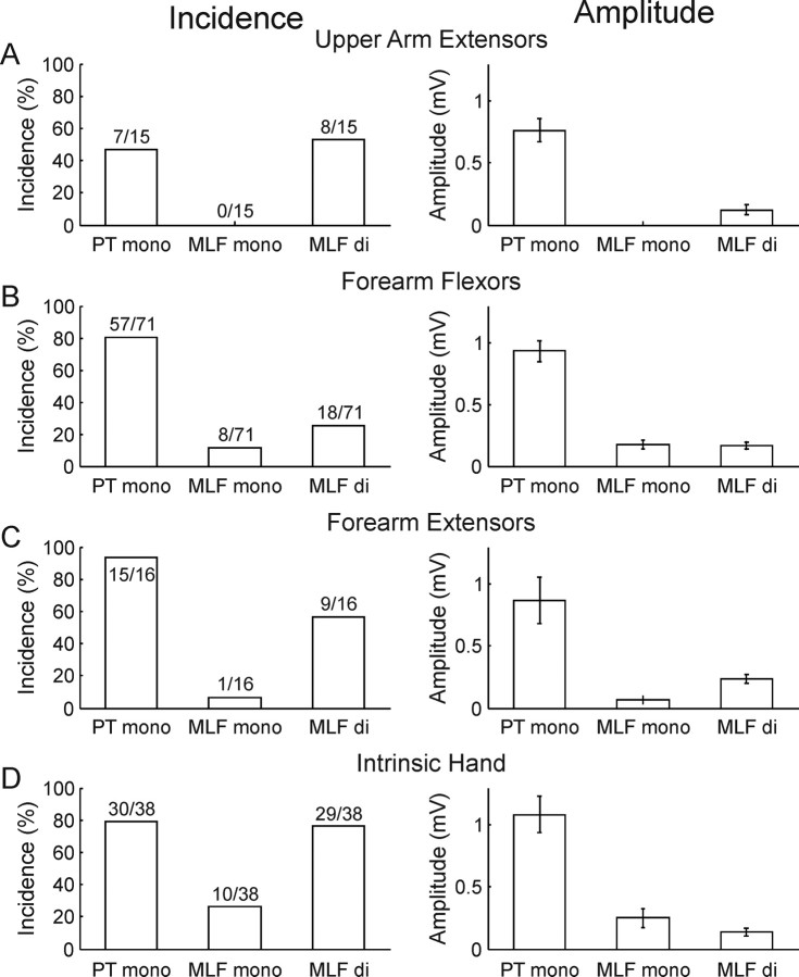
Although the reticulospinal tract is a major descending motor pathway in mammals, its contribution to upper limb control in primates has received relatively little attention. Reticulospinal connections are widely assumed to be responsible for coordinated gross movements primarily of proximal muscles, whereas the corticospinal tract mediates fine movements, particularly of the hand. In this study, we used intracellular recording in anesthetized monkeys to examine the synaptic connections between the reticulospinal tract and antidromically identified cervical ventral horn motoneurons, focusing in particular on motoneurons projecting distally to wrist and digit muscles. We found that motoneurons receive monosynaptic and disynaptic reticulospinal inputs, including monosynaptic excitatory connections to motoneurons that innervate intrinsic hand muscles, a connection not previously known to exist. We show that excitatory reticulomotoneuronal connections are as common and as strong in hand motoneuron groups as in forearm or upper arm motoneurons. These data suggest that the primate reticulospinal system may form a parallel pathway to distal muscles, alongside the corticospinal tract. Reticulospinal neurons are therefore in a position to influence upper limb muscle activity after damage to the corticospinal system as may occur in stroke or spinal cord injury, and may be a target site for therapeutic interventions.
Figures
Similar articles
-
Changes in descending motor pathway connectivity after corticospinal tract lesion in macaque monkey.Brain. 2012 Jul;135(Pt 7):2277-89. doi: 10.1093/brain/aws115. Epub 2012 May 11. Brain. 2012. PMID: 22581799 Free PMC article.
-
Corticospinal Inputs to Primate Motoneurons Innervating the Forelimb from Two Divisions of Primary Motor Cortex and Area 3a.J Neurosci. 2016 Mar 2;36(9):2605-16. doi: 10.1523/JNEUROSCI.4055-15.2016. J Neurosci. 2016. PMID: 26937002 Free PMC article.
-
Combined corticospinal and reticulospinal effects on upper limb muscles.Neurosci Lett. 2014 Feb 21;561:30-4. doi: 10.1016/j.neulet.2013.12.043. Epub 2013 Dec 31. Neurosci Lett. 2014. PMID: 24373988 Free PMC article.
-
Direct and indirect pathways for corticospinal control of upper limb motoneurons in the primate.Prog Brain Res. 2004;143:263-79. doi: 10.1016/S0079-6123(03)43026-4. Prog Brain Res. 2004. PMID: 14653171 Review.
-
Descending pathways in motor control.Annu Rev Neurosci. 2008;31:195-218. doi: 10.1146/annurev.neuro.31.060407.125547. Annu Rev Neurosci. 2008. PMID: 18558853 Review.
Cited by
-
An involuntary stereotypical grasp tendency pervades voluntary dynamic multifinger manipulation.J Neurophysiol. 2012 Dec;108(11):2896-911. doi: 10.1152/jn.00297.2012. Epub 2012 Sep 5. J Neurophysiol. 2012. PMID: 22956798 Free PMC article.
-
Mesencephalic Locomotor Region and Presynaptic Inhibition during Anticipatory Postural Adjustments in People with Parkinson's Disease.Brain Sci. 2024 Feb 15;14(2):178. doi: 10.3390/brainsci14020178. Brain Sci. 2024. PMID: 38391752 Free PMC article.
-
Cortical, Corticospinal, and Reticulospinal Contributions to Strength Training.J Neurosci. 2020 Jul 22;40(30):5820-5832. doi: 10.1523/JNEUROSCI.1923-19.2020. Epub 2020 Jun 29. J Neurosci. 2020. PMID: 32601242 Free PMC article.
-
Neural computational modeling reveals a major role of corticospinal gating of central oscillations in the generation of essential tremor.Neural Regen Res. 2017 Dec;12(12):2035-2044. doi: 10.4103/1673-5374.221161. Neural Regen Res. 2017. PMID: 29323043 Free PMC article.
-
The early release of actions by loud sounds in muscles with distinct connectivity.Exp Brain Res. 2014 Dec;232(12):3797-802. doi: 10.1007/s00221-014-4074-y. Epub 2014 Aug 21. Exp Brain Res. 2014. PMID: 25142152
References
-
- Alstermark B, Isa T, Ohki Y, Saito Y. Disynaptic pyramidal excitation in forelimb motoneurons mediated via C(3)-C(4) propriospinal neurons in the Macaca fuscata. J Neurophysiol. 1999;82:3580–3585. - PubMed
-
- Anderson ME, Yoshida M, Wilson VJ. Tectal and tegmental influences on cat forelimb and hindlimb motoneurons. J Neurophysiol. 1972;35:462–470. - PubMed
-
- Baldissera F, Hultborn H, Illert M. Integration in spinal neuronal systems. In: Brookhart JM, Mountcastle VB, editors. Handbook of physiology—the nervous system II. Bethesda, MD: American Physiological Society; 1981. pp. 509–595.
Publication types
MeSH terms
Grants and funding
LinkOut - more resources
Full Text Sources
Other Literature Sources