Comparative Study
. 2009 Jun;150(2):700-9.
doi: 10.1104/pp.109.136853. Epub 2009 Apr 15.

A comparative study of the involvement of 17 Arabidopsis myosin family members on the motility of Golgi and other organelles

Affiliations
Comparative Study

A comparative study of the involvement of 17 Arabidopsis myosin family members on the motility of Golgi and other organelles

Dror Avisar et al. Plant Physiol. 2009 Jun.

Abstract

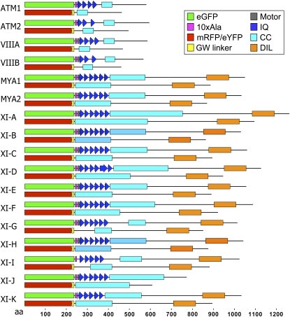
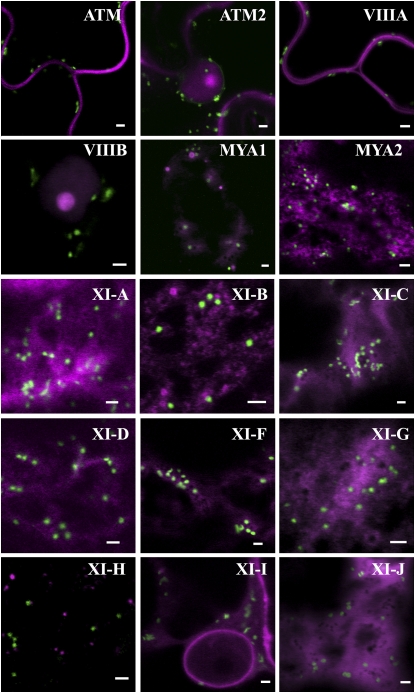
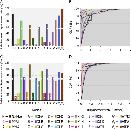
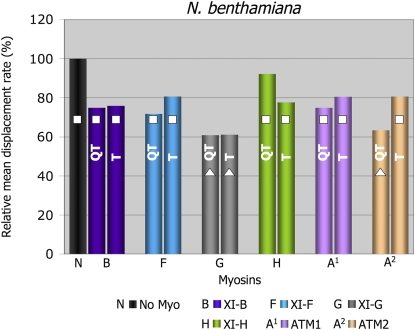
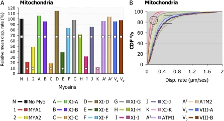
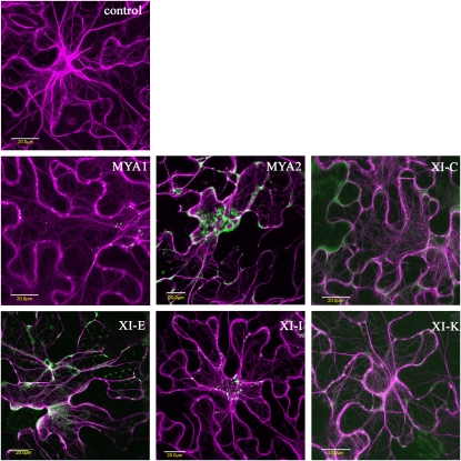
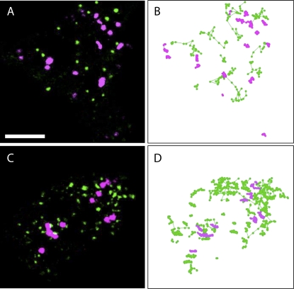
Gene families with multiple members are predicted to have individuals with overlapping functions. We examined all of the Arabidopsis (Arabidopsis thaliana) myosin family members for their involvement in Golgi and other organelle motility. Truncated fragments of all 17 annotated Arabidopsis myosins containing either the IQ tail or tail domains only were fused to fluorescent markers and coexpressed with a Golgi marker in two different plants. We tracked and calculated Golgi body displacement rate in the presence of all myosin truncations and found that tail fragments of myosins MYA1, MYA2, XI-C, XI-E, XI-I, and XI-K were the best inhibitors of Golgi body movement in the two plants. Tail fragments of myosins XI-B, XI-F, XI-H, and ATM1 had an inhibitory effect on Golgi bodies only in Nicotiana tabacum, while tail fragments of myosins XI-G and ATM2 had a slight effect on Golgi body motility only in Nicotiana benthamiana. The best myosin inhibitors of Golgi body motility were able to arrest mitochondrial movement too. No exclusive colocalization was found between these myosins and Golgi bodies in our system, although the excess of cytosolic signal observed could mask myosin molecules bound to the surface of the organelle. From the preserved actin filaments found in the presence of enhanced green fluorescent protein fusions of truncated myosins and the motility of myosin punctae, we conclude that global arrest of actomyosin-derived cytoplasmic streaming had not occurred. Taken together, our data suggest that the above myosins are involved, directly or indirectly, in the movement of Golgi and mitochondria in plant cells.


Figures

Figure 1.
Figure 1.
Schematic representation of the eGFP/mRFP-myosin fusions used in this work. A fragment containing the IQ tail domain of each of the 17 Arabidopsis annotated myosin cDNAs was fused to the C terminus of eGFP with a linker of 10×Ala. In some of the constructs, a few amino acids from the motor domain were included. The tail fusions did not contain the IQ domain and were fused using Gateway (GW) to either mRFP or eYFP. CC, Coiled coil; DIL, Dilute.
Figure 2.
Figure 2.
The subcellular localization patterns of Golgi and Arabidopsis myosin tail fragments in N.t. Myosin tail fragments fused to mRFP (magenta) were coexpressed with a Golgi marker (ST-GFP; green) in N.t. leaf epidermal cells. See Sparkes et al. (2008) for images of XI-E and XI-K tail fusions coexpressed with a Golgi marker. Bars = 2 μm.
Figure 3.
Figure 3.
Displacement rates of Golgi bodies in the presence of 17 Arabidopsis myosin tail fusions in N.t. (A and B) and myosin IQ tail fusions in N.b. (C and D). Using Volocity software, displacement rates were calculated for Golgi bodies in the presence of myosin tail fusions (A and B) or myosin IQ tail fusions (C and D). Values are expressed as percentages of the control displacement rate of Golgi bodies in each system. A and C, Plots of mean displacement. B and D, Cumulative distribution frequency (CDF) plots. Columns or curves overlaid with different shapes are statistically different at P < 0.05. (Data for XI-E and XI-K tail fusions in N.t. are from Sparkes et al. [2008].)
Figure 4.
Figure 4.
Displacement rates of Golgi in the presence of tail or IQ tail fusions in N.b. The myosin tail fusions that gave different results in N.t. compared with their IQ tail counterparts in N.b. were expressed in N.b., and the mean displacement rates were calculated. Columns overlaid with different shapes are statistically different at P < 0.05.
Figure 5.
Figure 5.
Displacement rates for mitochondria in the presence of IQ tail truncations of all 17 Arabidopsis myosins in N.b. leaves. All 17 myosin IQ tail fusions were coexpressed with mitochondria markers in N.b. leaves. Abaxial epidermal cells were analyzed by confocal microscopy 48 h after infiltration. Time-lapse image acquisition was performed, and organelle tracking was performed by Volocity. A, The mean displacement (disp.) rates were normalized to control organelle displacement in the absence of myosin mutant and plotted here as percentage of control (100%). B, Cumulative distribution frequency (CDF) plots showing the behavior of organelle populations in the presence of the indicated myosins.
Figure 6.
Figure 6.
Actin fibers are unaffected in the presence of the inhibitory eGFP-myosin mutants. An actin marker, DsRED-FABD2, was coexpressed with eGFP myosin fusions in N.b. leaves. Confocal images of abaxial epidermal cells are shown. Actin is not disrupted in the presence of the myosin mutants. Bars = 20 μm.
Figure 7.
Figure 7.
Tracking of both MYA1 and MYA2 punctae as well as Golgi stacks in the same cell. To demonstrate that arresting organelle motility was not accompanied by a general arrest of cytoplasmic streaming, both MYA1 or MYA2 and Golgi stacks were tracked in the same leaf. eGFP-IQ tail and the Golgi marker, ST-mRFP, were coexpressed in leaf abaxial epidermal cells of N.b. and observed by confocal microscopy 48 h after infiltration. A and B, MYA1 (green) and Golgi (magenta). C and D, MYA2 (green) and Golgi (magenta). A and C, The first frames of the time-lapse sequences. B and D, Tracks of Golgi (magenta) and eGFP-IQ tail puncta (green). While Golgi stacks are almost completely motionless, puncta of MYA1 and MYA2 are motile. Bar = 8 μm.

References

    1. Avisar D, Prokhnevsky AI, Dolja VV (2008. a) Class VIII myosins are required for plasmodesmatal localization of a closterovirus Hsp70 homolog. J Virol 82 2836–2843 - PMC - PubMed
    1. Avisar D, Prokhnevsky AI, Makarova KS, Koonin EV, Dolja VV (2008. b) Myosin XI-K is required for rapid trafficking of Golgi stacks, peroxisomes, and mitochondria in leaf cells of Nicotiana benthamiana. Plant Physiol 146 1098–1108 - PMC - PubMed
    1. Baluska F, Cvrckova F, Kendrick-Jones J, Volkmann D (2001) Sink plasmodesmata as gateways for phloem unloading: myosin VIII and calreticulin as molecular determinants of sink strength? Plant Physiol 126 39–46 - PMC - PubMed
    1. Baluska F, Samaj J, Hlavacka A, Kendrick-Jones J, Volkmann D (2004) Actin-dependent fluid-phase endocytosis in inner cortex cells of maize root apices. J Exp Bot 55 463–473 - PubMed
    1. Berg JS, Powell BC, Cheney RE (2001) A millennial myosin census. Mol Biol Cell 12 780–794 - PMC - PubMed

Publication types

MeSH terms