

Temporal bone imaging: comparison of flat panel volume CT and multisection CT
- PMID: 19369606
- PMCID: PMC7051536
- DOI: 10.3174/ajnr.A1560
Temporal bone imaging: comparison of flat panel volume CT and multisection CT
Abstract
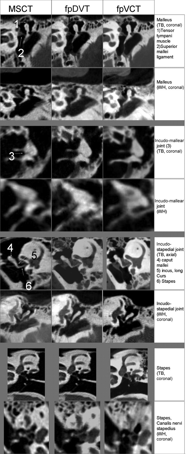
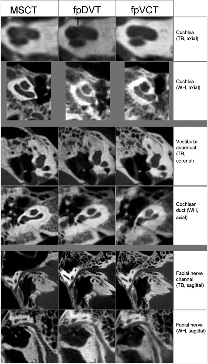
Background and purpose: A recent development in radiology is the use of flat panel detectors in CT to obtain higher-resolution images. This technique is known as flat panel volume CT (fpVCT). We sought to compare the image quality and diagnostic value of 2 different flat panel detector-equipped scanners: one is a prototype fpVCT scanner, and the other is a so-called flat panel digital volume tomography (fpDVT) scanner, which is routinely used in clinical setup with current state-of-the-art multisection CT (MSCT) scanners.
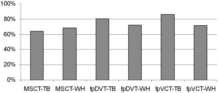
Materials and methods: Five explanted temporal bones and 2 whole-head cadaveric specimens were scanned with fpVCT, fpDVT, and MSCT scanners. The image series were blindly evaluated by 3 trained observers who rated 38 anatomic structures with regard to their delineation/appearance.
Results: Although the image quality obtained with fpVCT and fpDVT was rated significantly better compared with MSCT on isolated temporal bones, the differences were not significant when whole cadaveric heads were scanned.
Conclusions: Theoretic and practical advantages exist for flat panel detector-equipped scanners, including improved image quality. However, when imaging whole cadaveric heads, no significant difference could be demonstrated between them and standard-of-care MSCT.
Figures
References
-
- Kalender WA. Der Einsatz von Flachbilddetektoren für die CT-Bildgebung [The use of flat panel detectors for CT imaging]. Radiologe 2003;43:379–87 - PubMed
-
- Gupta R, Grasruck M, Suess C, et al. Ultra-high resolution flat panel volume CT: fundamental principles, design architecture, and system characterization. Eur Radiol 2006;16:1191–205 - PubMed
-
- Peltonen LI, Aarnisalo AA, Kortesniemi MK, et al. Limited cone-beam computed tomography imaging of the middle ear: a comparison with multislice helical computed tomography. Acta Radiol 2007;48:207–12 - PubMed
-
- Ross W, Cody DD, Hazle JD. Design and performance characteristics of a digital flat panel computed tomography system. Med Phys 2006;33:1888–901 - PubMed
Publication types
MeSH terms
LinkOut - more resources
Full Text Sources
Other Literature Sources
Medical