

Review
doi: 10.3174/ajnr.A1565.
Epub 2009 Apr 15.
Anterior-segment retinoblastoma mimicking pseudoinflammatory angle-closure glaucoma: review of the literature and the important role of imaging
Affiliations
- PMID: 19369612
- PMCID: PMC7051607
- DOI: 10.3174/ajnr.A1565
Item in Clipboard
Review
Anterior-segment retinoblastoma mimicking pseudoinflammatory angle-closure glaucoma: review of the literature and the important role of imaging
AJNR Am J Neuroradiol.
2009 Sep.
Abstract
A 7-year-old boy presented with angle-closure glaucoma, initially presumed to be idiopathic. A ciliary body mass was later detected on MR imaging, suggestive of medulloepithelioma but pathologically proved to be diffuse infiltrating retinoblastoma. We discuss the patient management and review the literature, with emphasis on the role of CT and MR imaging in evaluating pediatric angle-closure glaucoma and in influencing the management of patients with retinoblastoma and medulloepithelioma.
Figures
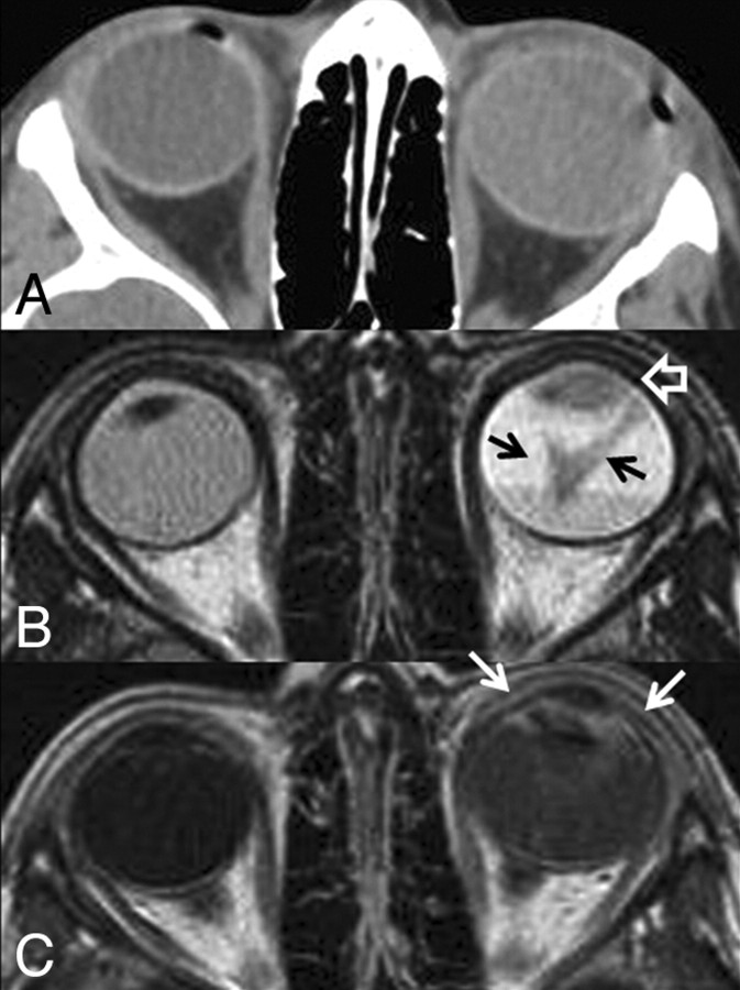
A, CT scan of the orbits. The left globe is asymmetrically enlarged and is hyperattenuating compared with the contralateral right globe, reflective of glaucoma and total retinal detachment, respectively. No calcifications are present. B, Axial T2-weighted MR image. There is total retinal detachment with a large fluid-fluid level. The detached retina is irregular and diffusely thickened (black arrows), and there is an irregular intermediate-signal-intensity mass involving the ciliary body and iris circumferentially (open white arrow). C, Axial T1-weighted postcontrast MR image. There is diffuse enhancement of the thickened detached retina and the anterior segment mass (white arrows). This constellation of findings is consistent with diffuse infiltrating retinal neoplasm extending into the anterior ocular segment. There is no evidence of optic nerve invasion or extrascleral extension.
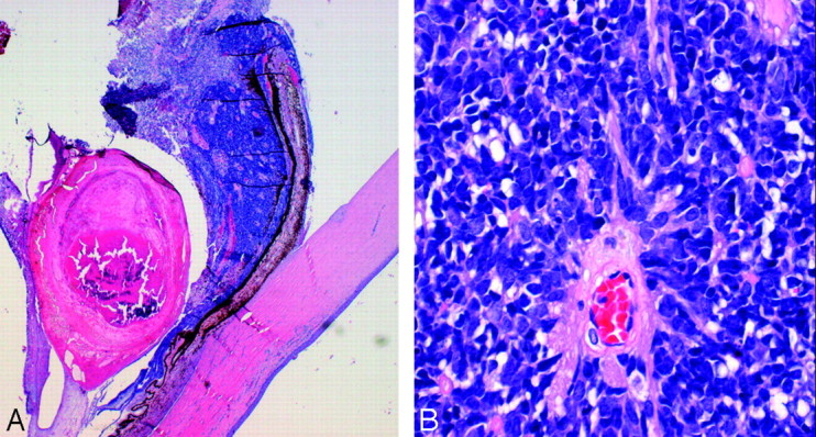
Histologic section of the enucleated left globe. A, Photomicrograph (hematoxylin-eosin [HE] stain) of the whole-mount specimen at original magnification ×2. The ciliary body is diffusely infiltrated with neoplastic cells with round basophilic nuclei. The cells form rosettes around central lumens, known as Flexner-Wintersteiner rosettes, which are characteristic of retinoblastoma. B, Photomicrograph (HE stain) of the whole-mount specimen at original magnification ×40. The neoplastic cells have round basophilic nuclei with scant cytoplasm and are arranged in a rosette around a central lumen.
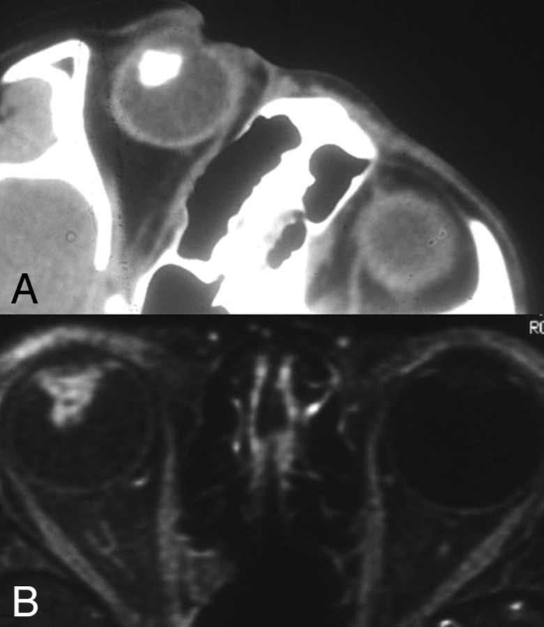
Teratoid medulloepithelioma. A, Noncontrast axial CT scan shows a densely calcified mass of the ciliary body in the anterior segment of the right globe. Teratoid medulloepitheliomas can contain dystrophic calcifications due to the presence of hyaline cartilage tissue elements. B, Axial T1-weighted fat-saturated postcontrast MR image shows an enhancing mass of the ciliary body corresponding to the area of calcification on the CT scan. The mass is confined to the ciliary body; therefore, medulloepithelioma is more likely than diffuse infiltrating retinoblastoma. Alternative diagnoses for a ciliary body mass in pediatric patients include pyogenic granuloma, juvenile xanthogranuloma, mesoectodermal leiomyoma, and ciliary body cyst.
Similar articles
-
Rare Anterior Segment Retinoblastoma Masquerading as Corneal Endotheliitis.Eye Contact Lens. 2016 Jan;42(1):e1-3. doi: 10.1097/ICL.0000000000000113. Eye Contact Lens. 2016. PMID: 25961779
-
Goniosynechialysis for the treatment of angle-closure glaucoma in retinoblastoma.Clin Exp Ophthalmol. 2023 Sep-Oct;51(7):739-742. doi: 10.1111/ceo.14271. Epub 2023 Jun 23. Clin Exp Ophthalmol. 2023. PMID: 37350374 No abstract available.
-
A magnetic resonance imaging diagnostic dilemma: diffuse infiltrating retinoblastoma versus Coats' disease.J Pediatr Ophthalmol Strabismus. 2010 Aug 23;47 Online:e1-3. doi: 10.3928/01913913-20100818-10. J Pediatr Ophthalmol Strabismus. 2010. PMID: 21158370
-
Angle closure glaucoma: a mechanistic review.Curr Opin Ophthalmol. 2011 Mar;22(2):96-101. doi: 10.1097/ICU.0b013e32834372b9. Curr Opin Ophthalmol. 2011. PMID: 21252671 Review.
-
[Diagnostic imaging of intraocular lesions in the child].Klin Monbl Augenheilkd. 1998 May;212(5):252-6. doi: 10.1055/s-2008-1034872. Klin Monbl Augenheilkd. 1998. PMID: 9677545 Review. German.
Cited by
-
Retinoblastoma: Recent trends A mini review based on published literature.Oman J Ophthalmol. 2011 Sep;4(3):108-15. doi: 10.4103/0974-620X.91265. Oman J Ophthalmol. 2011. PMID: 22279397 Free PMC article.
-
MRI of retinoblastoma.Br J Radiol. 2011 Sep;84(1005):775-84. doi: 10.1259/bjr/32022497. Br J Radiol. 2011. PMID: 21849363 Free PMC article. Review.
-
Bibliometric analysis on retinoblastoma literatures in PubMed during 1929 to 2010.Int J Ophthalmol. 2011;4(2):115-20. doi: 10.3980/j.issn.2222-3959.2011.02.01. Epub 2011 Apr 18. Int J Ophthalmol. 2011. PMID: 22553624 Free PMC article.
-
Correlation of apparent diffusion coefficient at 3T with prognostic parameters of retinoblastoma.AJNR Am J Neuroradiol. 2012 May;33(5):944-8. doi: 10.3174/ajnr.A2892. Epub 2012 Jan 12. AJNR Am J Neuroradiol. 2012. PMID: 22241394 Free PMC article.
-
Diffuse anterior retinoblastoma: current concepts.Onco Targets Ther. 2015 Jul 22;8:1815-21. doi: 10.2147/OTT.S79498. eCollection 2015. Onco Targets Ther. 2015. PMID: 26229489 Free PMC article. Review.
References
-
- Chung EM, Specht CS, Schroeder JW. Pediatric orbital tumors and tumorlike lesions: neuroepithelial lesions of the ocular globe and optic nerve. Radiographics 2007; 27: 1159– 86 - PubMed
-
- Vajaranant TS, Mafee MF, Kapur R, et al. Medulloepithelioma of the ciliary body and optic nerve: clinicopathologic, CT, and MR imaging features. Neuroimaging Clin N Am 2005; 15: 69– 83 - PubMed
-
- Shields JA, Eagle RC, Jr, Shields CL, et al. Congenital neoplasms of the nonpigmented ciliary epithelium (medulloepithelioma). Ophthalmology 1996; 103: - PubMed
-
- Karcioglu ZA, Abboud EB, Al-Mesfer SA, et al. Retinoblastoma in older children. J AAPOS 2002; 6: 26– 32 - PubMed
-
- Bhatnagar R, Vine AK. Diffuse infiltrating retinoblastoma. Ophthalmology 1991; 98: 1657– 61 - PubMed
Publication types
MeSH terms
LinkOut - more resources
Full Text Sources
Medical